Diagn Interv Radiol 2014; 20:65–71
© Turkish Society of Radiology 2014
Adrenal venous sampling for stratifying patients for surgery of adrenal nodules detected using dynamic contrast enhanced CT
Jin Young Kim, See Hyung Kim, Hee Jung Lee, Young Hwan Kim, Mi Jeong Kim, Seung Hyun Cho INTERVENTIONAL RADIOLOGY
ORIGINAL ARTICLE
PURPOSE
We aimed to assess the value of adrenal venous sampling (AVS) for diagnosing primary aldosteronism (PA) subtypes in patients with a unilateral nodule detected on adrenal com- puted tomography (CT) and scheduled for adrenalectomy.
MATERIALS AND METHODS
This retrospective study included 80 consecutive patients with PA undergoing CT and AVS. Different lateralization in- dices were assessed, and a cutoff established using receiv- er operating characteristic curve analysis. The value of CT alone versus CT with AVS for differentiating PA subtypes was compared. The adrenalectomy outcome was assessed, and predictors of cure were determined using univariate analysis.
RESULTS
AVS was successful in 68 patients. A cortisol-corrected al- dosterone affected-to-unaffected ratio cutoff of 2.0 and af- fected-to-inferior vena cava ratio cutoff of 1.4 were the best lateralization indices, with accuracies of 82.5% and 80.4%, respectively. CT and AVS diagnosed 38 patients with aldo- sterone-producing adenomas, five patients with unilateral adrenal hyperplasia, and 25 patients with bilateral adrenal hyperplasia. Of the 52 patients with a nodule detected on CT, subsequent AVS diagnosed bilateral adrenal hyperplasia in 14 patients (27%). Compared to the results of combining CT with AVS, the accuracy of CT alone for diagnosing aldoste- rone-producing adenomas was 71.1% (P < 0.001). The cure rate for hypertension after adrenalectomy was 39.2%, with improvement in 53.5% of patients. On univariate analysis, predictors of persistent hypertension were male gender and preoperative systolic blood pressure.
CONCLUSION
To avoid inappropriate surgery, AVS is necessary for diagnos- ing unilateral nodules with aldosterone hypersecretion de- tected by CT.
P rimary aldosteronism (PA) is the most common form of secondary hypertension, with a prevalence of 5%–11% (1–3). PA is due pri- marily to the hypersecretion of aldosterone by an aldosterone-pro- ducing adenoma (APA) or unilateral (primary) adrenal hyperplasia (UAH), which constitute 30%–40% of cases; the remainder are presumed to be secondary to idiopathic bilateral adrenal hyperplasia (BAH) (1, 4, 5). APA and UAH are two forms of unilateral aldosterone hypersecre- tion, and both are curable with adrenalectomy. BAH induces bilateral aldosterone hypersecretion, and anti-aldosterone drugs are used in its medical management (5–7).
The plasma aldosterone-to-renin ratio is used to screen for PA in pa- tients at high risk for PA (8). Recent guidelines recommend using com- puted tomography (CT) of the adrenal gland to categorize the subtype after confirming PA. However, CT cannot reliably visualize a microade- noma or distinguish between an incidentaloma or BAH and APA. It has been suggested that adrenal venous sampling (AVS) be performed to de- termine the subtype of PA and to differentiate between unilateral and bilateral production of aldosterone preoperatively (9). AVS to measure the adrenal vein aldosterone and cortisol is the gold standard for later- alizing aldosterone secretion (10). Lateralization is defined using several ratios. In patients with APA or UAH, a unilateral adrenalectomy results in a complete cure or improved hypertension and potassium normalization in approximately 30% of patients, with reported rates up to 86% (11–15).
This study assessed several lateralization ratios to establish the most predictive of unilateral disease. We also compared the CT results with those of bilateral AVS for differentiating the PA subtype, with the as- sumption that AVS is necessary before surgery, even in patients with nodules <10 mm detected with CT. Finally, we assessed the outcomes of adrenalectomy in our patients to identify preoperative predictors of a good outcome.
Materials and methods Patient population
The records of consecutive patients referred to Keimyung University Dongsan Hospital, Endocrinology Department for suspected PA between January 2004 and June 2012 were reviewed retrospectively. A preliminary diagnosis of PA was based on clinical suspicion, including severe hyper- tension (blood pressure [BP] >180/110 mmHg despite drug treatment or drug resistance), hypertension with hypokalemia (serum potassium <3.6 mmol/L), or hypertension with an incidental adrenal nodule (9). Diuretics, beta-blockers, and antagonists of the renin-angiotensin system were with- held for two weeks, and aldosterone antagonists were stopped six weeks
From the Department of Radiology (J.Y.K. kseehdr@dsmc.
or.kr, S.H.K., H.J.L., Y.H.K., M.J.K.), Dongsan Hospital, Keimyung University College of Medicine, Daegu, Republic of Korea;
the Department of Radiology (S.H.C.), Kyungpook National University School of Medicine, Daegu, Republic of Korea.
Received 2 April 2013; revision requested 14 May 2013; revision received 5 June 2013; accepted 10 June 2013.
Published online 13 September 2013.
DOI 10.5152/dir.2013.13144
before screening for PA. All patients un- derwent a saline suppression test after withdrawing interfering medications.
A serum aldosterone >137 pM after in- fusing 2 L of 0.9% saline confirmed PA (16). All patients who were candidates for surgery underwent CT and AVS.
Definition of primary aldosteronism To interpret the results of AVS, an abnormal adrenal gland was defined based on the absolute aldosterone lev- el or the cortisol-corrected aldosterone (Aldo/Cort). For CT, lateralization was defined as a unilateral adenoma (≥10 mm) with a completely normal con- tralateral gland, based on the possible cutoff for adrenalectomy without the use of AVS (15, 17).
Surgery was indicated when patients had clear lateralization on AVS and concordant CT. However, before mak- ing this decision, an affected-to-unaf- fected aldosterone ratio >2.2 with an unaffected-to-inferior vena cava (IVC) ratio <1.7 were used to define lateral- ization (18). Concordance was defined as CT showing a normal gland contra- lateral to the aldosterone lateralization.
The gland to which the aldosterone lateralizes might have an abnormali- ty of any size or might indeed appear normal, but be harboring a very small adenoma undetected by CT.
Based on the CT and AVS results, the PA was classified, and APA was defined using concordant results between the unilateral nodule on CT and later- alized aldosterone production as as- sessed by AVS. UAH was defined as a normal gland or unilateral hyperplasia on CT and lateralized aldosterone pro- duction as assessed by AVS. BAH was defined by normal glands or bilateral CT abnormalities and bilateral aldoste- rone secretion on AVS, or bilateral CT abnormalities and lateralized aldoste- rone secretion on AVS.
After adrenalectomy, the patients were grouped by their BP as either cured, improved, or no improvement.
Cure was defined as a BP <140/90 mmHg with no antihypertensives. Im- provement was defined as a BP <140/90 mmHg on the same or a reduced dose of antihypertensives. Those with no improvement met neither of these two criteria. Outcome was assessed as the best BP measured between 120 and 365 days after the adrenalectomy (19).CT
Adrenal CT was performed using various multidetector scanners (Sensa- tion 16, 64; Somatom Definition Flash, Siemens, Erlangen, Germany). CT was performed at 3–5 mm collimation, 3–5 mm reconstruction intervals, 120 kVp, 250 mA, and a 1:1 table pitch. A max- imum of 150 mL of nonionic contrast medium was injected intravenously at 2–3 mL/s with a power injector. Unen- hanced and enhanced CT was acquired at 60 s and 15 min, respectively. The adrenal CT appearance was described as normal, hyperplasia when the gland volume was high or if its edges had lost their concave shape, and nod- ular when a circular adrenal lesion was detected. An adrenal adenoma was defined on CT using the follow- ing criteria: a nodular lesion that was negative or 10 Hounsfield units less on unenhanced CT and a homogeneous enhancing nodular lesion with an ab- solute percentage loss of enhancement
>60% (20, 21).
Adrenal venous sampling
After obtaining informed consent, bilateral AVS was performed by one ex- perienced vascular radiologist over the 10-year period using a percutaneous right transfemoral vein approach. Var- ious 4 or 5 F glide catheters were used to catheterize both adrenal veins, and the catheter placement was confirmed fluoroscopically before sampling by injecting a small amount of contrast.
Samples for measuring the serum al- dosterone and cortisol were collected from both adrenal veins and the IVC.
Accurate catheterization of the adrenal veins was assumed when the ratio of the cortisol levels in the adrenal vein to the IVC was >2.0.
Hormone assays
The serum and urine aldosterone concentrations were measured by ra- dioimmunoassay (Abbott Architect, Maidenhead, UK) with a coefficient of variation of 9.5%. The plasma renin activity was measured by immunoche- miluminometry (Liaison, DiaSorin, Dartford, UK) with a coefficient of variation of 6%. The plasma cortisol was measured using immunochemilu- minometry (Cobas-6000, Indianapo- lis, Indiana, USA) with a coefficient of variation of 4.5%.
Statistical analysis
All data are shown as the mean±- standard deviation. Patients with uni- lateral (APA and UAH) and bilateral (BAH) findings were compared using an unpaired Student’s t test. Categor- ical variables were compared using the chi-square test.
A receiver operating characteris- tic (ROC) curve analysis of published lateralization ratios and novel ratios was used to assess the efficacy of each method and to establish appropriate cutoff values. The ratio of the affect- ed to unaffected side and unaffect- ed-to-peripheral ratio were analyzed.
The sensitivity, specificity, and pre- dictive values of CT alone for the diag- nosis of APA were calculated and com- pared to results combining CT with AVS. The positive predictive values ac- cording to nodule size were compared using Fisher’s exact test. The accuracy of CT alone and CT combined with AVS was compared using McNemar’s test. The statistical power for detecting a difference with 72.4%–100% accura- cy using a sample of 68 paired observa- tions exceeded 99%.
Univariate analysis was performed on all possible variables of hyperten- sive cure using the paired Student’s t test and chi-square test for continuous and categorical variables, respectively.
Statistical analyses were per-formed using a computer software (Statistical Package for Social Sciences, Version 17.0, SPSS Inc., Chicago, Illinois, USA).
P values that were less than 0.05 was deemed to indicate statistical signifi- cance.
Results
Baseline patient characteristics
Eighty patients with PA were identi-
fied, all of whom underwent adrenal
CT and AVS. The mean patient age
was 46 years (range, 22–70 years) and
52 were males (65%). The mean systol-
ic and diastolic BP were 155±5.7 and
93±2.4 mmHg, respectively. Drug-re-
sistant hypertension was observed in
48 patients (60%), and there were no
local or general complications during
or after AVS. Cortisol levels were de-
tectable in 68 patients with bilateral
AVS (85%), while the results were in-
complete in 12 patients (15%) due to
difficulty catheterizing the right adre-
nal vein; the latter were treated medi- cally. The diagnosis was APA in 38 pa- tients, presumed BAH in 30, and UAH in five.
No significant differences in age, gender, systolic or diastolic BP, dura- tion of hypertension, family history of hypertension, plasma renin activity, or urinary aldosterone were observed in patients with APA, UAH, or BAH (Table 1). The potassium levels were slightly lower in the APA group, al- though this was not significant (APA/
UAH, 2.9±0.1 mmol/L vs. BAH, 3.1±0.1 mmol/L; P = 0.085). The plasma al- dosterone level and plasma aldoste- rone-to-renin ratio did not differ sig- nificantly between the two groups.
Lateralization ratios
Only those patients with successful bilateral adrenal sampling were in- cluded. We compared the published lateralization ratios in patients with unilateral and bilateral disease and preformed ROC curve analysis for each ratio (Table 2). Only those ratios in which the area under the curve (AUC) was highly significant were included in further analysis. The AUC was high- est for Aldo/Cort A:U (AUC=0.882, P = 0.0007) and Aldo/Cort U:IVC (AUC=0.851, P = 0.0009). The opti- mum cutoff for lateralization was con- sidered for these two ratios. The cutoff values for Aldo A:IVC (P = 0.825) and Aldo/Cort A:IVC (P = 0.093) were not significant.
The ROC curve for Aldo/Cort A:U had a cutoff >2.0, giving the optimum sensitivity with minimal loss of speci- ficity. Using cross-tabulation, the sen- sitivity was 100% (43/43 APA or UAH), specificity 56.0% (14/25 BAH), and diagnostic accuracy 82.5%. For Aldo/
Cort U:IVC, a cutoff <1.4 gave an op- timum sensitivity of 95.3% (41/43 APA or UAH), specificity of 48.0% (12/25 BAH), and diagnostic accuracy of 80.2%.
CT findings of the adrenal gland
Fifty-two patients (76.4%) were diag- nosed with a unilateral nodular abnor- mality, of whom 30 patients (55.6%) had a nodule at least 10 mm, four (5.8%) had unilateral hyperplasia, six (8.8%) had bilateral abnormalities, and six (8.8%) had a normal CT.
Diagnosis of aldosterone-producing adeno- ma using CT and adrenal venous sampling Using the CT and AVS results, APA was diagnosed in 38 patients (55.8%) (Table 3, Fig.). Of these, 28 APA under- went an adrenalectomy, and adenomas were confirmed in 25 patients histo- logically. In patients 6, 11, and 28, the histology showed adrenal hyperplasia.
Patients 6 and 11 had a nonsignificant Aldo/Cort A:U of 1.6 and 1.4 and an Aldo/Cort U:IVC of 2.1 and 2.6, re- spectively. Nevertheless, they request- ed surgery to try to cure hypokalemia.
Patient 28 had severe, drug-resistant hypertension, which warranted radi- cal treatment despite the small nodule size and hyperplastic nature of the ad- renal gland as assessed by CT. Twelve patients with APA preferred medical treatment and an annual follow-up to immediate surgery.
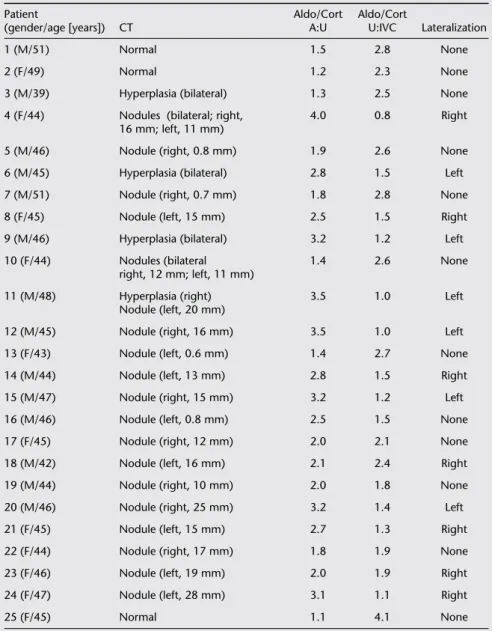
Diagnosis of unilateral adrenal hyperplasia using CT and adrenal venous sampling
Five patients (7.4%) were diagnosed with UAH, three of whom had a nor- mal CT, and two had unilateral adre- nal hyperplasia (Table 3, Fig.). All three patients have been treated medically in our institution, with annual CT and laboratory follow-up.
Diagnosis of bilateral adrenal hyperplasia using CT and adrenal venous sampling
Benign adrenal hyperplasia was di- agnosed in 25 patients (36.7%). The mean Aldo/Cort A:U was significantly higher in APA patients than in BAH pa- tients (APA 3.2±0.5 vs. BAH 2.1±0.4, P
< 0.001), and the Aldo/Cort U:IVC was significantly lower (APA 0.9±0.2 vs.
BAH 1.9±0.3, P < 0.001). Three patients had normal CT and six had bilateral abnormalities (Table 4).
Table 1. Demographics of patients with unilaterality and bilaterality in primary aldosteronism
APA/UAH (n=43) BAH (n=25) P
Age (years) 46.3±1.8 48.2±1.5 0.086
Gender (male/female) 25/18 14/11 0.863
Systolic BP at referral (mmHg) 153.2±3.3 156.8±3.0 0.158 Diastolic BP at referral (mmHg) 95.4±2.5 92.5±2.1 0.330 Duration of hypertension (years) 7.25±0.8 8.0±1.0 0.072 Family history of hypertension 15/27 (55.5%) 10/20 (50.0%) 0.834
Plasma potassium (mmol/L) 2.9±0.1 3.1±0.1 0.085
Plasma renin activity (ng/L) 6.0±1.5 7.0±1.5 0.081
Plasma aldosterone (ng/100 mL) 26.0±4.5 22.5±3.5 0.076
Plasma aldosterone-to-renin ratio 112.5±34.7 88.5±23.8 0.061
Urinary aldosterone (mg/day) 22.5±3.5 20.3±2.8 0.090
APA, aldosterone producing adenoma; BAH, bilateral adrenal hyperplasia; BP, blood pressure; UAH, unilateral adrenal hyperplasia.
Data are given as mean±standard deviation or n (%).
Table 2. Results of ROC curve analysis of different lateralization indices
Lateralization index AUC P
Aldo A:U 0.705 0.019
Aldo U:IVC 0.740 0.007
Aldo A:IVC 0.470 0.825
Aldo/Cort A:U 0.882 < 0.001
Aldo/Cort U:IVC 0.850 < 0.001
Aldo/Cort A:IVC 0.652 0.093
A, affected adrenal gland; Aldo, aldosterone; Aldo/Cort, cortisol corrected aldosterone; AUC, area under
the curve; IVC, inferior vena cava; ROC, receiver operating characteristic; U, unaffected adrenal gland.
Sixteen (64%) patients had a unilat- eral nodular CT abnormality. Of these, 12 (75%) had nodules ≥10 mm, and four patients had nodules <10 mm.
Twelve patients had Aldo/Cort A:U
>2.2 and Aldo/Cort U:IVC <1.7, but bilateral CT abnormalities in three pa- tients or a contralateral CT nodule in nine patients led to a diagnosis of BAH.
Assessment of unilateral adrenalectomy using CT and the adrenal venous sampling results
Fifty-two patients (76.4%) had a unilateral nodule on CT. Of the pa- tients undergoing AVS, 38 (73.0%) were confirmed to have APA, and 14 (27.0%) were diagnosed with BAH due to bilateral or contralateral aldosterone production. Fifteen of these 18 pa- tients (83.3%) had a nodule ≥10 mm on CT. The patients were referred for a unilateral adrenalectomy using the above criteria. Compared to CT com- bined with AVS (Cort A:U >2.0, Aldo/
Cort U:IVC <1.4) to diagnose APA, CT alone had 100% sensitivity (38/38), 42.8% (6/14) specificity, 65.6% pos- itive predictive value (21/32), 100%
negative predictive value (20/20), and 71.1% (37/52) diagnostic accuracy for APA, which differed significantly (P
< 0.001) from the 98.0% (51/52) di- agnostic accuracy obtained when CT was combined with AVS. Overall, most patients would have been classified ap- propriately as having either unilateral or bilateral disease.
A unilateral adenoma ≥10 mm with a completely normal contralateral gland has been suggested as a criterion for surgery (22). In comparison, the pos- itive predictive value of CT alone for diagnosing APA with nodules ≥10 mm was no different from that for small nodules detected using CT (65% vs.
75%; P = 0.900).
Outcome after adrenalectomy
Of the 38 patients with unilateral disease, 35 had long-term outcome data. Of the remaining three patients, follow-up records were unavailable for two, while the remaining patient awaits review after a recent adrenalec- tomy. After adrenalectomy, the hyper- tension was cured in 39.2% (11/28) of the patients and improved in 53.5%
(15/28). Two patients did not meet Table 3. Characteristics of aldosterone-producing adenoma and unilateral adrenal hyper-
plasia using CT and adrenal venous sampling
Patient Aldo/Cort Aldo/Cort
(gender/age [years]) CT A:U U:IVC Lateralization
Aldosterone-producing adenoma
1 (M/39) Nodule (right, 6 mm) 2.8 1.2 Right
2 (F/40) Nodule (right, 10 mm) 3.0 1.1 Right
3 (M/42) Hyperplasia (left, 7 mm) 3.2 1.0 Left
4 (F/44) Nodule (left, 16 mm) 3.5 0.8 Left
5 (M/47) Hyperplasia (right) 3.1 1.1 Right
6 (M/48) Nodule (left, 8 mm) 1.6 2.1 Left
7 (M/38) Nodule (right, 15 mm) 5.5 0.4 Right
8 (F/42) Nodule (left, 8 mm) 2.5 1.4 Left
9 (M/43) Nodule (left, 10 mm) 3.8 0.7 Left
10 (M/41) Nodule (left, 25 mm) 6.2 0.1 Left
11 (F/49) Nodule (right, 18 mm) 1.4 2.6 Right
12 (M/44) Nodule (left, 15 mm) 5.2 0.3 Left
13 (F/38) Nodule (right, 7 mm) 3.5 0.7 Right
14 (M/41) Nodule (left, 16 mm) 4.2 0.5 Left
15 (M/40) Nodule (right, 15 mm) 2.4 1.6 Right
16 (F/42) Nodule (left, 22 mm) 5.8 0.3 Left
17 (M/45) Nodule (left, 24 mm) 6.2 0.2 Left
18 (F/46) Nodule (left, 14 mm) 2.5 1.5 Left
19 (M/44) Nodule (right, 12 mm) 2.9 1.4 Right
20 (F/43) Nodule (left, 8 mm) 3.5 1.2 Left
21 (M/41) Nodule (left, 16 mm) 4.2 0.7 Left
22 (F/38) Nodule (left, 25 mm) 7.5 0.1 Left
23 (M/50) Nodule (right, 26 mm) 2.5 1.4 Right
24 (M/49) Nodule (left, 32 mm) 2.4 1.5 Left
25 (F/47) Nodule (left, 28 mm) 2.8 1.4 Left
26 (F/44) Nodule (left, 7 mm) 3.5 1.1 Left
27 (M/42) Nodule (left, 15 mm) 3.2 1.2 Left
28 (M/41) Nodule and hyperplasia (left, 4 mm) 4.0 1.0 Left 29 (M/40) Nodule and hyperplasia (left, 6 mm) 4.2 1.0 Left
30 (F/44) Nodule (left, 18 mm) 3.5 1.2 Left
31 (F/45) Nodule (left, 24 mm) 3.1 1.3 Left
32 (F/44) Nodule (right, 28 mm) 3.8 1.4 Right
33 (M/43) Nodule (left, 26 mm) 2.4 1.6 Left
34 (F/42) Nodule (left, 24 mm) 5.2 0.9 Left
35.M/45 Nodule (right, 20 mm) 6.5 0.3 Right
36 (M/46) Nodule (left, 10 mm) 2.3 1.5 Left
37 (F/48) Nodule (left, 15 mm) 4.5 0.7 Left
38 (M/45) Nodule (right, 16 mm) 4.2 0.9 Right
Unilateral adrenal hyperplasia
1 (F/44) Normal 4.5 0.5 Left
2 (M/43) Hyperplasia (left) 5.8 0.3 Left
3 (M/41) Normal 4.0 0.8 Right
4 (M/42) Hyperplasia (right) 2.4 1.4 Right
5 (F/45) Normal 3.5 1.2 Left
A, affected adrenal gland; Aldo, aldosterone; Aldo/Cort, cortisol corrected aldosterone; F, female; IVC,
inferior vena cava; M, male; U, unaffected adrenal gland.
the criteria for cured or improved hy- pertension, although they had postad- renalectomy BPs of 140/90 mmHg on three antihypertensives, which was a decrease from 175/115 mmHg preop- eratively on five agents. The potassium normalized in all patients from a mean of 3.0±0.1 to 4.8±0.1 mmol/L (P <
0.001). BP improved substantially from 152.2/93.4±3.2/2.4 preoperatively to 128.1/82.2±1.7/1.2 postoperatively (P
< 0.001), with a reduction in the num- ber of antihypertensives taken. The predictors of persistent hypertension in the univariate analyses are summa- rized in Table 5. Significant predictors included male gender (P = 0.025) and preoperative systolic BP (P = 0.012).
Discussion
In this study, AVS showed bilateral or contralateral aldosterone production in 27% of patients with PA with a unilater- al nodule on CT. Focusing on patients with nodules ≥10 mm in diameter, 35%
had similar bilateral or contralateral al- dosterone production. Therefore, the accuracy of CT alone was inadequate for diagnosing APA, even for large nodules.
Consequently, we examined the effi- ciency of AVS for reliably categorizing nodules detected by CT as PA and iden- tifying the need for surgery.
A number of studies have applied various lateralization methods to pa- tients in whom bilateral AVS was suc- cessful. In our series, Aldo/Cort A:U
>2.0 and Aldo/Cort U:IVC <1.4 gave the best diagnostic accuracy of 82.5%
and 80.2%, respectively, with 56.0%
and 48.0% specificity. This is similar to the cutoff based on the Endocrinon- olgy Society guidelines (9, 22–24). The absolute aldosterone concentration was not as useful as the cortisol-cor- rected values, which correct for the dilutional effects of the inferior phren- ic vein flow into the left adrenal vein and for IVC blood when sampling at or near the orifice of the right adrenal vein (12–14, 25). In addition, the con- tralateral-gland-to-IVC ratio was accu- rate, suggesting that not all patients with unilateral disease have complete contralateral aldosterone suppression.
We found that adrenal CT was also insufficient for differentiating PA sub- types. In our study, CT correctly cate- gorized 71.1% of the patients. A recent Table 5. Univariate analysis of preoperative predictors of postadrenalectomy hypertension
Cure (n=11) No cure (n=17) P
Age (years) 43.8±2.7 46.2±2.5 0.131
Gender (male/female) 3/8 12/5 0.025
Preoperative systolic BP (mmHg) 155.2±4.5 158.8±3.6 0.01 Preoperative diastolic BP (mmHg) 93.4±4.3 95.5±2.7 0.630
Size of nodules (mm) 2.0±0.3 1.4±0.2 0.142
Family history of hypertension 3 9 0.248
Duration of hypertension (years) 4.17±1.3 6.4±0.9 0.196
Plasma potassium (mmol/L) 3.0±0.1 2.7±0.1 0.158
BMI (kg/m
2) 27.1±1.5 28.8±1.4 0.208
Aldo postsaline suppression test
a648.5±109.1 528.6±78.5 0.309
Number of antihypertensives 2.1±0.3 2.5±0.3 0.311
a