하악골비대칭 환자의 형태학적 특징에 관한 연구
1)한양대학교 의과대학 치과학 교실 대학원생
2)한양대학교 의과대학 치과학 교실 조교수
이 태 희1)․유 임 학2)
M orphological characteristics of the mandibular asymm etry in adult patients
Tae-Hee Lee, Eem-Hak Yoo
Department of Dentistry, College of Medicine, Hanyang University 17, Haengdang-Dong, Sungdong-Gu, Seoul 133-792, Korea
Morphological characteristics of the asymmetric mandible in 135 adult male patients were investigated. Panoramic X-ray images were recorded. Age, ramus height, antegonial notch height, condylar height, coronoid process height, ramus body height and condylar neck angle were calculated and measured. Statistical analysis of unpaired two-tailed t-test and correlation coefficients was performed to find the morphologic differences between short ramus height side and the other.
Condylar height and ramus body height in short ramus height side were smaller than those of the other side. Antegonial notch height, coronoid process height, condylar neck angle showed no differences. Condylar neck height was correlated with condylar neck angle.
Key words : mandibular asymmetry, morphological characteristics.
Ⅰ 서. 론
안면비대칭(facial asymmetry)은 안모를 이루는 두 개(calvarium), 두개저(skull base), 악안면골의 위치 및 형태 연조직의 두께 차이 등에 의해 발생된다, 1. 안면비대칭의 원인으로는 hemifacial microsomia, hemifacial hypertrophy, juvenile rheumatoid arthritis, condylar hyperplasia, cleft lip and palate, holoprosen- cephaly, neurofibromatosis, mandibular fracture 등이 (condylar fracture), drifting and tipping of teeth 보고되었다2.
대칭성이 두부에서의 가장 명백한 형태학적 특징 이라 보고되었고 인체의 구조적 기능적 비대칭은, 자연적 현상이며 인체의 기본상태라 하였다, 3-6. 최 초의 비대칭에 관한 연구는 1887년에 예술가인 에 의해 발표되었으며 초기 그리스의 조각품 Hasse
을 연구한 결과 이들이 다양한 정도의 안면비대칭, 을 나타낸다고 기술하였다7. 이집트 왕가의 두개골 을 직접 계측한 연구에서는 인간의 두개 안면부의 비대칭이 존재한다고 보고되었고 뇌의 우측반구의 발생이 우세하여 전반적으로 안모의 우측이 좌측보 다 크다고 하였다8. 또한 우측 두개골이 더 크며 상 악과 하악의 정중선을 맞추기 위하여 비대칭이 발 생된다는 보고도 있다9. 근신경계의 기능적 요구에 의해 안면 비대칭이 발생될 수 있다 기능적인 이유. 에 의한 뇌의 비대칭적인 발달이나 편측 저작과 같 은 비대칭적인 근육습관에 의해 안면비대칭이 발생 될 수 있으며 대부분의 사람이 왼쪽보다는 주로 오 른쪽으로 저작하기 때문에 우측이 더 크게 발달된
다고 보고되었다10,11.
안면비대칭은 거의 모든 경우 하악골의 위치와 형태가 원인이 되어 나타난다 그것은 안면비대칭. 의 대부분의 경우가 하악골 자체의 비대칭이 원인 이 되어 발생되기 때문이기도 하지만 상악골 상부 의 안면골에 조그만 비대칭이 나타난다 하더라도 하방으로 올수록 비대칭 양상이 더욱 증폭되어 나 타나기 때문이다12.
하악골 비대칭의 원인으로는 fracture, congenital factor, internal derangement, tumors, dysplastic
등이 있다
overgrowth 13. 측두하악관절에 부하되는 저작력의 양과 하악 과두의 크기와의 관련에 의해 기능적 기계적 하악 비대칭이 발생한다는 보고가, 있으며, 형태학적 비대칭이 제시되기도 하였다
4,5,6,14
이러한 하악 비대칭에 관한 연구는 최근에 이 .
르러bone scan 혹은computerized assessment를 위한 의 개발 등에 의해 더욱더 정밀한 하악골 program
비대칭에 관한 진단이 가능하게 되었다15,16. 하악골 비대칭에 대한 여러 항목의 형태학적 연 구에서 하악지의 수직적 높이(mandibular ramus
가 줄어들면 전악각 함요
vertical height) (antegonial 가 깊어지고 과두의 길이가 짧아지며 이렇게
notch) , ,
됨으로써 과두가 점차 후방으로 위치한다고 기술되 었으며 이런 경우, 과두가 짧아지면 오훼돌기 는 상대적으로 상방으로 재위치 (coronoid process)
될 뿐만 아니라 오훼돌기 자체가 신장된다고 보고 되었다17. 그러나 이러한 보고는 임상적 관찰에 근 거한 것으로 종합적인 통계학적 비교분석의 자료가 없는 실정이다 이에 저자는 상기한 현상들이 보편.
하악골비대칭 환자의 형태학적 특징에 관한 연구
1)한양대학교 의과대학 치과학 교실 대학원생
2)한양대학교 의과대학 치과학 교실 조교수
이 태 희1)․유 임 학2)
좌 우측 하악지와 과두 오훼돌기 전악각 함요 등, , , 을 통계학적으로 비교 분석하여 하악골 비대칭의, 형태학적 특징을 알아보고자 본 연구를 시행하였 다.
Ⅱ 연구재료 및 방법.
1. 연구재료
한양대학교 병원 치과에 내원한 환자들 중 파노 라마 X선 사진 상에서 좌 우측의, ramus height가 이상 차이를 보이는 세 이상의 성인 남성
5mm 18
환자 135명을 연구대상으로 하였다.
Fig. 1.
Table 1. Mean, standard deviation and unpaired T-test of control and test group
variables control test
probability
mean±S.D mean±S.D
age 34.3±13.9 34.3±13.9
ramus height(mm) 84.0±5.9 76.6±6.6
antegonial notch height(mm) 3.1±1.5 2.9±1.3 0.3574
condylar height(mm) 24.1±4.5 22.2±4.3 0.0005 *
coronoid process height(mm) 13.3±3.6 13.1±3.3 0.6014
ramus body height(mm) 58.5±6.1 53.5±8.0 0.0001 *
condylar neck angle(degree) 68.8±5.9 68.3±6.7 0.5109
significant at the level of p<0.05
치과용 방사선 촬영기(Siemens 社, 독일 를 사용) 하여 파노라마 사진을 촬영하였다 촬영시 환자의. 을 지면과 평행이 되도록 Frankfort horizontal plane
하였으며 촬영된 필름은 자동현상기(Flat 社, 일본) 를 사용하여 현상하였다.
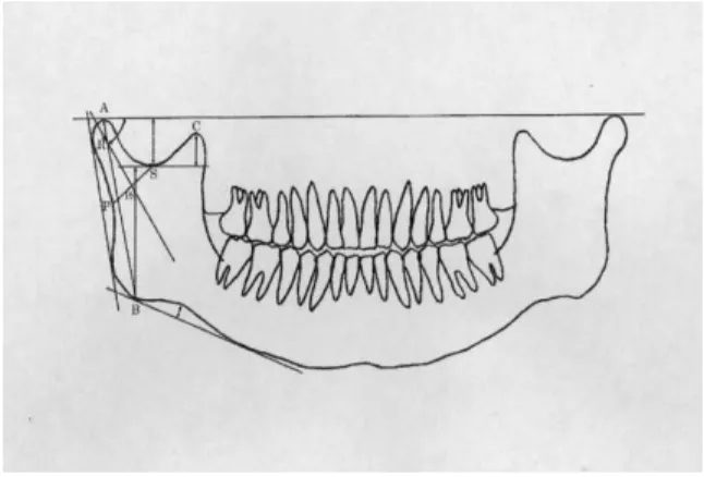
현상된 파노라마 사진 상에서 하악골의 투사도를 작성하고 양측 하악 과두의 상방에 접선을 그어 라 정의하고 하악 과두와의 접점을 라 하였
line A A
다 하악 하연에 접선을 그어 후방 접점을 라 하고. B 같은 쪽의A-B간의 거리를ramus height라 정의하였 으며 하악 하연의 접선에서 하악 하연의 가장 깊은 곳까지의 거리를 antegonial notch height라 하였다. 이후 sigmoid notch의 최하방점을 S라하고 S에서
까지의 최단거리를 라 정의하
line A condylar height
였다. Coronoid process의 최상방점을 C라하고 를S 지나며line A에 평행한 선을 그어 이 선에서 까지C 의 최단거리를 coronoid process height, B까지의 최 단거리를 ramus body height라 하였다 하악 후연에. 접선을 그어 가장 깊은 곳을 라 하고 에서 로P P S 직선을 그어 그 중점을PS라 하였다. Condylar head 의 중점을 이라 하고R PS에서 로 직선을 그은 후R
와 이루는 예각을 이라 하
line A condylar neck angle 였다(Figure 1).
환자의 ramus height가 긴 쪽을 대조군으로, 5mm 이상 짧은 쪽을 실험군으로 하여 위의 항목에 대6
한 계측을 실시하였다 투사도 작성 및 계측은 동일. 인이 시행하였으며 실험군 및 대조군의 각각의 계 측항목에 대한 평균 및 표준편차를 구하였다 이후. StatView II프로그램(Abacus Concepts, Ins., Berkeley, 으로 대조군과 실험군간의 형태학적 차 CA, 1987))
이점을 알아보기 위해unpaired two tailed t-test를 시 행하였으며 각 계측항목간의 correlation coefficient 를 구하였다 이 때. probability가0.05이하인 경우를 통계학적 유의성이 있는 것으로 판정하였다.
Ⅲ 연구 성적.
및 는 실험군에
Condylar height ramus body height
서 작게 나타났으나 antegonial notch height, coronoid 에서는 두 군간의 process height, condylar neck angle
차이점이 발견되지 않았다(Table 1). Ramus height는
및 와 통계학적으
condylar height ramus body height
로 유의한 상관관계를 보였다 또한. condylar height 가 감소함에 따라condylar neck angle도 감소하였다 (Table 2).
Ⅳ 총괄 및 고안.
안면비대칭에 관한 문헌은 1836년 Rheumatoid 를 동반하는 하악 과두 과성장 환자에 대한 arthritis
최초 보고가 있었으며 과성장된 과두에 대해 외과, 적 절제술이 처음 시행되었다18,19. 그 후 안면비대칭 에 관한 수많은 논문들이 발표되었으나 거의 대부, 분 논문이 기형이 가장 현저하게 나타나는 과두부
에 관하여 기술되고 있다1. 악골 기형으로 일어나는 안면비대칭의 대부분은 하악골의 비대칭적 성장에 의해 진행된다고 보고되었으며 하악골 형태를 기, 준으로 한 안면비대칭 분류가 보고되고 있다20-22. 두부방사선 계측사진을 이용하여 정상적인 안모에 서도 골격적 비대칭이 존재함이 밝혀졌다8,11,23. 이 러한 골격적 비대칭에서 연조직의 보상에 의해 현 저한 비대칭이 관찰되지 않는 경우도 보고되어있다
11. 심미적 기능적 문제를 동반하지 않은 미약한 안, 면비대칭은 흔히 관찰된다.
이번 연구에서 평균 연령은 34.3±13.9세 이고 연 령 변화에 따른 가지의 계측항목의 변화는 관찰되6 지 않았다. Ramus height는 대조군에서 84.0±
실험군에서 로 나타났다
5.9(mm), 76.6±6.6(mm) . 는 대조군에서
Antegonial notch height 3.1±1.5(mm), 실험군에서 2.9±1.3(mm)이었으나 두 군간의 통계 학적 유의차가 관찰되지 않았고 이는 이전 연구와, 는 다른 결과를 보인다17. 김 등의 논문에서 보면 이 전의 연구 결과와는 달리 측두하악장애 환자에서 좌 우측 하악지의 비대칭이 존재할 때 전악각 함요, 의 변화는 유의성 있게 나타나지 않아 이 결과를 뒷 받침해 주고있다24. Condylar height는 대조군에서
실험군에서 으로 두
24.1±4.5(mm), 22.3±4.3(mm)
군간의 통계학적 유의차가 관찰되었으며 ramus 와 유의한 상관관계가 있음이 밝혀졌다 측두
height .
하악관절장애 환자군과 정상군과의 좌 우측 하악과, 두 높이에 있어서 통계학적인 유의차가 발견되었으 며 하악지 높이에 대한 하악과두 높이 비율의 차이, 또한 유의차가 있다고 보고한 논문이 있어 본 논문 Table 2. Correlation coefficient and those probability(test+control)
age ramus height antegonial notch height
condylar height
coronoid process height
ramus body height
condylar neck angle
age 1
ramus height -0.086(0.1578) 1
antegonial notch height 0.024(0.6895) -0.002(0.9691) 1
condylar height -0.221(0.0003) 0.474(0.0001) 0.063(0.2998) 1
coronoid process height -0.016(0.7958) 0.153(0.0119) -0.163(0.0071) -0.066(0.2813) 1
ramus body height 0.046(0.4548) 0.648(0.0001) -0.099(0.1033) -0.12(0.0487) 0.166(0.0063) 1
condylar neck angle -0.149(0.0141 0.308(0.0001) -0.061(0.3204) 0.518(0.0001) -0.063(0.2991) -0.013(0.8214) 1
이고 두 군간에 유의차가 없는 것으로 나타 3.3(mm)
났으며 이는 과두의 길이가 짧아지면 오훼돌기가 신장 또는 재위치 된다는 이전 연구결과와 다른 결 과를 보인다. Ramus body height는 대조군에서 58.5
실험군에서 이고 두 군간
±6.1(mm), 53.5±8.0(mm)
의 통계학적 유의차가 있는 것으로 나타났으며 와의 유의한 상관관계가 관찰되었다
ramus height .
은 대조군에서
Condylar neck angle 68.8±5.9
실험군에서 이고 두 군간
(degree), 68.3±6.7(degree)
의 유의차가 없는 것으로 나타났다 주목할 것은 상. 관관계 분석 결과 condyle height가 줄어들수록 도 감소한다는 것이다 이는 과 condylar neck angle .
두의 길이가 줄어들수록 과두가 후방으로 위치한다 는 Farrar의 내용과 상반된 결과를 보인다.
이상의 내용으로 미루어 볼 때135명의 하악골 비 대칭 환자를 조사한 결과 condylar height 및 ramus
는 실험군에서 작게 나타났으며
body height , ramus
는 및 와 통계
height condylar height ramus body height 학적으로 유의한 상관관계를 보인다 또한. condylar
와 은 양의 상관관계를 가
height condylar neck angle 지고 있다.
V. 결 론
명의 하악골비대칭 환자를 대상으로 조사한 135
결과 다음과 같은 결론을 얻었다.
실험군에서 및
1. condylar height ramus body height 의 감소가 관찰되었다.
2. antegonial notch height, coronoid process height, 에서는 두 군간 차이점이 발견 condylar neck angle
되지 않았다
는 과 유의한
3. Condylar height condylar neck angle 상관관계를 보였다.
참 고 문 헌
이충국 장현호 안모비대칭의 진단 및 처치 대한구
1. , : .
강악안면외과학회지 1991;17:1-10.
2. Van Steenbergen E, Nanda R : Biomechanics of orthodontic correction of dental asymmetries. Am J
4. Mills L : Eyedness and handedness. Am J Opthal 1925;8:933-941.
5. Schwarz R : New cephalometric method and apparatus and its application to orthodontia. Int J Orthodon 1925;11:989-1017.
6. Thompson JR : Asymmetry of the face. J Am Dent Assoc 1943;30:1859-1871.
7. Hasse C : Uber Gesichtasymmetrien, Arch. Anat. V.
Physical anat. Abteil, pp 1887;119-125.
8. Vig PS, Hewitt AB : Asymmetry of the facial skeleton.
Angle Orthod 1967;37:205-211.
9. Arne Bj rk, Lise Bj rk : Artificial deformation and craniofacial asymmetry in ancient peruvians. J Dent Res 1964;43:353-362.
10. Chierici G, Havold EP, Dowson WJ : Primate experi- ments on facial asymmetry. J Dent Res 1970;49:
847-851.
11. Shah SM, Joshi MR : An assesment of asymmetry in the normal craniofacial complex. Angle Orthod 1978;48:
141-148.
김일현 이충국 구조적 및 구성적 분석방법에 의한
12. , :
한국 정상 성인의 두개안면부 형태에 관한 연구.
대한구강악안면외과학회지 1991;17:33-45.
13. Westesson PL, Tallents RH, Katzberg RW, Guay JA : Radiographic assessment of asymmetry of the mandible.
Am J Neuroradiol 1994 May;15:991-9.
14. Costa RL Jr : Asymmetry of the mandibular condyle in Haida Indians. Am J of Phys Anthropol 1986;70:119- 123.
15. EldlerR, Wertheim D, Greenhill D : Clinical and computerized assessment of mandibular asymmetry. Eur J Orthod 2001;23:485-494.
16. Samman N, Cheung LK, Tideman H : Bone scans in the diagnosis and management
of mandibular asymmetry. Int Dent J 1995;45:304.
17. William F, William L Mccarty Jr : A clinical outline of temporomandibular joint diagnosis and treatment.
Normandie, 1983.
18. Adams R : A treatment on rheumatic gout or chronic rheumatic arthritis of all the joints. Churchill, 1873.
19. Humphrey GM : Excision of the condyle of the lower jaw. Assoc Med J 1856;160:61-62.
20. Gottlieb O : Hyperplasia of the mandibular condyle. J Oral Surg 1951;9:118.
21. Rushton MA : Growth at the mandibular condyle in relation to some deformities. Br Dent J 1944;76:57.
22. Rowe NL : A etiology, clinical features, and treatment of mandibular deformity. Br Dent J 1960;108:45-64.
23. Letzer GM, Kronman JH : A posteroanterior cephalo- metric evaluation of craniofacial asymmetry. Angle Orthod 1967;37:205-211.
이영수 김동언 유임학 우순섭 심광섭 성인 만성
24. , , , , :
측두하악장애 환자에서 전악각 함요 및 하악지 고경
의 변화에 대한 연구 대한악기능교합학회지. 2001;
17:8-12.
윤귀현 최순철 파노라마 선 사진을 이용한 측두하
25. , : X
악관절 장애환자의 수직적 하악비대칭에 관한 연구.
대한구강악안면방사선학회지 1993;23:315-321.
25. Habets LLMH, Bezuur JN, Jimenez LV, Hansson TL : The Orthopantomogram, an aid in diagnosis of the temporomandibular joint problems. Ⅱ. The vertical symmetry. J Oral Rehabil 1988;15:465-471.