Received: 9 August, 2017 Revised: 21 August, 2017 Accepted: 22 August, 2017
Ⓒ The Korean Society of Mycology
This is an Open Access article distributed under the terms of the Creative Commons Attrib- ution Non-Commercial License (http://creative- commons.org/licenses/by-nc/4.0/) which permits unrestricted non-commercial use, distribution, and reproduction in any medium, provided the original work is properly cited.
Kor. J. Mycol. 2017 September, 45(3): 229-233 https://doi.org/10.4489/KJM.20170027
pISSN : 0253-651X eISSN : 2383-5249
OPEN ACCESS
RESEARCH NOTE
철쭉의 뿌리에서 분리된 두 종의 국내 미기록 내생균
박 혁, 엄안흠*
한국교원대학교 생물교육과
Two Novel Species of Endophytic Fungi Isolated from Roots of Rhododendron schlippenbachii in Korea
Hyeok Park, Ahn-Heum Eom
*Department of Biology Education, Korea National University of Education, Cheongju 28173, Korea
*Corresponding author: [email protected]
Abstract
We isolated endophytic fungal strains from roots of Rhododendron schlippenbachii growing in Minjujisan mountain, Korea. The two strains were identified as Engyodontium album and Phaeosphaeria microscopica based on their morphological characteristics and sequence analysis of the internal transcribed spacer (ITS) and large subunit regions of the ribosomal DNA. These two species, E. album and P. microscopica, have not been previously reported in Korea.
Keywords: Endophytes, Engyodontium album, Phaeosphaeria microscopic, Rhododendron schlippenbachii
철쭉(Rhododendron shlippenbachii Maxim.)은 주로 산에서 자라는 낙엽성 관목으로 진
달래과(Ericaceae)에 속하는 식물이다. 진달래과에 속하는 식물은 Helotiales에 속하는 자낭
균과 공생하여 진달래균근(ericoid mycorrhizas)을 형성하므로 다른 식물들이 살기 어려운
무기양분이 부족한 산성의 토양에서 자랄 수 있다[1]. 균근과 달리 식물 내생균(endophytic
fungi) 은 식물체 내에서 살며 병증을 유발하지 않는 균류의 통칭이다[2, 3]. 내생균은 식물에
게 다양한 환경 스트레스에 대한 내성을 갖게 해주며, 포식자, 병원체 등 외부의 위협으로부
터 식물을 보호하는 것으로 알려져 있다[2]. 내생균과 숙주 식물 사이의 특이적인 관계에 대
해서는 아직까지 명확히 밝혀지지는 않았으나 내생균의 전파가 식물 간의 특이성에 기반한
수직적인 전달을 통해 이루어진다는 보고가 있으며[4], 내생균에는 숙주 식물에 대한 특이성
이 다소간 존재하는 것으로 보여진다[5]. 따라서 내생균에 대한 연구는 다양한 숙주 식물을
대상으로 이루어져야 국내 미기록 균주의 확보와 균류의 발굴 및 균류 자원 확보에 도움이 될
수 있을 것이라고 생각된다. 본 연구에서는 진달래과 식물인 철쭉의 뿌리에서 2종의 국내 미 기록 내생균을 분리하여 그 특징을 보고하고자 한다.
2016 년 9월 충북 영동군의 민주지산(N 36° 2′ 49″, E 127° 46′ 36″)에서 철쭉의 뿌리를 수 집하였다. 실험실로 운반한 뿌리는 흐르는 물로 흙을 제거한 후, Ahlich와 Sieber [6]의 방법 에 따라 뿌리에서 균을 분리하였다. 뿌리를 증류수로 세척한 후 70% EtOH와 3% NaClO 용 액을 차례로 처리하고, 항생제인 streptomycin 용액을 처리하여 표면을 살균하였다. 멸균된 거름종이로 물기를 제거한 후 뿌리를 약 5 mm 길이로 잘라 4개 조각을 1% water agar 배지에 치상하였고, 25°C 암소에서 배양한 후 균사가 뻗어 나오면 potato dextrose agar (PDA)와 malt extract agar (MEA) 배지에 계대하여 순수 분리하였다. 분리된 균주는 형태학적 특징을 관찰하였으며, 균사와 분생포자(conidia)를 관찰하기 위하여 25°C 암소에서 7일간 슬라이드 배양법을 이용하여 배양하였다. 시료는 lactophenol cotton blue 용액으로 염색하여 광학현 미경(AXIO Imager A1; Carl Zeiss, Oberkochen, Germany) 하에서 관찰하였다. 균 특이적 primer 인 ITS1F와 ITS4를 이용하여[7] ribosomal DNA의 internal transcribed spacer (ITS) 지역과 large subunit (LSU) 지역을 ITS1F와 ITS4 프라이머와 LR0R과 LR16을 각각 이용하 여[8] 증폭하였다. DNA 염기서열은 SolGent (Daejeon, Korea)에 의뢰하여 분석하였다.
DNA 염기서열은 National Center for Biotechnology Information (NCBI)에서 basic local alignment search tool (BLAST) 을 이용하여 GenBank 데이터베이스의 염기서열과 비교하 였다. 종 간의 유연 관계를 파악하기 위해 MEGA6 프로그램을 이용하여[9] neighbor- joining 방법으로 계통수를 작성하였다. 두 개의 균주 16E002와 16E007은 국립생물자원관 (NIBR) 에 기탁하여 기탁번호 NIBRFG0000499916과 NIBRFG0000499918을 부여 받았 다. 또한 각 균주의 염기서열은 GenBank에 기탁하였다(MF576267, MF576268).
Engyodontium album (Limber) de Hoog, Persoonia 10: 53 (1978) 16E002
≡ Tritirachium album Limber. 1940
PDA 배지에서 7일간 배양된 균총의 직경은 19~20 mm 정도이고, 앞면은 흰색이고 뒷면은
연어살 색이다(Table 1, Fig. 1). 집락의 고도는 가장자리에서 납작하게 붙어있고 중앙부는 볼
록 융기되어 있으며, 집락의 모양은 불규칙한 타원형에 가깝다. MEA 배지에서 7일간 배양된
균총의 직경은 19 mm 정도이고, 형태적 특성은 PDA에서 배양한 균주와 대부분 일치하는 것
을 확인할 수 있었다. 나뭇가지 모양의 분생포자경(conidiophore)에서 쌀알처럼 생긴 짧은 타
원형의 분생포자를 형성하며 크기는 2.8~3.6 × 1.2~1.8 µm이다. 본 균주의 ITS 지역의 DNA
염기서열의 BLAST 결과 E. album KJ767113.1과 99%의 상동성을 보였으며, LSU 지역은
E. album HM214541.1 과 99%의 유사도를 나타내었다. 계통수를 이용한 분석결과 비교 균
주와 100% bootstrap 값으로 같은 계통으로 나타났다(Fig. 2). 이 종은 자낭균의 Hypocreales,
Cordyciitaceae 에 속하는 종으로 Engyodontium속은 현재 4개의 종이 속해 있으며 윤생
(verticillate) 하는 분생포자경에서 작고, 투명한 단포자를 형성하는 형태적 특징을 갖는다
[10]. 이 균은 토양이나 해양의 퇴적물 등 다양한 곳에 서식하고 있으며, 다양한 식물의 잎이
나 뿌리에서 내생균으로 존재하는 것으로도 보고되고 있다[11-13]. 또한 이 균은 단백질과 케
라틴의 분해에 대한 활성을 갖고 있어 인간에 기회성 병원균으로 간주되고 있다[14].
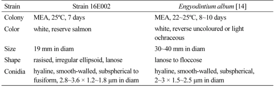
Table 1. Morphological characteristics of fungal strains isolated from this study
Strain Strain 16E002 Engyodintium album [14]
Colony MEA, 25ºC, 7 days MEA, 22~25ºC, 8~10 days
Color white, reserve salmon white, reverse uncoloured or light ochraceous
Size 19 mm in diam 30~40 mm in diam
Shape rasised, irregular ellipsoid, lanose lanose to floccose Conidia hyaline, smooth-walled, subspherical to
fusiform, 2.8~3.6 × 1.2~1.8 µm in diam
hyaline, smooth-walled, subspherical, 2~3 × 1.5~2.5 µm in diam
MEA, malt extract agar.
Fig. 1. Colonies of strain 16E002 (Engyodontium album) grown on PDA (A) and MEA (B).
Colonies of strain 16E007 (Phaeosphaeria microscopica) grown on PDA (C) and MEA (D).
Conidia of 16E002 (E) (scale bar = 10 µm). PDA, potato dextrose agar; MEA, malt extract agar.
Fig. 2. Neighbor-joining phylogenetic tree based on a combined alignment of internal transcribed
spacer (ITS), large subunit (LSU) sequences alignment. Mucor variisporus was used as an
outgroup. Numbers on branches indicate bootstrap values (1,000 replicates). Fungal strains
isolated in this study are in bold.
Phaeosphaeria microscopica (P. Karst.) O.E. Erikss. Arkiv før Botanik 6: 426 (1967) 16E007
≡ Leptosphaeria microscopica P. Karst. 1872
PDA 배지에서 7일간 배양된 균총의 직경은 33~36 mm 정도이고, 앞면은 흰색이고 뒷면 은 붉은빛이 살짝 도는 베이지색이다(Table 2, Fig. 1). 집락의 고도는 주변부에서는 평평하 게 융기되어 있고 중앙부는 볼록 솟은 파도형에 가깝다. 가장자리 부분은 미세한 섬유상으 로 균사가 뻗어 있다. MEA 배지에서 7일간 배양된 균총의 직경은 26 mm이고, 형태적 특성 은 PDA 배지에서 배양된 균주와 비슷하다. 본 균주의 ITS 지역 염기서열 BLAST 결과 P.
microscopica FN386274.1 과 98%의 유사성을 확인하였으며, LSU 지역의 분석 결과 P.
microscopica FN386274.1 과 99%의 유사성을 확인할 수 있었다. 계통수를 이용한 분석결과 비교 균주와 100% bootstrap 값으로 같은 계통으로 나타났다(Fig. 2). 이 종은 자낭균의 Pleosprales 목에 속하는 종으로 다양한 초본식물의 잎에 병증을 나타내는 것으로 보고되고 있으며[15, 16], 일부 식물의 잎에서 내생균으로도 보고되고 있다[12].
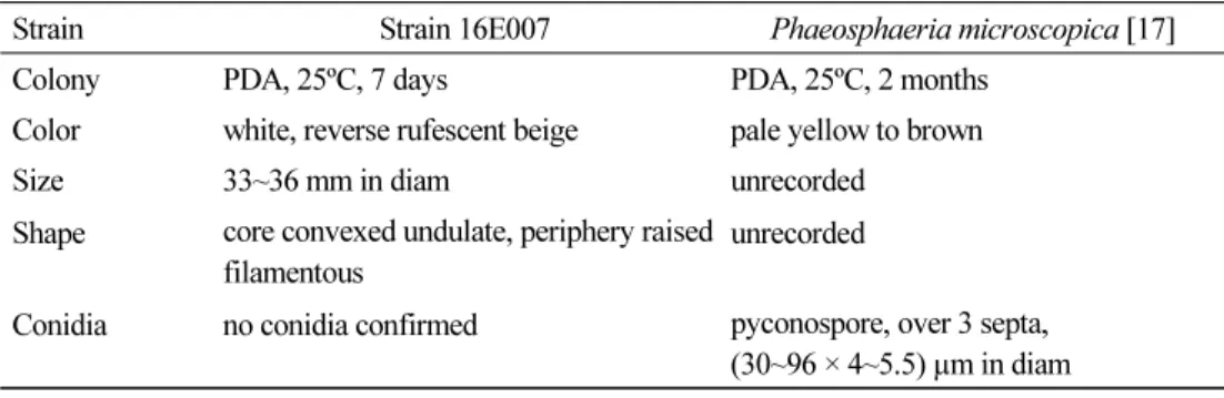
Table 2. Morphological characteristics of fungal strain 16E007 isolated from this study
Strain Strain 16E007 Phaeosphaeria microscopica [17]
Colony PDA, 25ºC, 7 days PDA, 25ºC, 2 months
Color white, reverse rufescent beige pale yellow to brown
Size 33~36 mm in diam unrecorded
Shape core convexed undulate, periphery raised filamentous
unrecorded
Conidia no conidia confirmed pyconospore, over 3 septa, (30~96 × 4~5.5) µm in diam
PDA, potato dextrose agar.
적 요
본 연구에서는 민주지산에 서식하는 철쭉(Rhododendron schlippenbachii)의 뿌리에서 내 생균을 분리하였다. 형태적 특성의 확인 및 ribosomal DNA의 internal transcribed spacer (ITS) 지역, large subunit (LSU)지역의 분자적 분석을 통해 국내 미기록종인 Engyodontium album, Phaeosphaeria microscopica 2 종을 동정하였다. 연구 과정에서 확인된 미기록종 내 생균 2종의 형태적, 분자적 특성을 여기에 기술하였다.
Acknowledgements
This work was supported by the project on Survey and Discovery of Indigenous Species
of Korea funded by NIBR of the Ministry of Environment (MOE), Republic of Korea.
REFERENCES