S.-C. Kang ( )
세명대학교 자연약재과학과
(Department of Natural Medicine Resources, Semyung University, Jecheon 390-711, Korea)
e-mail: [email protected]
자가식세포작용: 천연물항암제로서의 신규작용기전
강세찬
Autophagy: Noble target mechanisms in natural medicines as anti- cancer agents
Se-Chan Kang
Received: 24 February 2010 / Accepted: 3 March 2010
ⓒ Korean Society for Plant Biotechnology
Abstract Programmed cell death systems are important for an active type of cell deaths. Among them, a type of pro- grammed cell death, autophagy is activated in cancer cells in response to multiple stresses and has been demonstrated to promote tumor cell survival and drug resistance. Thus, in the area of cancer, over the time frame form around the 1940s to date, of the 155 small molecules, 73% are other than
“synthetic”, with 47% actually being either “natural prod- ucts” or “directly derived therefrom”. Autophagy has multiple physiological functions in multicellular organisms, including protein degradation and organelle turnover. Genes and pro- teins that constitute the basic machinery of the autophagic process were first identified in the yeast system and some of their mammalian orthologues have been characterized as well. Numerous oncogenes, including Akt1, Bcl-2, NF1, PDPK1, class I PI3K, PTEN, and Ras and oncosuppressors, inculuding Bec-1, Bif-1, DAPK-1, p53 and UVRAG sup- press or promote the autophagy pathway. Regulation of autophagy in tumors is governed by similar principles of the normal cells, only in a much more complicated manner, given the frequently observed abnormal PI3K activation in cancer and the multitude of interactions between the PI3K/
AKT/mTOR pathway and other cell signaling cascades, often also deregulated in tumor cells. Autophagy induction by some anticancer agents underlines the potential utility of
its induction as a new cancer treatment modality of devel- opment for natural medicines.
서 론
천연물의약품은 식물, 동물, 미생물 및 세포내용물과 광물 등 자연계에 존재하는 물질을 의약품으로 사용하는 것을 말하며, 광범위하게 정의되는 첨단 바이오기술을 포함하는 바이오의약품의 일부로 정의될 수 있다. 이러 한 천연물 중 특히 식물에는 다양한 성분들이 서로 상호 작용하면서 혼합되어 있는 형태로 다양한 질환에 대한 조합치료 (combination therapy)에 이용될 수 있으며, 생체 내에서 다중 타겟에 영향을 미치므로 천연물에 함유되어 있는 다양한 성분이 함유되어 있는 조추출물 (crude extract), 용매분획물 (solvent fraction)을 의약품으로 이용하거나, 이로부터 분리된 단일성분 화합물 또는 이에 대한 유도 체를 합성하여 새로운 의약품에 대한 개발이 시도되고 있다. 특히, 1981년부터 2005년까지 개발된 의약품을 분 석한 보고에 따르면, 단일화합물 847개의 의약품 중 5%
는 천연물에서 분리된 저분자 물질로 부터 개발된 의약 품이며, 약 27%가 천연물에서 분리된 화합물로 부터 유 도체를 합성하여 개발되었으며, 나머지 572개의 제품은 화학적으로 합성된 의약품으로 보고된바 있다 (Newman and Cragg 2007).
이 중에서 항암제의 분야는 1940대 이후 개발된 155개 의 저분자 화합물 중 73%이상이 화학적으로 합성되지 않았으며, 이 중 47%는 천연추출물을 그대로 이용하거나 천연물로부터 유도된 합성물질인 것으로 파악된다. 2010 년 현재 바이오의약 시장은 70조원 이상으로 예상되고 있으며, 암 치료제에 집중되어 있는 상황이다 (Kwon et DOI:10.5010/JPB.2010.37.1.057
Review
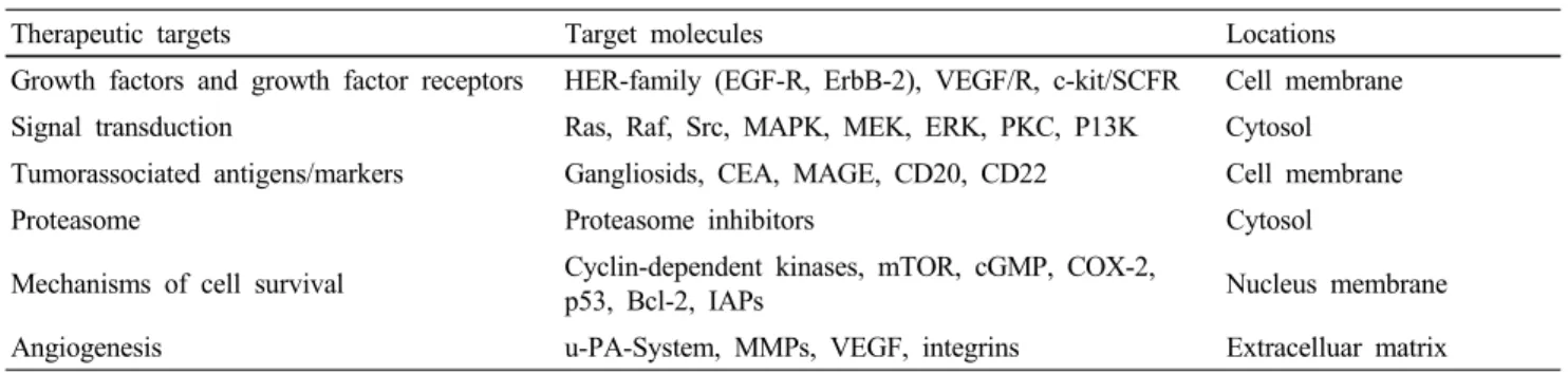
Table 1 Overview on major target molecules for early clinical trials
Therapeutic targets Target molecules Locations
Growth factors and growth factor receptors HER-family (EGF-R, ErbB-2), VEGF/R, c-kit/SCFR Cell membrane Signal transduction Ras, Raf, Src, MAPK, MEK, ERK, PKC, P13K Cytosol Tumorassociated antigens/markers Gangliosids, CEA, MAGE, CD20, CD22 Cell membrane
Proteasome Proteasome inhibitors Cytosol
Mechanisms of cell survival Cyclin-dependent kinases, mTOR, cGMP, COX-2,
p53, Bcl-2, IAPs Nucleus membrane
Angiogenesis u-PA-System, MMPs, VEGF, integrins Extracelluar matrix
al. 2009). 최근 몇십년간 항암제를 개발하기 위한 분자마 커 (molecular marker)가 빠르게 개발되고 있으며 이미 항 암제 타겟으로 이용되고 있거나, 이용가능성이 있는 것 으로 growth factor, growth factor receptor, signal transduction 과 관련된 생체분자, tumor-associated antigen, proteasome inhibitor, cell cylcle 과 cell death 조절자, 종양의 invasion, metastasis 및 신생혈관형성과 관련된 인자 등이 보고되었 다 (Green 2004; Green 2003; Jonathan and Green 2002; Rogers et al. 2000; Liu et al. 2002; Festuccia et al. 2005).
세계 항암제 시장은 2000년 기준 북미 47%, 아시아, 아 프리카, 호주가 26%, 유럽이 26%를 차지 하고 있으며, 세 계항암제 시장규모는 2000년 18조에서 매년 8~10%, 국내 항암제 시장규모는 2000년 900억에서 년평균 20%이상 성장하고 있다. 천연물 유래항암제는 taxol이 가장 시장 규모가 크며, 현재 전체 의약품 시장에서 천연물의약품 및 천연물유래 의약품이 약 60%를 차지 하고 있는 것을 감안하면 천연물 항암제의 시장은 지속적으로 발전할 것 으로 기대된다. 따라서 기존에 개발되어 임상에 사용되 고 있는 target을 대상으로 천연물로부터 항암제 개발이 우선 진행될 것으로 보여지며, 이후 천연물 특유의 작용 기전이 밝혀 질 수 있을 것으로 기대된다. 현재 임상에서 항암제로서의 주요 target은 Table 1과 같으며, 항암효과 뿐 만 아니라 암환자의 고통을 줄여주거나 암환자의 골 손실을 억제해주는 zoledronic acid 등도 포함될 수 있다 (Allgayer and Fulda 2008).
천연물 항암제의 target 또한 Table1의 초기 항암제의 target에 대한 연구로 시작되어 현재까지 합성항암제와 함께 계속되어 발전하고 있다. 그러나, 대부분의 항암요 법은 암세포의 apoptosis와 밀접하게 연관되어 있다 (Fulda and Debatin 2004). 많은 항암제들이 임상에서 사용될 때 apoptosis program과 관련된 target molecule을 억제할 경우 내성암이 유발되어 항암연구의 새로운 시도로 내성암 억 제에 대한 연구가 진행되고 있다 (Johnstone et al. 2002).
또한 apoptosis를 타겟으로하는 항암연구분야에서는 다양 한 의약품이 개발되고 있으며, 특히 화학적 항암요법과 방사선조사에서도 항암보조제로 많은 연구가 현재 진행
되고 있다 (Fulda and Debatin 2006). 이러한 Programmed cell death와 관련된 항암제의 연구는 apopotis와 관련된 signal transduction에 관여하는 인자에 대한 연구가 대부분 이며, Programmed cell death 연구분야의 또 다른 핵심 작 용기전인 autophagy system을 이용한 항암연구는 최근에 새롭게 조명되어 항암제 연구의 새로운 패러다임으로 대 두되고 있다.
현재까지 항암제 개발의 새로운 target으로 autophagy system과 관련된 작용기전과 target molecule이 밝혀지고 있으나, 이를 이용한 천연물의약품 분야 특히, 항암제 개 발과 관련된 임상연구는 이루어지지 않고 있으며, 천연 물 유래의 autophagy inducible compound가 보고된 바 없 다. 따라서, 본 연구에서는 autophagy와 관련된 다양한 항 암제 개발에 대한 시도와 현재까지 보고된 target molecule 에 대하여 새롭게 조명하여 항암제 개발에 있어 특히, 천 연물신약의 새로운 target으로 autophagy system에 대한 항 암제 개발의 가능성에 대하여 알아보고자 하였다.
Programmed cell death의 비교
1960대에 세포사멸에 대한 세포구조 변형의 차이가 관 찰되어 Programmed cell death의 용어가 necrosis와 구분되 어 사용되어 오다 1972년 Kerr 등 (1972)에 의하여 Pro- grammed cell death의 유사명으로 apoptosis의 용어가 제안 되어 Programmed cell death를 총칭하여 사용되어 왔다. 그 러나, 1990년 Clarke (1990)에 의하여 세포사멸에 대한 mor- phology를 기준으로 하여 4가지 type의 cell death가 제안되 어 현재까지 Programmed cell death를 세분하여 연구되고 있다 (Gozuacik and Kimchi 2004).
첫번째 type으로 type I 또는 apoptosis로 일컬어 지고 있 는 것으로 cell shrinkage, chromatin condensation, nucleosomal DNA degradation 및 fragmentation의 대표적인 세포형태가 나타나는 이른바 ‘apoptotic bodies’의 형성으로 capase family 에 의하여 조절되는 것으로 보고되고 있다.
두번째 type으로 type II 또는 autophagic cell death로 일
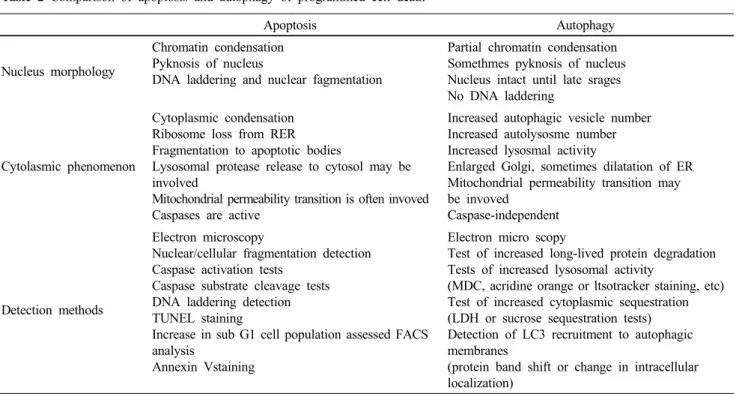
Table 2 Comparison of apoptosis and autophagy of programmed cell death
Apoptosis Autophagy
Nucleus morphology
Chromatin condensation Pyknosis of nucleus
DNA laddering and nuclear fagmentation
Partial chromatin condensation Somethmes pyknosis of nucleus Nucleus intact until late srages No DNA laddering
Cytolasmic phenomenon
Cytoplasmic condensation Ribosome loss from RER Fragmentation to apoptotic bodies
Lysosomal protease release to cytosol may be involved
Mitochondrial permeability transition is often invoved Caspases are active
Increased autophagic vesicle number Increased autolysosme number Increased lysosmal activity
Enlarged Golgi, sometimes dilatation of ER Mitochondrial permeability transition may be invoved
Caspase-independent
Detection methods
Electron microscopy
Nuclear/cellular fragmentation detection Caspase activation tests
Caspase substrate cleavage tests DNA laddering detection TUNEL staining
Increase in sub G1 cell population assessed FACS analysis
Annexin Vstaining
Electron micro scopy
Test of increased long-lived protein degradation Tests of increased lysosomal activity
(MDC, acridine orange or ltsotracker staining, etc) Test of increased cytoplasmic sequestration (LDH or sucrose sequestration tests) Detection of LC3 recruitment to autophagic membranes
(protein band shift or change in intracellular localization)
Modified from Gozuacik and Kimchi 2004.
컬어 지고 있으며, 세포내에서 autophagic vesicle을 형성 하여 lysosomal system과 유사하게 세포내 macromolecule 을 degradation하는 특징을 나타낸다. 세포내에 autophagic vesicle (autophagosome)의 형성이 촉진되면 부분적인 chro- matin condensation이 apoptosis와 유사하게 나타나나, DNA fragmentaion은 관찰되지 않으면서 세포사를 유도하여 self- eating의 뜻을 나타내는 autophagy의 용어로 정립되었다.
세번째 type으로 type III가 있으며 이는 nonlysosomal vesiculate degradation으로 정의 되고 있다. Type III pro- grammed cell death는 다시 type IIIA와 type IIIB로 구분되 는데 nonlysosomal degradation과 cytoplasmic type of degener- ation으로 나타낼 수 있다 (Clarke 1990).
이 가운데 type I과 type II인 apoptosis와 autophagy의 nucleus 및 cytoplasm의 형태변화 및 현재 주로 사용되고 있는 시험방법이 Table 2에 나타나 있다. Nucleus의 특징 적인 차이점은 apoptosis의 경우 DNA laddering과 nuclear fragmentation 및 chromatin condensation이 관찰되는 반면 autophagy에서는 DNA laddering이 관찰되지 않으며 chromatin 의 경우 부분적인 condensation이 관찰되는 것으로 보고되 어 있다 (Gozuacik and Kimchi 2004). 현재까지 autophagy system을 평가하기 위한 방법으로 현미경을 통한 세포형 태의 관찰을 비롯하여 long-lived protein에 대한 degradation assay, monodencylcadaverine (MDC) 등의 방법이 개발되었 으며, 이들 assay법으로 부터 천연자원식물의 autophagy system 유도물질을 규명할 수 있을 것으로 사료된다 (Table 2).
Autophagy와 암세포
1970년대에서 1980년대까지 autophagy 활성과 세포의 malignant transformation과의 연관성에 대한 연구가 진행 되었다. 특히 정상세포에 비하여 암세포에서는 autophagy 의 활성이 매우 약하게 발현되고 있는 것으로 보고되고 있으며, 암세포의 성장환경 중 세포가 매우 밀집되거나 serum 및 아미노산이 결핍되었을 경우 autophagy의 활성 이 증가되어 세포사를 유도할 수 있을 것으로 보고된 바 있다. 즉 transformed cell의 경우 nutrient 및 serum의 결핍 상태에서 autophagy가 활성화되어 protein이 degradation되 어 세포사가 나타나는 것으로 많은 연구자에 의하여 관 찰되었다 (Gunn et al. 1977; Lockwood and Minassian 1982;
Cockle and Dean 1984; Gronostajski and Pardee 1984; Knecht et al. 1984).
Carcinogenesis에 대한 동물시험에서 또한 autophagy 능 력이 억제됨이 확인되었다. Chemical carcinogen으로 유도 된 Preneoplastic 간조직에서 분리된 세포 및 primary hepato- celluar carcinomas에서 정상세포에 비하여 autophagy 형성 이 억제됨이 보고되었으며, hepatocelluar carcinomas로부 터 분리된 autophagic vesicle에서 lysosomal enzyme의 활성 이 억제되어 있음이 보고되었다 (Schwarze and Seglen 1985;
Canuto et al. 1993; Kisen et al. 1993; Ahlberg et al. 1987;
Yucel et al. 1989). 이상의 연구보고에서 autophagy 활성의
억제는 암의 발달과정에서 매우 중요한 인자로 판단되어
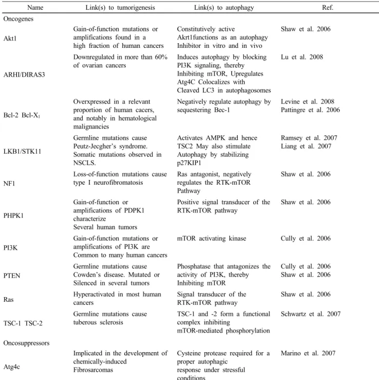
Table 3 Examples of autophagy regulation by oncoproteins and tumor sumor suppressor proteins
Name Link(s) to tumorigenesis Link(s) to autophagy Ref.
Oncogenes
Akt1
Gain-of-function mutations or amplifications found in a high fraction of human cancers
Constitutively active
Akrt1functions as an autophagy Inhibitor in vitro and in vivo
Shaw et al. 2006
ARHI/DIRAS3
Downregulated in more than 60%
of ovarian cancers
Induces autophagy by blocking PI3K signaling, thereby Inhibiting mTOR, Upregulates Atg4C Colocalizes with
Cleaved LC3 in autophagosomes
Lu et al. 2008
Bcl-2 Bcl-X
1Overxpressed in a relevant proportion of human cacers, and notably in hematological malignancies
Negatively regulate autophagy by sequestering Bec-1
Levine et al. 2008 Pattingre et al. 2006
LKB1/STK11
Germline mutations cause Peutz-Jecgher’s syndrome.
Somatic mutations observed in NSCLS.
Activates AMPK and hence TSC2 May also stimulate Autophagy by stabilizing p27KIP1
Ramsey et al. 2007 Liang et al. 2007
NF1
Loss-of-function mutations cause type I neurofibromatosis
Ras antagonist, negatively regulates the RTK-mTOR Pathway
Shaw et al. 2006
PHPK1
Gain-of-function or amplifications of PDPK1 characterize
Several human tumors
Positive signal transducer of the RTK-mTOR pathway
Shaw et al. 2006
PI3K
Gain-of-function mutations or amplifications of PI3K are Common to many human cancers
mTOR activating kinase Cully et al. 2006
PTEN
Germline mutations cause Cowden’s disease. Mutated or Silenced in several tumors
Phosphatase that antagonizes the activity of PI3K, thereby Inhibiting mTOR
Cully et al. 2006 Shaw et al. 2006
Ras Hyperactivated in most human
cancers
Signal transducer of the RTK-mTOR pathway
Shaw et al. 2006
TSC-1 TSC-2
Germline mutations cause tuberous sclerosis
TSC-1 and -2 form a functional complex inhibiting
mTOR-mediated phosphorylation
Schwartz et al. 2007
Oncosuppressors
Atg4c
Implicated in the development of chemically-induced
Fibrosarcomas
Cysteine protease required for a proper autophagic
response under stressful conditions
Marino et al. 2007 지며, 세포배양을 통한 연구에서도 정상세포에 비하여
암세포에서 autophagy활성 및 autophagic vesicle의 형성이 억제됨이 보고되었다 (Schwarze and Seglen 1985).
Oncogenesis에서의 autophagy의 역할
최근의 연구에서 기존에 잘 알려져 있는 oncogene과 tumor suppressor 유전자들의 발현조절에 따라 autophagy활 성이 조절되어 암이 발달되거나 억제됨이 보고되었다.
특히, 인체세포에서 autophagy 억제 조절자로 잘 알려져 있는 mTOR (mammalian target of rapamycin) kinase는 auto- phagic cascade의 개시단계에 필요한 단백질에 hyperphosphoryla- tion을 유지함으로써 autophagy 활성을 억제하며, 반대로 영양분 결핍 등의 스트레스 조건에서 mTOR 발현이 억제 되어 autophagy활성이 증가됨이 보고되었다 (Galluzzi et al.
2008). 이러한 mTOR 발현을 조절하는 oncogene으로 PI3K, Ras, PDPK1, NF1등이 알려져 있다 (Table 3).
Oncogene으로 잘 알려져 있는 Akt1은 in vivo 및 in vitro
시험에서 autophagy를 억제하여 암발현을 촉진하는 것으
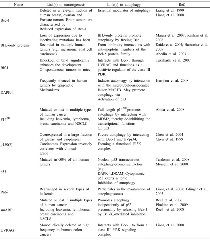
Table 3 Examples of autophagy regulation by oncoproteins and tumor sumor suppressor proteins (continued)
Name Link(s) to tumorigenesis Link(s) to autophagy Ref.
Bec-1
Deleted in a relevant fraction of human breast, ovarian and Prostate tumors. Brain tumors are characterized by
Reduced expression of Bec-1
Essential modulator of autophagy Liang et al. 1999 Liang et al. 2008
BH3-only proteins
Loss of expression due to inactivating mutations has been Recorded in multiple human tumors (e,g., melanoma, enal cell carcinoma)
BH3-only proteins promote autophagy by freeing Bec_1 From inhibitory interactions with anti-apoptotic members of the Bcl-2 protein family
Maiuri et al. 2007; Rashmi et al.
2008
Daido et al. 2004; Hamacher et al.
2007
Abedin et al. 2007
Bif-1
Knockout of bif-1 significantly enhances the development Of spontaneous tumors in mice
Interacts with Bec-1 through UVRAC and functions as a positivie regulator of the class III PI3K
Takahashi et al. 2007
DAPK-1
Frequently silenced in human tumors by epigenetic
Mechanisms
Induces autophagy by interaction with the microtubule-assocciated factor MAP1B. May promote autophagy via
Activation of p53
Harrison et al. 2008
P14
ARFMutated or lost in multiple types of human cancer
Including leukemia, lymphoma, breast carcinoma and NSCLC
Full length p14
ARFpromotes autophagy by interacting with MDM2, thereby de-inhibiting the transcriptional functions
Of p53
Abida et al. 2008
p150(?)
Overexpressed in a large fraction of gastric and esophageal Carcinomas. Expression inversely correlates with clinical
grade
Favors autophagy by interacting with Bec-1 and hVps34,
Forming a functional PI3K complex
Chen et al. 2004 Chen et al. 1999
p53
Mutated in>50% of all human tumors
Nuclear p53 transactivates autophagy-promoting factors (e.g.,
DAPK-1,DRAM).Cytoplasmic p53 exerts a tonic
Inhibition of autophagy
Tasdemir et al. 2008 Morselli et al. 2008
Rab7 Rearranged in several types of leukemia
Participates in the mauturation of autophagosomes
Liang et al. 2008; Edinger et al., 2003
smARF
Mutated or lost in multiple types of human cancer
Including leukemia, lymphoma, breast carcinoma and
NSCLS
Promotes autophagy independently of p53,
presumably by releasing Bex-1 by Bcl-X
L-mediated inhibition
Reef et al. 2006 Pimkina et al. 2009 Reef et al. 2008
UVRAG
Monoallelically deleted at high frequency in human colon cancers
Interacts with Bec-1 to from a class III PI3K signaling complex
Liang et al. 2008
로 보고 되었으며, 난소암의 60% 이상에서 억제되어 있는 것으로 보고된 Ras-related protein인 ARHI/DIRAS3은 PI3K signaling을 억제하여 mTOR을 negative하게 조절하여 auto- phagy를 활성화 할 것으로 보고되어 있다 (Shaw et al. 2006;
Lu et al. 2008). 대부분의 인체암에서 높게 발현되고 있는 Ras는 RTK-mTOR pathway의 signal transducer로 autophagy
를 억제하지만 Ras의 antagonist인 NF-1 (neurofibromin 1)는 RTK-mTOR pathway를 억제하여 autophagy를 증가시키는 것으로 예측되나, 이 부분에 대한 명확한 기전은 현재까 지 보고되어 있지 않다 (Shaw et al. 2006).
Oncosupperssor인 Bec-1은 autophagy의 주요한 modulator
로 Bec-1의 발현이 촉진되면 smARF이 p53을 조절하여
Fig. 1 Autophagy regulation pathway. Iilustration was modifided from Chen and Karantza-Wadsworth, 2009
RTK-mTOR pathway와는 독립적으로 autophagy를 활성화 시키는 것으로 예상되고 있다 (Ling et al. 1999; Liang et al. 2008; Tasdemir et al. 2008; Reef and Kimchi 2008). 또한, knockout 되었을 때 mouse tumor를 일으키는 Bif-1, UVRAG (UV radiation resistance associated gene) 등은 autophagy 의 modulator인 Bec-1과 상호작용하여 class III PI3K signaling complex에 의하여 p53, RTK-mTOR pathway와 서로 다른 경로로 autophagy를 조절하는 것으로 보고되고 있다 (Liang et al. 2008; Edinger et al. 2003; Takahashi et al. 2007). 따라 서, oncogene과 oncosupperssor들은 p53, mTOR 및 class III PI3K signaling 등에 의하여 autophagy를 조절하고 있는 것 으로 요약될 수 있으며, 이들 유전자 및 단백질의 발현 및 signaling에 대한 inhibitor 또는 activator에 대한 연구를 통하여 autophagy를 조절함으로써 새로운 항암성분을 규 명할 수 있을 것으로 사료된다 (Table 3).
Cancer cell에서의 autophagy의 작용기전
앞에서 설명된 바와 같이 mTOR의 발현에 따라 autophagy 활성이 조절되어 암세포의 세포사가 조절된다. 즉, mTOR 의 발현이 증가될 경우 autophagic cascade에 관여하는 단 백질의 phosphorylation이 크게 증가하여 autophagy가 억제 되며, 반대로 발현이 억제될 경우 phosphorylation이 감소 하여 autophagy system이 증가되어 암세포의 세포사가 유 도된다. 이러한 mTOR의 downstream에서 nutrient의 sensor 단백질인 PI3K의 phosphorylation이 조절되어 세포의 영양 분과 growth factor의 사용이 조절되어 cancer의 proliferation 이 촉진되고 autophagy system이 억제된다. 따라서, 이러 한 조절 메커니즘은 PI3K/AKT/mTOR pathway에 의하며 인체 암세포의 일반적인 조절 작용기전으로 밝혀지고 있 다 (LoPiccolo et al. 2008). Fig. 1은 PI3K/AKT/mTOR path- way에 따른 조절, 즉 AKT의 overexpression, PI3K의 발현
조절에 따른 암발생 및 암전이과정에 관여하는 oncogene 과 autophagy system조절자에 대하여 나타내고 있다 (Arico et al. 2001; Ueno et al. 2008).
즉, growth factor signaling은 PI3K/AKT/mTOR pathway의 활성을 촉진시킴으로써 결론적으로 autophagy를 억제한 다. 여기에 관여되는 단백질 및 ligand 중 GPCR (G-protein coupled receptor) antagonist들은 growth factor receptor를 억 제하며, lithium과 carbamazepine은 class I PI3K를 억제, perifostine과 AKT/PKB signaling inhibitor-2 (API-2)는 AKT 활성을 억제한다. 또한, rapamycin은 직접적으로 mTOR complex를 억제하여 결과적으로 autophagy를 촉진하여 암 세포 사멸을 유도하는 것으로 보고된 바 있다 (Degenbardt et al. 2006; Karantza-Wadsworth et al. 2007; Mathew et al.
2007). 이와 대조적으로 class III PI3K는 autophagy를 유도 하며, 따라서 class III PI3K의 inhibitor로 알려져 있는 3-MA (methyl-adenine), worthmannin 등은 autophagy를 억제하는 것으로 판단된다 (Fig. 1).
Cancer therapeutic target으로서의 autophagy
Autophagy의 암과 관련된 작용기전과 역할은 앞에 살 펴본 바와 같이 매우 다양한 경로에 의하며, 아직까지 autophagy를 target으로 한 의약품이 개발되지 않은 상황 에서 신규타겟 항암제 개발에 대한 가능성은 무한하다고 판단된다. Autophagy는 주로 암의 hypoxia 상태 및 stress 상태에서 활성화되므로 다양한 항암제와 autophagy 활성 화 물질을 병용하여 사용시 더욱 효과가 극대화 될 것으 로 예측된다 (Degenhardt et al. 2006; Karantza-Wadsworth et al. 2007). 따라서, 현재 미국에서는 항말라리아제인 HCQ 의 autophagy 활성을 이용하여 HCQ와 bortezomib, temozolo- mide, ixabepilone, carboplatin, paclitaxel 및 bevacizumab와의 병용요법을 암치료 임상에서 시도하고 있다 (Chen and Karantza-Wadsworth 2009). 현재 본 연구실에서도 cisplatin 등 기존의 세포독성 항암제로 유도된 암세포의 stress상 태에서 autophagy를 활성화 시키는 천연식물추출물을 처 리하였을 때 암세포의 세포성장 억제가 크게 증가됨을 확인한 바 있다 (data not shown).
또한, 앞서 기술된 autophagy system을 억제하는 조절자 에 대한 inhibitor를 이용할 경우 다양한 항암보조제의 개 발이 가능할 것으로 판단된다. 최근의 연구결과에 따르 면 class III PI3K의 inhibitor로 알려져 있는 chloroquine계 화합물, 3-methyladenine (3-MA)과 histone deacetylase inhibitor 인 SAHA (suberoylanilide hydroxamic acid)의 혼합투여로 인한 CML (chronic myelogenous leukemia) 세포에서의 암 세포 성장저해 연구가 보고된 바 있다 (Carew et al. 2007).
또한 기존의 유방암치료제인 tamoxifen 또는 4-hydroxy-
Fig. 2 Sample of autophagy inducible a natural product. A. MDC (monodencylcadaverine) staining. B. Relative autophagy activity by n-hexnae layer of 80% EtOH extract of a natural medicine resources
tamoxifen (4-OH-T)을 이용한 연구에서 여성호르몬 수용 체 양성세포인 MCF-7 및 T-47D 세포주에서 autophagy이 활성이 증가됨이 보고된바 있다 (Qadir et al. 2008; Sama- ddar et al. 2008). 최근에는 방사선조사에 내성을 나타내 는 암세포에서 autophagy가 억제되어 있음이 나타나 방사 선내성암에도 autophagy를 활성화 시키는 유효물질을 처 리할 경우 내성암 억제효과가 기대되고 있다 (Apel et al.
2008).
Autophagy 유도 식물자원의 탐색 및 항암제 개발
Autophagy 활성을 유도하였을 때 기존의 항암제에 대 한 효과가 더욱 뚜렷이 나타내고 있는 임상시험결과와 화학요법 항암제 및 방사선조사에 의한 stress 유발 및 autophagy 억제조절자에 대한 inhibitor assay를 통하여 다 양한 autophagy의 항암보조제 및 암예방 및 치료효과 연 구에도 불구하고 현재까지 천연자원식물로부터 autophagy 활성을 증가시키는 유효성분의 규명연구가 미흡한 실정 이다. 이에 본 연구자는 국내 유통되고 있는 한약재 및 자생식물 라이브러리로부터 수 종의 autophagy억제 천연 식물추출물을 탐색한 바 있으며, 이로부터 유효성분 규 명 및 작용기전을 연구하고 있다. 이 중 Fig. 2에 나타낸
바와 같이 한약재 고련피 추출물에서 autophagosome에 대 한 MDC staining을 통하여 autophagy 활성증가를 나타내 는 n-hexane 분획을 통하여 유효성분을 규명하고 있다 (Fig. 2).
Autophagy를 조절하는 다양한 oncogene 및 oncosuppressor 에 대한 inhibitor 또는 activator를 천연자원식물로부터 탐 색하여 새로운 항암타겟의 천연식물추출물을 개발하거 나 이에 대한 유효성분의 규명 또는 이에 대한 유도체의 합성을 통하여 새로운 개념의 항암제 및 항암보조제로서 천연물신약의 개발이 가능할 것으로 판단된다.
결 론
Autophagy는 programmed cell death의 하나의 작용 메커 니즘으로 apoptosis와 유사하지만 apoptosis와는 세포의 형 태 및 작용기전 면에서 차별화되고 있다. 특히, autophagy 의 작용기전으로 Mtor pathway, p53에 의한 조절, class III PI3K 등의 경로를 통하여 조절되고 있으며, 여기에 관여 하는 inhibitor로서의 oncogene 및 activator로서의 oncosup- pressor들이 다양하게 밝혀지고 있다. 따라서, 이러한 inhibitor 및 activator로 작용하는 oncogene, oncosuppressor 및 작용 기전에 관여하는 modulator들을 조절할 수 있는 물질을 규명하여 기존의 항암제에 대한 보조요법이나 새로운 작 용기전의 항암제 개발이 가능할 것이다. 또한, 암세포에 대한 세포독성을 유발하거나 stress상태의 암세포에 auto- phagy 활성을 촉진시킬 경우 암세포 성장이 크게 저해됨 이 보고되고 있으며, 방사선조사시의 내성암에도 autophagy 활성 촉진자에 의하여 내성이 억제될 수 있는 가능성이 보고되고 있다. 따라서, autophagy활성을 유도하는 성분 에 대한 연구는 새로운 항암제 개발에 대한 가능성을 시 사하고 있다. 현재까지 기존에 알려져 있는 화학적 합성 성분 및 타질환에 이용되고 있는 성분의 autophagy활성 촉진 특성을 이용하여 기존의 세포독성 항암제와 병용투 여시의 임상효과에 대한 연구에 한정되어 있어 새로운 항암제 및 항암보조제로서의 autophagy system은 매우 유 용할 것이라 판단되며, 특히 국내의 풍부한 천연자원식 물을 활용하여 고부가가치의 신규타겟 항암제의 개발이 매우 중요하다고 판단된다.
인용문헌
Abedin MJ, Wang D Mcdonnell MA, Lehmann U, Kelekar A (2007) Autophagy delays apoototic death in breast cancer cells following DNA damage. Cell Death Differ 14:500-510 Abida WM, Gu W (2008) p53-Dependent and p53-independent
activation of autophagy by ARF. Cancer Res 68:352-357 Ahlberg J, Yucel T, Eriksson L, Glaumann H (1987) Charac-
terization of the proteolytic compartment in rat hepatocyte nodules. Virchows Arch B Cell Pathol Incl Mol Pathol 53:79-88
Allgayer H, Fulda S (2008) An introduction to molecular targeted therapy of cancer. Advances in medical sciences 53:130-138 Apel A, Herr I, Schwarz H, Rodemann HP, Mayer A (2008) Block- ed autophagy sensitizes resistant carcinoma cells to radiation therapy. Cancer Res 68:1485-1494
Arico S, petiot A, Bauvy C, Dubbelhuis PF, Meijer AJ, Codogno P, Ogier-Denis E (2001) The tumor suppressor PTeN positively regulates macroautophay by inhibiting the phosphatidylino- ditol 3-kinase/protein kinase Bpathway. J.Biol, Chem 276:
35243-35246
Barbara MS, David MR, Peter EL, Ilya R (2007) Revisiting the anient concept of botanical therapeutics. NATURE CHEMICAL BIOLOGY 3:360-366
Canuto RA, Tessitore L, Muzio G, Autelli R, Baccino FM (1993) Tissue protein turnover during liver carcinogenesis. Carcino- genesis 14:2581-2587
Canuto RA, Tessitore L, Muzio G, Autelli R, Baccino FM, (1993) Tissue protein turnover during liver carcinogenesis. Carcino- genesis 14:2581-7
Carew JS, Nawrocki ST, Kahue CN, Zhang H, Yang C, Chung L, Houghton JA, Huang P, Giles FJ, Cleveland JL (2007) Tar- geting autophagy auments the anticancer activity of the histone deacetylace inhibitor SAHA to overcome Bcr-Abl- mediated drug resistance. Blood 110:313-322
Chen G, Burger MM (2004) p150 overexpression in gastric car- cinoma: the addociation with p53. Apopotosis and cell pro- liferation. Int. J. Cancer 84:95-100
Chen N, Karantza-Wadsworth V (2009) Role and regulation of autophagy in cancer. Biochimica et Biophysica Acta 1793:
1516-1523
Clarke PG (1990) Developmental cell death: morphological diver- sity and multiple mechanisms. Anat Embryol (Berlin) 181:
195-213
Cockle SM, Dean RT (1984) Distinct proteolytic mechanisms in serum-sufficient and serum-restricted fibroblasts. Transformed 3T3 cells fail to regulate proteolysis in relation to culture density only during serum-sufficiency. Biochem J 221:53-60 Cully M, You H, Levine AJ, Mak TW (2006) Byond PTEN
mutations: the PI3K pathway as an integrator of multiple inputs during tumorigenesis. Nat. Rev. Cancer 6:184-192 Daido S, Kanzawa T, Yamamoto A, Takeuchi H, Kondo Y, Kondo
S (2004) Pivotal role of the cell death facor BNIP3 in ceramide-induced autophagic cell death in malignant glioma cells. Cancer Res 64:4286-4293
Dedenhardt K, Mathew R, Beaudoin B, Bray K, Anderson D, Chen G, Mukherjee C, Shi Y, Gelinas C, Fan Y, Nelson DA, Jin S, White E (2006) Autophagy promotes tumor cell survival and restricts necrosis, inflammation, and tumorigenesis. Cancer cell 10:51-64
Degenhardt K, Mathew R, Beaudoin B, Bray K, Anderson D, Chen G, Mukherjee C, Shi Y, Gelinas C, Fan Y, Nelson DA, Jin S, Whtie E (2006) Autophagy promotes tumor cell surval and
restricts necrosis, inflammation, and tumorigenesis. Cancer Cell 10:51-64
Edinger AL, Cinalli RM, Thompson CB (2003) Rab7 prevents growth factor independent survival by inhibiting cell-automous nutrient transporter expression. Dev. Cell 5:961-967 Eugenia M, Lorenzo G, Oliver K, Jose-Miguel V, Algredo C,
Maria Chiara M, Guido K (2009) Anti- and pro-tumor func- tions of autophay. Biochimcal et Biophysica Acts 1793:
1524-1532
Festuccia C, Angelucci A, Gravina GL, Biordi L, Millimaggi D, Muzi P, Vicentini C, BoloGna M (2005) Epidermal growth factor modulates prostate cancer cell invasiveness regulating urokinase-type plasminogen activator activity. EGF-receptor inhibition may prevent tumor cell bissemination. Thromb Haemost 93:964-75
Fulda S, Debatin KM (2004) Targeting apoptosis pathways in can- cer therapy. Curr Cancer Durg Targets 4:569-76
Fulda S, Debatin KM (2006) Extrinsic versus intrinsic apoptosis pathways in anticancer chemotherapy. Ocecogene 25:8543-67 Galluzzi L, Vicencio JM, Kepp O, Tasdemir E, Maiurl MC,
Kroemer G (2008) To die or not to die: that is the autophagic question. Curr 8:78-91
Gozuacik D, Kimchi A (2004) Autophagy as a cell death and tumor suppressor mechanism. Oncogene 23:2891-2906
Greem JR (2003) Antituomor effects of bisphosphonates. Cancer 97:840-7
Green JR (2004) Bisphosphonates: preclinical review. Oncologist 4:3-13
Gronostajski RM and Pardee AB (1984) Protein degradation in 3T3 cells and tumorigenic transformed 3T3 cells. J Cell Physiol 119: 127-32
Gunn JM, Clark MG, Knowles SE, Hopgood MF, Ballard FJ (1977) Reduced rates of proteolysis in transformed cells.
Nature 266: 58-60
Hamacher-Brady A, Brady NR, Logue SE, Sayen MR, Jinno M Kirshenbaum LA, Gottlieb RA, Gustafsson AB (2007) Re- sponse to myocardial ischemia/reperfusion injury involves Bnip3 and autophagy. Cell Death Differ 14:146-157 Harrison B, Kraus M, Burch L, Stevenc C, Craig A, Gordon-Weeks
P, Hupp TR (2008) DAPK-1 binding to a linar peptide motif in MAP1B stimulates autophagy and membrane blebbing. J.
Biol. Chem 283:9999-1014
Johnstone RW, Ruefli AA, lowe SW (2002) Apoptosis: alink be- tween cancer therapy. Cell 108:153-64
Jonathan R, Green JR (2002) Pharmacologic profile of zoledronic acid: a highly potent inhibitor of bone resorption. Durg Dev Res 55:210-24
Karantza-Wadsworth V, Patel S, Kravchuk O, Chen G, Mathew R, Jin S, White E (2007) Autophagy mitigates metabolic stress and genome damage in mammary tumorigenesis. Genes Dev 21:1621-1635
Kisen GO, Tessitore L, Costelli P, Cordon PB, Schwarze PE, Baccino FM, Seglen PO (1993) Reduced autophagic activity in primary rat hepatocellular carcinoma and ascites hepatoma cells. Carcinogenesis 14:2501-2505
Knecht E, Hernandez YJ, Grisolia S (1984) Regulation of lysoso- mal autophagy in transformed and non-transformed mouse fibroblasts under several growth conditions. Exp Cell Res 154:224-232
Kwon JY, Cheon SH, Lee HR, Han JY, Kim DI (2009) Production of biopharmaceuticals in transgenic plant cell suspension sultures. J Plant Biotechnol 36:309-319
Levine B, Sinha S, Kroemer G (2008) Bcl-2 family members: dual regulators of apoptosis and autophagy. Autophagy 4:600-606 Liang C, Lee JS, Inn KS, Gack MU, Li Q, Roberts EA, Vergen I,
Deretic V, Feng P, Akazawa C, Jung JU (2008) Beclin1-binding UVRAG targets the class C Vps Complex to coordinate autophagosome maturation and endocytic trafficking, Nat.
Cell Bio 10:776-787
Liang J, Shao SH, Xu ZX, Hennessy B, Ding Z, Larrea M, Kondo S, Dumont DJ, Gutterman JU, Walker CL, Sligerland JM, Mills GB, (2007) The energy sensing LKB1-AMPK pathway regulates p27(kip 1) phosphorylation mediating the decision to enter autophagy or apoptosis. Net. Cell Biol 9:218-224 Ling C, Lee JS, Inn KS, Gack MU, Li Q, Roberts EA, Vergne I
Deretic V, Feng P, Akazawa C, Jung JU (2008) Beclin1- binding UVRaG targets the class C Vps complex to coordinate autophagosome maturation and endocytic trafficking. Net Cell Biol 10:776-787
Ling XH, Jackson S, Seaman M, Brown K, Kempkes B, Hibshoosh H, Levine B, (1999) Induction of autophagy and inhibiton of tumorigenesis by beline 1. Nature 402:672-676
Liu D, Aguirre Ghiso J, Estrada Y, Ossowski L (2002) EGFR is a transducer of the urokinase receptor initiated signal that is required for in vivo growth of a human carcinoma. Cancer Cell 1:445-57
Lockwood TD, Minassian IA (1982) Protein turnover and pro- liferation. Failure of SV-3T3 cells to increase lysosomal pro- teinases, increase protein degradation and cease net protein accumulation. Biochem J 206:251-258
Lopiccolo Y, Blumenthal Gm, Bernstenin WB, Dennis PA (2008) Targeting the PI3K/ Akt/mTOR pathway: effective combi- nations and clinical considerations, Drug Resist. Updat 11:
32-50
Lu Z, Luo RZ, Lu Y, Zhang X, Yu Q, Khare S, Kondo S, Kondo Y, Yu Y, Mills GB, Liao WS, Bast Jr RC (2008) The tumor sup- pressor gene ARHI regulates autophagy and tumor dormancy in human ovarian cancer cells. J. Clin invest 118: 3917-3929 Maiuri MC, Le Toumelin G, Criollo A, Rain JC, Gautier F, Juin P,
Tasdemir E, Functional G, Troulinaki K, Tavernarakis N, Hickman JA, Geneste O, Kroemer G (2007) Functional and physical interaction between Bcl-X(L) and a BH30like do- main in Belin-1. EMBO J 26:2527-2539
Marino G, Salvador-Montoliu N, Fueyo A, Knecht E, Mizushima N, Lopezotine C (2007) Tissue-specific autophagy alterations and increased tumorigenesis in mice deficient in Atg4C/ auto- phagun-3. J.Biol Chem 282:18573-18583
Mathew R, Kongara S, Beaudoin B, Karp CM, Bray K, Degenhardt K, Chen G, Jin E, White E (2007) Autophagy suppresses tumor progression by limiting chromosomal instability. Genes Dev
21:1367-1381
Morselli E, Tasdemir E, Maiuri MC, GAlluzzi L, Kepp O, criollo A, Vicencio JM, Soussi T, Kroemer G (2008) Mutant p53 protein localized in the cytoplasm inhibits autophagy. Cell Cycle 7:3056-3061
Nan C, Jayanta D (2010) Autophagy and tumorigenesis. FEBS Letters 1-8
Newman DJ, Cragg GD (2007) Natual Products as Sources of New Drug over the Last 25 Years J. Nat. Prod 70:461-477 Pattingre S, Levine B (2006) Bcl-2 inhibition of autophagy: a new
route to Cancer? Cancer Res. 66:2885-2888
Qadir MA, Kwok B, Dragowska WH, To KH, Le D, Bally MB, Gorski SM (2008) Macroautophgy inhibition sensitizes ta- moxifen-resistant breast cancer cells and enhances mitochon- drial depolarization, Breast Cancer Res. Treat.
Ramsey HJiMR, Hayes DN, Fan C, Kozlowski MP, Torrice C, Wu DC, Shimamura T, Prerea SA, Liang MC, Cai D, Naumov GN, Bao L, Contreras CM, Li D, Chen L, Krishnamurthy J, Koivunen J, Chirieac LR, Padera RF, Bronson RT, Lindeman NI, Christiani DC, Lin X, Shapiro GI, Janne PA, Johnson BE, Meyerson M, Kwiatkowski DJ, Castrillon DH, Bardeesy N, Sharpless NE, Wong KK (2007) LKB1 modulates lung cancer differentiation and metastasis. Nature 448: 807-810
Rashmi R, Pillai SG, Vijayalingam S, Ryerse J, Chinnadurai G (2008) BH3-only protein BIK induces caspase-independent cell death with autophagic features in Bcl-2 null cells. Onco- gene 27:1366-1375
Reef S, Kimchi A(2008) Nucleolar p19ARF, unlike mitochondrial smRFA, is incapable of inducing p53-independent autophagy.
Autophagy 4:866-869
Reef S, Zalckvar E, Shifman O, Bialik S, Sabanay H, Oren M, Kimchi A (2006) Ashort mitochondrial form of p19ARF induces autophagy and caspase- independent cell death. Mol.
Cell 22:463-475
Rogers MJ, Grodon S, Benford HL, Coxon FP, Luckman SP, Monkkonen J, Frith JC (2000) Cellular and molecular mech- anisms of action of biphosphonates. Cancer 88:2961-78 Samaddar JS, Gaddy VT, Duplantier J, Thandavan SP, Shah M,
Smith MJ, Browning D, Rawson J, Smith SB, Barrett JT, Schoenlein PV (2008) A role for macroautophagy in pro- tection against 4-hydroxytamoxifen-induced cell death and the dwvwlopment of antiestrogen resistance, Mol. Cancer Ther 7:2977-2987
Schwartz RA, Fernandez G, Kotulska K, Jowiakk S (2007) Tu- berous sclerosis complex: advances in diagnosis, genetics, and management. J. Am. Acad. Mermatol 57:189-202 Schwarze PE and Seglen PO (1985) Reduced autophagic activity,
improved protein balance and enhanced in vitro survival of hepatocytes isolated from carcinogen-treated rats Exp Cell Res 157: 15-28
Shaw RJ, Cantley LC (2006) PI(3)K and mTOR signaling controls tumour cell growth. Nature 441: 424-430
Takahashi Y, Coppola D, Matsushita N, Cualing HD, Sum M, Sato Y, Liang C, Jung JU, Chenh JQ, Mul JJ, Pledger WJ, Wang HG, (2007) Bif-1 interacts with Beclin 1 through UVRAG and
regulates autophagy and tumorigenesis. Net, Cell Biol 9:
1142-1151
Tasdemir E, Maiuri MC, Galluzzi L, Vitale I, Djavaheri-Mergny M, D’amelio M, Criollo A, Morselli E, Zhu C, Harper F, Nannmark U, Samara C, Pinton P, Szabadkai G, Pierron G, Blomgren K, Tavernarakis N, Codogno P, Cecconi F, Kroemer G (2008) Regulation of autophagy by cytoplsdmic p53. Nat. Cell Biol 25:1025-1040
Ueno T, Sato W, horie Y, Komatsu M, Tanida I, Yoshida M, Ohshima S, Mak TW, Watanabe S, Kominami E (2008) Loss of Pten, a tumor suppressor, csuses the strong inhibition of autophagy without affecting LC3 lipidation. Autophagy 4:692-700
Yucel T, Ahlberg J, Glaumann H (1989) Overall proteolysis in perfused and subfractionated chemically induced malignant hepatoma of rat: effects of amino acids. Exp Mol Pathol 50:
38-49