REMOVAL TORQUE OF BICORTICALLY STABILIZED RBM(RESORBABLE BLAST MEDIA) PIN IMPLANTS IN RABBIT TIBIA
Kwon-Sik Kim, D.M.D., M.S.D., Ph.D.1, Kyu-Won Suh, D.D.S., M.S.D., Ph.D.2 Richard Leesungbok, D.M.D., M.S.D., Ph.D.3, Jae-Jun Ryu, D.D.S., M.S.D., Ph.D.4
1Major in Dentistry, Department of Medical Science, Korea University, Seoul, Korea
2Professor, Department of Prosthodontics, Korea University Medical Center, Korea University, Seoul, Korea
3Professor, Department of Prosthodontics, School of Dentistry, Kyung-Hee University, Seoul, Korea
4Associate Professor, Department of Prosthodontics, Korea University Medical Center Korea University, Seoul, Korea
Statement of problem.The use of small diameter implants having less than 3 mm in diameter were restricted because of lack of bonding strength to bone.
Purpose.The purpose of this study was to observe how much resorbable blast media pin implants increase the binding force to the bone compared to machined transitional pin implants by measuring removal torque, and whether they can be used as final implants for replacement of small diameter teeth.
Material and method. Fifteen rabbits were used in this study. Two kinds of implants (resorbable blast media pin implants and machined transitional pin implants) were inserted in each tibia bicortically. After healing time of 2, 4 and 8 weeks, the removal torque values were recorded and the rabbits were sacrificed for histological analysis. Linear finite element method analyses were conducted to compare bicortical fixation with monocortical fixation.
Result and conclusion. Within the limitation of this in vivo study, the following conclusions were drawn:
1) The removal torque value of RBM pin implants showed statistically significant increase compared to machined pin implants at 2, 4, and 8 weeks respectively (p<0.05).
2) The removal torque value of RBM pin implants at 2, 4, and 8 weeks was increased statistically significantly with time (p<0.05).
3) Bicortical fixation showed better stress distribution compared with monocortical fixation in a linear finite element method analysis.
4) RBM pin implants are not recommended as transitional implants because they showed a lot of bone fracture in histologic specimens.
Key Words
RBM(Resorbable blast media), Pin implant, Removal torque, Rabbit. Histological Analysis J Korean Acad Prosthodont : Volume 44, Number 6, 2006
D
ental implants have been successfully used for rehabilitation of partially or fully edentu- lous patients. Since Branemark suggested osseoin- tegration, direct anchorage between implant and bone, it has been supported through many lab- oratory and clinical reports.1-3Complete bone healing usually needs 3 to 6 months according to Branemark’s original pro- tocol, depending on bone quality, and any load- ing on the implants was prohibited for this peri- od. Premature loading to the implants can disturb bone healing, and lead to fibrointegration and implant failure.1,4,5
For this above mentioned reason, functional and esthetic problems for patients were inevitable until bone healing was completed and restoration was inserted. To solve these problems, many attempts to shorten bone healing time and pros- thesis insertion time have been made. Of the many ways to solve this discomfort, transitional implants were introduced as a good solution.6-9 Transitional implants can support immediate overdenture or fixed prosthesis after final implants placement, and the patient’s diet difficulties and unaesthetic appearance can be improved imme- diately. Transitional implants are usually small- er in diameter than final implants, and were contrived to be maintained in the bone for a short period and to be removed after bone heal- ing is completed. However, some of them could not be removed in some cases because they bonded to the bone strongly if they were retained for a longer time than planned.9
Transitional implants having less than 3 mm in diameter had been considered too weak to with- stand occlusal forces for a long time, which was the reason why small diameter implants had been used only for temporary use. Small diameter implants showed a low level of osseointegra-
tion because of reduced contact surfaces between implant and bone. However, there were trials to use them as final abutments in restricted areas like mandibular incisors or maxillary lateral incisors where tooth diameter is narrow, or ful- ly edentulous atrophic mandible where it lacks the bone amount for regular sized implants.10-13
To adapt small diameter implants for final abutments, primary stability and high bonding strength of the implants in the bone are required for implant success. There have been many attempts to secure the primary stability. A rough implant surface demonstrates a stronger biome- chanical anchorage with the surrounding bone than a machined implant surface. Initial mechanical interlocking prevents micromotion and is a pre- requisite for direct bone apposition. A rough- ened implant is known to show more bone implant contact and higher resistance to torque removal in animal studies as compared to a machined implant.14-16
Another attempt is to insert implants into the bone bicortically. Bone quality is thought to be as the most important factor affecting osseointe- gration. Higher success rates for titanium implants have been reported for mandibles than maxillae because the mandible is denser bone than the max- illa. It is assumed that bicortical fixation would enhance primary stability of the implant easily by engaging more compact bone. In addition, using a long implant to engage two cortical layers also presents a biomechanical advantage.17,18
The purpose of this study was to observe how much RBM(resorbable blast media) pin implants increase the binding force to the bone compared to machined transitional pin implants by measuring removal torque, and whether they can be used as final implants for replacement of small diameter teeth.
MATERIALS AND METHODS
1) Animals
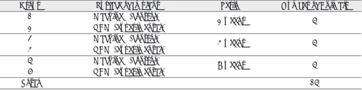
Fifteen adult New Zealand rabbits of both sex- es, weighing approximately 3.5 kg at the start, were used in this study. The rabbits were divided into six groups, each consisting of five animals that were planned to be sacrificed at 2, 4, and 8 weeks after the insertion of the implants. The animals were kept in standard cages and fed standard diet (Table I).
2) Implants
Implants manufactured from grade 5 com- mercially pure titanium were used in this study.
The implants had a length of 10 mm and an out- er diameter of 2.4 mm and had a hexagonal impression on the top that fitted to the removal equipment. A total of ninety implants were divid- ed into two groups. Half of them were RBM Slimplant (YK-pros, Seoul, Korea) and the others were machined Slimplant (YK-pros, Seoul, Korea) (Fig. 1).
3) Implant surgery
The rabbits were anesthetized with intramuscular injections of Xylazine (Rompun, Bayer, Korea, 5mg/kg body weight) and Ketamine (Ketara,
Yu?han, Korea, 35 mg/kg body weight), and local anesthesia was injected into each implantation site with 2 % Lidocaine (with epinephrine 12.5 μg/ml, Yu han, Korea). Each 3 mm long inci- sion was made to expose the bone, and both skin and periosteum were retracted with cus- tom made wire retractors. The preparation of the bone site was done with burs under generous saline irrigation. Three RBM pin implants were inserted in every left tibia and three machined pin implants were placed in every right tibia in a way that they engaged two cortical layers. The implants were placed more than 5 mm apart from each other. After the implants insertion, the soft tissues were sutured to cover the implants.
Table I. Classification of experimental animals
Group Surface of Implant Period Number of Animals
1 Machined (control)
2 weeks 5
2 RBM (experimental)
3 Machined (control)
4 weeks 5
4 RBM (experimental)
5 Machined (control)
8 weeks 5
6 RBM (experimental)
Total 15
RBM: resorbable blast media
Fig. 1. Photograph of machined and RBM pin implants.
Post operatively, the animals received antibi- otics intramuscularly for three days.
4) Research protocol
Animals were anesthetized with the same pro- tocol at two, four, and eight weeks respectively after implant surgery, and the soft tissue of the tibia was exposed. Any bone covering the implant top was gently removed not to disturb removal torque measurement. The necessary force to unscrew the implants from the tibia was measured with a Tonichi torque gauge instrument (Tonichi, Tokyo, Japan) (Fig. 2).
5) Preparation of specimens
After measurements of removal torque, the animals were euthanized with intravenous over- dose of pentobarbital. The implants were unscrewed from the bone and the remaining bone was harvested and fixed in 10 % neutral buffered formaldehyde. Then, the bone samples were dehydrated with EDTA solution and sliced.
Specimens were stained with hematoxylin eosin, and observed under an Olympus BX 51 (Olympus Co., Tokyo, Japan) microscope.
Two implants had fractured above the bone level when removal torque was applied. The
fractured implants and bone were made non demineralization samples. The implants includ- ing surrounding bone were harvested en bloc and fixed in 10% paraformaldehyde. Subsequently, the samples were dehydrated in ascending ethanol, soaked into the Technovit 7200 VLC (Kultz, Germany) light cured resin for 5 days, and cured under the visual light for 16 hours. For microscopic observation, cuttings were made with diamond discs to a thickness of 200 μm.
Their preparation was completed by abrasion to a thickness of 25 μm. The cuttings were stained with hematoxylin eosin and observed with an Olympus BX 51 (Olympus Co., Tokyo, Japan) microscope.
6) Statistical analysis.
Mean removal torque measurements and stan- dard deviation were calculated with SPSS version 10.0 (SPSS Inc., Chicago, U.S.A.). Differences in mean peak torque values of RBM pin implants ver- sus machined pin implants were compared for sig- nificance with a paired student t test. One way analysis of variance (ANOVA) and Tukey HSD were used to compare significance differences among two, four, and eight weeks results.
7) Finite element analysis
Linear finite element analysis was conducted for comparison of bicortical implant fixation with monocortical implant fixation using 3G (Plasso tech Co., U.S.A.) software. Two kinds of implants with different lengths (5 mm and 10 mm) were used. For the bicortical fixation, the implant tip was anchored in the lower cortical bone. The bone and implant elements were treated as fully bonded, mimicking complete osseointegration. The implant and their components were modeled on the basis of drawings provided by Osstem (Seoul, Korea).
The mesh was constructed using Tetra hedral Fig. 2. Tohnichi torque gauge(Tohnichi MFG, Tokyo,
Japan).
elements. Three dimensional models of dental implants and their surrounding bone were applied for studying the bone loading patterns for 50 Ncm vertical and 50 Ncm oblique (30 degree angle) forces respectively. Bone tissue presents a wide variety of data regarding the mechanical properties of bone. For this study of the influence of implant fixation, the Young’s modulus was cho- sen 3 GPa for cancellous bone and 13.7 GPa for cor- tical bone. The Young’s modulus of titanium implant was 120 GPa. Poisson’s ratios of 0.3 for titanium implant and 0.33 for cortical and can- cellous bone was applied(Table II).
RESULTS
1) Removal torque
RBM pin implants displayed higher removal torque value compared to machined pin implants.
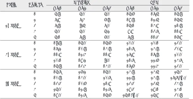
Removal torque values are presented in table III. The mean removal torque for machined pin implants at 2, 4, and 8 weeks (n= 15 for each time point) were 6.4 ± 1.3 Ncm, 14.6 ± 3.6 Ncm, and 16.4 ± 5.0 Ncm, respectively. The torque measurements for machined pin implants ranged from 5.0 to 26.2 Ncm. The mean removal torque for RBM pin implants at 2, 4, and 8 weeks (n= 15 for each time point) were 14.7 ± 3.6 Ncm, 22.5 ± 3.5 Ncm, and 26.4 ± 3.3 Ncm, respectively (Table IV.). The torque measurements for RBM pin implants ranged from 7.8 to 34.8 Ncm. Two RBM pin implants of 8 weeks group were fractured
while unscrewing and the values at the point of fracture were recorded. Considering the results of machined and RBM groups, the torque mea- surements were increased in both groups with time. The removal torque measurements yielded statistically significant differences between machined and RBM pin implants at all time peri- ods (p<0.05). Machined pin implants did not show significant difference at 8 weeks, com- pared to 4 weeks (p= 0.35). Statistically significant differences were seen at all time intervals for RBM pin implants(p<0.05)(Fig. 3).
2) Histologic findings
Microscopically, newly formed bone paralleling to the implant surfaces was observed inside cor- tical bone. New bone appeared to be formed mainly from the cortical bone because greater amount of new bone was observed near cortical bone. New bone surrounding implants surfaces showed in the form of implant threads and many osteoblasts producing an osteoid matrix were observed. Remaining bone showed implants were in contact with predominantly upper and lower cortical bones while the threads in the bone marrow were in contact with rather newly formed bone or with normal marrow tissue.
New bone in the form of threads was observed at 2 weeks; however, its maturity or density was low.
Maturity and amount of newly formed bone increased with time. A greater quantity of new- ly formed bone was observed in RBM pin implants Table II. Material value of linear FEM(finite element method) analysis of the implants
Part Material Elastic Modulus (GPa) Poisson’s Ratio
Screw Titanium 120 0.3
Bone Cortical bone 13.7 0.33
Cancellous bone 3.0 0.33
GPa: giga pascal
samples when they were compared with machined pin implants. Bone close to the interface with implants when they were removed from the bone showed crack or fracture tendency. Cracks or fractures were increased at 8 weeks than 2 or 4 weeks. RBM pin implants showed more fracture tendency than machined pin implants at any time periods(Fig. 4-6). Non mineralization sam- ples showed intimate contact between bone and RBM pin implants and newly formed bone along the RBM pin implant threads. The implant threads appeared mostly surrounded by the newly formed bone(Fig. 7).
Fig. 3. Comparison of removal torque between RBM and machined Pin Implants. RBM pin implants displayed higher removal torque value compared to machined pin implants.
Table IV. Group statistics of the implants (mean ± SD) (Ncm)
Surface type N 2weeks 4weeks 8weeks
Machined 15 6.4 ± 1.3 14.6 ± 3.6 16.4 ± 5.0
RBM 15 14.7 ± 3.6 22.5 ± 3.5 26.4 ± 3.3
RBM: resorbable blast media, N: number
Table III. Removal torque measurements (Ncm) of the implants
Period Animal Machined RBM
No.1 No.2 No.3 No.1 No.2 No.3
1 5.8 5.0 5.0 15.6 17.5 15.8
2 7.9 7.4 6.8 19.8 12.5 15.5
2weeks 3 7.8 8.5 7.0 15.1 14.9 21.8
4 5.0 5.0 5.2 9.9 13.7 11.9
5 5.1 7.8 6.0 7.8 11.3 16.9
1 18.8 15.0 15.6 20.0 20.1 23.0
2 17.2 10.8 14.8 21.7 24.8 30.9
4weeks 3 11.9 12.4 11.9 27.9 17.8 20.0
4 20.1 19.2 8.0 21.7 22.6 24.7
5 15.8 13.4 14.0 17.6 22.4 23.0
1 15.7 26.2 15.0 24.8 24.5 25.4
2 10.8 14.0 20.7 22.8 24.8 27.7(fx.)
8weeks 3 13.3 10.6 21.9 23.3 23.5 34.8
4 25.0 12.8 12.7 29.3 29.1 24.1
5 19.0 12.7 15.6 25.1(fx.) 25.9 30.8
RBM: resorbable blast media, fx.: implant fracture
2weeks 4weeks 8weeks 30
25 20 15 10 5 0
Fig. 4. RBM pin implant at 2 weeks (H-E stain, ×40).
Bone close to the interface with implants when they were removed from the bone showed a very small amount of crack or fracture tendency.
Fig. 5. RBM pin implant at 4 weeks (H-E stain, ×40).
Bone close to the interface with implants when they were removed from the bone showed a moderate amount of crack or fracture tendency.
Fig. 7. Undecalcified experimental specimen of RBM pin implant at 8 weeks(H-E stain, ×12.5). Non-mineral- ization samples showed intimate contact between bone and RBM pin implants and newly formed bone along the RBM pin implant threads. The implant threads appeared mostly surrounded by the newly formed bone.
Fig. 6. RBM pin implant at 8 weeks (H-E stain, ×40).
Bone close to the interface with implants when they were removed from the bone showed a large amount of crack or fracture tendency. Cracks or fractures were increased at 8 weeks than 2 or 4 weeks.
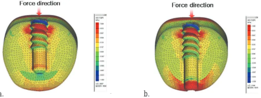
3) Finite element analysis
Maximum equilibratory stresses were record- ed as 17.9 MPa under vertical load and 106.3 MPa under 30�oblique load in monocortical fix- ation, and 19.0 MPa under vertical load and 115.8 MPa under 30�oblique load in bicortical fix- ation. Regarding the figures, stress patterns were showed as contour lines with different colors. The
effects of bicortical and monocortical fixation are illustrated in Fig. 8 for 50 Ncm vertical force and Fig. 9 for 50 Ncm oblique force, respectively.
In the pictures, compressive stress was shown in upper cortical bone and tensile stress in lower cor- tical bone in the bicortical models, which explains that engagement of two cortical bones which has more advantage in stress distribution.
DISCUSSION
Removal torque measurement has been used as a biomechanical measure of endosseous inte- gration of implants since Johansson and Albrektsson19firstly demonstrated the positive cor- relation between the removal torque of implants and the amount of bone to implant contact. They observed an increase of the amount of bone formed at the interface in proportion to an increase of removal torque. The removal torque
is defined as the amount of torque required to unscrew an implant from bone and is deter- mined by the total degree of contact between the implant surface and bone. It has been con- sidered as a reasonable method to assess the rel- ative amount of bone and implant contact though it has been regarded as a rough measure of osseointegration. The greater removal torque values may be interpreted as the higher osseoin- tegration.20
It is well known that surface modification can
a. b.
Fig. 9. Principal stress distribution.
a. Oblique load on a short implant with monocortical fixation.
b. Oblique load on a long implant with bicortical fixation.
Fig. 8. Principal stress distribution.
a. Vertical load on a short implant with monocortical fixation.
b. Vertical load on a long implant with bicortical fixation.
a. b.
enhance bone integration of titanium implants, which was observed as higher bone to implant con- tact ratio and greater resistance to removal torque compared to machined surfaces. Modifying topo- graphic characteristics can be achieved by plas- ma spray, abrasion, blasting, etching, or sintering.21,22 Surface roughness can increase surface area of implant adjacent to bone and improve cell attach- ment to the implant surface, resulting in increased biomechanical interaction of the implant with bone. Davies hypothesized that improved wet- tability and increased clot retention observed at acid etched implant surfaces resulted in improved osseointegration, perhaps through mechanisms that promote osteoconduction at the titanium surface.23
Blasted implants demonstrated a higher removal torque and interfacial bone contact than machined implants.16However, surface coatings or associ- ated debris from surfaces could be displaced into bone on implant placement. Recently, a new surface treatment called resorbable blast media (RBM) has been introduced as a surface treatment of implants. RBM involves blasting the implant sur- faces with coarsely ground calcium phosphate (par- ticle size, 180um × 425um), which gives the implant a coarse surface without leaving any residues. The calcium phosphate is a resorbable material that is not permanently imbedded into the surface of the implant primarily because of the passivation method used. Comparing RBM implants with machined implants, recent studies showed a significantly higher bone implant con- tact percentage and higher removal torque results in the RBM implants. According to the studies, the RBM surfaces could be considered more osteo- conductive than the machined surfaces.24-26
Implants placed in compact bone are more likely to ensure initial stability and, hence, better able to sustain against stresses. The cortical lamel- lar bone may heal with little interim woven bone
formation, ensuring good bonding strength while healing with endosteal implants.27,28Therefore, engagement of as much cortical bone as possible seems logical for improvement of implant stability.
Ivanoff17suggested that the bicortical implant fixation showed more favorable osseointegra- tion than monocortical fixation because most bone implant contacts were observed within the cortical bones. Engaging two cortical layers demonstrated a positive effect on osseointegration of titanium implants and resulted in higher removal torque values. In his study, the bicorti- cal implants required approximately twice as much of removal torque as the monocortical implants did in rabbit’s tibia. Gapski et al27 also suggested increasing contact surface with corti- cal bone through bicortical fixation was helpful for initial fixation and enabling immediate loading.
The bicortical implants and a higher bone to implant contact percentage showed high values of removal torque. Biomechanically, the concept of bicortical placement is certainly valuable since the higher initial stability of the fixture can be eas- ily achieved. Van Oosterwyck and colleagues29, using finite element analysis, showed that bicor- tical anchorage did not reduce the risk of marginal bone loss when implants were submitted to lat- eral forces. Most authors have consistently point- ed out that bicortical anchorage enhances implant stability.
From finite element analysis of this study, equi- libratory stress distribution showed that the stress on cervical cortex was greater in monocortical fixation than bicortical fixation because two cor- tical layers could distribute stress and lower cor- tical layer seemed to be helpful for additional fix- ation. Stress distribution was considered as an important factor because there was not much difference in comparison of maximum equili- bratory stress values between two groups. The cor- responding color scales suggested that stress
and strain were decreased in bicortical fixation in the cervical cortex as well as in the cancellous bone.
Simon and Caputo9reported that transitional nar- row implants having 1.8mm in diameter could be firmly osseointegrated and not be removed.
According to their study, the removal torque values were 16.1 ± 4.8 Ncm in the maxilla and 24.0
± 7.3 Ncm in the mandible. The mean torque val- ues were significantly higher in the mandible than in the maxilla, increasing with time. Several transitional implants were fractured while being removed because they were strongly integrated to the bone. Removal torque values of fractured implants were recorded 27�35 Ncm at 10 months after insertion.
In this study 2.4 mm in diameter RBM pin implants recorded 23�35 Ncm at 8 weeks and two of them were fractured while unscrewing. It explained narrow RBM pin implants could attain high bonding strength with bone through the blasting treatment.
Small diameter implants cause minimal dis- ruption to the periosteum. As a result, the reduced damage to insertion areas can contribute to expe- ditious tissue recovery, potentially decreased crestal bone loss, increased osseointegration rate, and ultimately more favorable patient accom- modations.30The use of small diameter implants is recommended in some cases where the avail- able bone is narrow for standard diameter implants. It could minimize peri implant tissue and bone damage through single minimally inva- sive placement procedure because the wider the implant, the greater the number of steps are required establishing the sites.31Moreover, repeat- ed surgical steps could result in over instru- mentation, directional changes, and loss of primary stability. Lack of interdental space is a common finding in cases of congenitally missing anterior teeth, closure of space after extractions, and after extraction of narrow diameter teeth such as the
lower and upper lateral incisors.10,13A small diam- eter implant may be indicated where the mesiodis- tal space available is less than 7mm because minimum of 1.5 mm of bone, cortical and can- cellous, is needed circumferentially throughout the length of the implant for favorable bone healing. The use of small diameter implants can prevent the need for bone reconstruction like bone grafting or other augmentation proce- dures. 32-35
Severely resorbed mandibles could result in mandibular fracture even with slight force. It is a serious complication that needs to be consid- ered, particularly in persons with an atrophic mandible, especially decreased bone mass.
Osteoporosis also makes an already atrophic mandible even weaker. The use of small diame- ter implants can decrease the risk of jaw fracture if they are used in edentulous patients. Likewise, it is recommended that implants be engaged to the inferior cortex of atrophic mandibles to obtain max- imum stabilization.36-38
In the present study, that RBM pin implants showed high removal torque values, implying increased bonding strength to bone, compared with machined pin implants. Increased bonding strength implies the possibility of adaptation of RBM pin implants in some limited area where occlusal force is not strong as final abutments. In addition, increased bone fracture tendency shown in specimens implies that RBM pin implant should not be used as transitional implants.
CONCLUSION
Within the limitation of this in vivo study, the following conclusions were drawn:
1) The removal torque value of RBM pin implants showed statistically significant increase compared to machined pin implants at 2, 4, and 8 weeks respectively (p<0.05).
2) The removal torque value of RBM pin implants at 2, 4, and 8 weeks was increased statistically significantly with time (p<0.05).
3) Bicortical fixation showed better stress dis- tribution compared with monocortical fixa- tion in a linear finite element method analy- sis.
4) RBM pin implants are not recommended as transitional implants because they showed much bone fracture in histologic specimens.
REFERENCES
1. Branemark P, Hansson B, Adell R, Breine U, Linstrom J, Hallen O et al. Osseointegrated implants in the treatment of the edentulous jaw. Experience from a 10 years period. Scand J Plast Reconstr Surg 1977;16:1-132.
2. Adell R. Tissue integrated prostheses in clinical den- tistry. Int Dent J 1985;35:259-65.
3. Albrektsson T, Dahl E, Enbom L, Engevall S, Engquist B, Eriksson AR, Feldmann G, Freiberg N, Glantz PO, Kjellman O. and et, al. Osseointegrated oral implants. A Swedish multicenter study of 8139 consecutively inserted Nobelpharma im- plants. J Periodontol 1988; 59:287-96.
4. Schnitman PA, Wohrle PS, Rubenstein JE.
Immediate fixed interim prostheses supported by two-stage threaded implants: Methodology and results. J Oral Implantol 1990;16:96-105.
5. Sagara M, Akagawa Y, Nikai H, Tsuru H. The effects of early occlusal loading on one-stage ti- tanium alloy implants in beagle dogs: A pilot study. J Prosthet Dent 1993;69:281-288.
6. Lee SB. Immediate temporary prosthesis and loading by using transitional pin implant. J Kyung Hee Medical. Vol.16 No2. 2000:126-135.
7. Froum S, Emtiaz S, Bloom MJ, Scolnick J, Tarnow DP. The use of transitional implants for immedi- ate fixed temporary prostheses in case of implant restoration. Pract Periodontics Aesthet Dent 1998;10:737-746; quiz 748.
8. Petrungaro PS. Fixed temporization and bone- augmented ridge stabilization with transitional im- plants. Pract Periodontics Aesthet Dent 1997;9:1071- 1078; quiz 1080.
9. Simon H, Caputo AA. Removal torque of imme- diately loaded transitional endosseous implants in human subjects. Int J Oral Maxillofac Implants 2002;17:839-845.
10. Mazor Z, Steigmann M, Leshem R, Peleg M. Mini- implants to reconstruct missing teeth in severe ridge deficiency and small interdental space: a 5-year case series. Implant Dent. 2004 Dec;13(4):336-41.
11. Nazarian A. Mini dental implants: immediate gratification for patient and provider. Dent Today.
2005 Oct;24(10):110, 112.
12. Zinsli B, Sagesser T, Mericske E, Mericske-Stern R.
Clinical evaluation of small-diameter ITI implants:
a prospective study. Int J Oral Maxillofac Implants.
2004 Jan-Feb;19(1):92-9.
13. Wojcik MS, Pokorny PH. Use of a one-stage nar- row-diameter implant to replace incisors. J Mich Dent Assoc 2006;88:42, 44-9.
14. Carlsson L, Rostlund T, Albrektsson B, Albrektsson T. Removal torques for polished and rough titanium implants. Int J Oral Maxillofac Implants 1988;3:21- 24.
15. Tjellstrom, Jacobsson M, Albrektsson T. A Removal torque of osseointegrated craniofacial implants: A clinical study. Int J Oral Maxillofac Implants 1988;3:287-289.
16. Wennerberg A, Albrektsson T, Andersson B, Krol JJ. A histomorphometric and removal torque study of screw-shaped titanium implants with three different surface topographies. Clin Oral Implants Res 1995;6:24-30.
17. Ivanoff CJ, Sennerby L, Lekholm U. Influence of mono- and bicortical anchorage on theintegra- tion of titanium implants. A study in the rabbit tib- ia. Int J Oral Maxillofac Surg 1996;25:229-235.
18. Pierrisnard L, Renouard F, Renault P, Barquins M.
Influence of implant length and bicortical an- chorage on implant stress distribution. Clin Implant Dent Relat Res 2003;5:254-62.
19. Johansson C, Albrektsson T. Integration of screw implants in the rabbit: A 1-year follow-up of removal torque of titanium implants. Int J Oral Maxillofac Implants 1987;2:69-75.
20. Cho SA, Park KT. The removal torque of titanium screw inserted in rabbit tibia treated by dual acid etching. Biomaterials 2003;24:3611-7.
21. Sennerby L, Dasmah A, Larsson B, Iverhed M.
Bone tissue responses to surface-modified zirconia implants: A histomorphometric and removal torque study in the rabbit. Clin Implant Dent Relat Res 2005;7 Suppl 1:S13-20.
22. Li D, Ferguson SJ, Beutler T, Cochran DL, Sittig C, Hirt HP, Buser D. Biomechanical comparison of the sandblasted and acid-etched and the machined and acid-etched titanium surface for dental implants.
J Biomed Mater Res 2002;60:325-32.
23. Davies JE. Mechanisms of endosseous integra- tion. Int J Prosthodont 1998;11:391-401.
24. Gonshor A, Goveia G, Sotirakis E. A prospective, multicenter, 4-year study of the ACE surgical re- sorbable blast media implant. J Oral Implantol 2003;29:174-180.
25. Piattelli M, Scarano A, Paolantonio M, Iezzi G, Petrone G, Piattelli A. Bone response to machined and resorbable blast material titanium implants: An experimental study in rabbits. J Oral Implantol 2002;28:2-8.
26. Sanz A, Oyarzun A, Farias D, Diaz I. Experimental study of bone response to a new surface treat- ment of endosseous titanium implants. Implant Dent 2001;10:126-31.
27. Gapski R, Wang HL, Mascarenhas P, Lang NP.
Critical review of immediate implant loading.
Clin Oral Implants Res 2003;14:515-527.
28. Friberg B, Sennerby L, Linden B, Grondahl K, Lekholm U. Stability measurements of one-stage Branemark implants during healing in mandibles.
A clinical resonance frequency analysis study.
Int J Oral Maxillofac Surg 1999;28:266-72.
29. Van Oosterwyck H, Duyck J, Vander Sloten J, Van der Perre G, De Cooman M, Lievens S, Puers R, Naert I. The influence of bone mechanical prop- erties and implant fixation upon bone loading around oral implants. Clin Oral Implants Res 1998;9:407-418.
30. Cehreli MC, Akca K. Narrow-diameter implants as terminal support for occlusal three-unit FPDs:
a biomechanical analysis. Int J Periodontics Restorative Dent 2004;24:513-9.
31. English C, Bahat O, Langer B, Sheets CG. What are the clinical limitations of wide-diameter (4 mm or greater) root-form endosseous implants? Int J Oral Maxillofac Implants 2000;15:293-6.
32. Comfort MB, Chu FC, Chai J, Wat PY, Chow TW.
A 5-yearprospective study on small diameter screw-shaped oral implants. J Oral Rehabil 2005
;32:341-5.
33. Davarpanah M, Martinez H, Tecucianu JF, Celletti R, Lazzara R. Small-diameter implants: indica-
tions and contraindications. J Esthet Dent 2000;
12:186-94.
34. Hallman M. A prospective study of treatment of se- verely resorbed maxillae with narrow nonsub- merged implants: results after 1 year of loading. Int J Oral Maxillofac Implants 2001;16:731-6.
35. Barber HD, Seckinger RJ. The role of the small-di- ameter dental implant: a preliminary report on the Miniplant system. Compendium 1994;15:1390, 1392.
36. Laskin DM. Nonsurgical management of bilater- al mandibular fractures associated with dental implants: report of a case. Int J Oral Maxillofac Implants 2003;18:739-44.
37. Goodacre CJ, Kan JY, Rungcharassaeng K. Clinical complications of osseointegrated implants. J Prosthet Dent 1999;81:537-52.
38. Murata T, Yamashita Y, Kurokawa H, Takahashi T. Dental rehabilitation using an implant-sup- ported overdenture after repair of a fracture in a se- verely resorbed edentulous mandible: a case report.
Int J Oral Maxillofac Implants 2004;19:749-52.
Reprint request to:
RICHARDLEESUNGBOK, D.M.D., M.S.D., PH.D.
DEPARTMENT OFPROSTHODONTICS, SCHOOL OF DENTISTRY, KYUNG HEEUNIVERSITY
#1, HOEGI-DONG,DONGDEAMOON-GU,SEOUL130-702, KOREA [email protected]