MICROPATTERNED GROOVES AND ACID- ETCHING ON TITANIUM SUBSTRATA ALTER
VIABILITY AND GENE EXPRESSION OF ADHERED HUMAN GINGIVAL FIBROBLASTS: A PILOT STUDY
Suk-Won Lee, D.D.S., M.S.D.1, Su-Yeon Kim, M.S., Ph.D.2, Keun-Woo Lee, D.D.S., M.S.D., Ph.D.3
1Department of Dentistry, College of Medicine, The Catholic University of Korea
2Research Institute of Medical Science, St. Vincent’s Hospital
3Department of Prosthodontics, Collage of Dentistry, Yonsei University
Statement of problem.Prior to determining an optimal width of micropatterned grooves pro- vided on titanium substrata, we have done a pilot study using surface topographies in com- bined microm and submicrom levels.
Purpose.The purpose of this study was twofold 1) to assess the proliferation and 2) to ana- lyze the expression of genes encoding the intracellular signaling proteins involved in cell-sub- stratum adhesions and adhesion-dependent G1 phase cell cycle progression of human gingival fibroblasts plated on smooth and microgrooved/acid-etched titanium substrata.
Material and methods.Three groups of titanium discs as NE0 (smooth Ti substrata), E15 (Ti substrata with microgrooves of 15 μm of spacing and 3.5 μm in depth and with further acid- etching), and E30 (Ti substrata with microgrooves of 30 μm spacing and 3.5 μm in depth and with further acid-etching) served as the human gingival fibroblasts’substrata. Viability and proliferation of fibroblasts were determined using an XTT assay. Gene expressions of fibronectin, α5 integrin, CDK4, and p27kipwere analyzed in RT-PCR. Cell-substratum interactions were analyzed in SEM.
Results.From the XTT assay at 24 h incubation, the mean optical density (OD) value of E15 was significantly greater than the values of E30 and NE0. At 48 and 96 h however, the mean OD values of E30 were significantly greater than the values of E15 and NE0. No differences in the expression of PCR transcripts at 96 h incubations were noted between groups, whereas at 48 h, an unexpected increase in the expression of all the transcripts were noted in E15 compared with other two groups. Fibroblasts were observed to orient and adhere inside the microgrooves.
Conclusion. Micropatterned grooves and acid-etching on Ti substrata alter viability and gene expression of adhered human gingival fibroblasts.
Key Words
Microgrooves, Acid-etching, Fibroblast, Viability, Gene expression
J Korean Acad Prosthodont : Volume 45, Number 3, 2007
※This study was supported by St. Vincent’s Hospital Research Grants in 2006.
P
rovision of microtopography such as micropat- terns or microgrooves on titanium substrata has been introduced in special relation to studying cell- substratum interactions of soft tissue cells around titanium oral implants.1,2In the studies reporting the changes in cell shape in relations to micro- grooves, human fibroblasts grown on such sub- strata compared to those on the smooth ones were reported to be significantly elongated and orientated along the grooves, leading to an increase in the amount of fibronectin3or alterations in the expression of numerous genes responsible for various biological activities.4As a pilot study using surface topographies in combined microm and submicrom levels, the purpose of this study was twofold1) to assess viability and proliferation and 2) to ana- lyze the expression of genes encoding the intra- cellular signaling proteins involved in the events of cell-substratum adhesions and adhesion- dependent G1 phase cell cycle progression of human gingival fibroblasts plated on smooth and microgrooved/acid-etched titanium sub- strata.
MATERIAL AND METHODS CELL CULTURE
Healthy gingival tissues were obtained from patients who underwent oral surgery for remov- ing impacted wisdom teeth at St. Vincent’s Hospital Department of Dentistry. In all cases, tis- sues were obtained from subjects following informed consent as prescribed in an approved St.
Vincent’s Hospital Institutional Review Board (IRB) protocol. Obtained connective tissues were cut into small pieces and placed in Petri dishes (direct explant method) in Dulbecco’s modified Eagle’s medium (DMEM, Gibco BRL, Grand
Island, NY, USA) supplemented with antibiotics and were kept overnight at 4℃.
Cells or explants were suspended in DMEM supplemented with 10 % fetal bovine serum (FBS, Sigma-Aldrich, Co., St. Louis, MO, USA) and antibiotics. When cells reached 80% confluence (about once per week), they were removed and sus- pended using a trypsin-EDTA solution (0.25%
trypsin and 0.1% glucose dissolved in 1 mM of EDTA-saline, Sigma-Aldrich Co., Louis, MO, USA), washed, centrifuged and resuspended. Finally, human gingival fibroblasts were seeded for sub- culture at a cell population density of 2×10⁴ cells/ml in 6-well plastic culture dishes in DMEM supplemented with 10% FBS and antibiotics. In all experiments in this study, the culture medium was changed every second day after seeding.
FABRICATION OF TITANIUM SUBSTRATA
Commercially pure titanium (Ti) discs were mechanically polished (Ra ≤ 0.06 μm) and used as the culture substrata in the control group, NE0 (smooth Ti substrata with neither micropat- terned grooves nor acid-etching), in this study. Ti discs with continuous 15 and 30 μm-wide grooves were fabricated with photolithography (MEMSware, Kwangju, Gyeonggi, Korea). The widths of the microgrooves were chosen on the basis of the results from our previous unpublished study, in which the cells are allowed to settle themselves. Fabricated surfaces of the discs were further acid-etched using 1% hydrofluoric acid (HF) for approximately 30 sec. Prepared Ti discs were used as the culture substrata in the two experi- mental groups, E15 (micropatterned Ti substra- ta with continuous grooves of 15 μm of spacing and 3.5 μm in depth and with further acid-etching) and E30 (micropatterned Ti substrata with continuous grooves of 30 μm spacing and 3.5 μm in depth and with further acid-etching), in this study. Floors of
24-well tissue culture plates were removed and the remaining cylinders were attached onto the fab- ricated surfaces of the prepared Ti substrata using a silicone adhesive.
TIME-LINE ANALYSIS ON VIABILITY AND PROLIFERATION
Fibroblasts were trypsinized from subculture, plated on the Ti substrata at a cell population den- sity of 1×10⁴cells/ml and incubated in DMEM supplemented with 10% FBS and antibiotics for 24, 48, 72, and 96 h according to the previously described time line of cultured cells’proliferation in microenvironments.5The viability and pro- liferation of fibroblasts were determined by XTT assay.6In brief, an XTT labeling mixture was prepared by mixing 50 μl of XTT labeling reagent and 1 μl of electron coupling reagent. 50 μl of XTT labeling mixture was added per well and incubated for 2 h in a humidified incubator at 37℃ with 5%
CO2in 95% air. Absorbance (optical density, OD) of produced formazan transferred to 96- well plates was measured using ELISA analyzer (Spectra MAX 250, Molecular Devices Co., Sunnyvale, CA, USA) at 470 nm with a refer- ence wavelength at 650 nm. An additional purpose of the XTT analysis to the assessment of viabili- ty and proliferation was to determine the optimal time of incubation at which fibroblasts plated on microgrooved/acid-etched Ti substrata were expected to show significant alterations in gene expression compared with those on smooth Ti sub-
strata. Experiments were repeated independently in triplicate. Differences in the mean optical den- sity (OD) values between groups were analyzed using one-way ANOVA.
ANALYSIS ON GENE EXPRESSION
Cultured human gingival fibroblasts (3rd-4th pas- sage) were trypsinized and plated on the Ti sub- strata of NE0, E15, and E30 at a cell population den- sity of 1×10⁴cells/ml in DMEM supplemented with 10% FBS and antibiotics. At 48 and 96 h plating and incubation, expression of fibronectin, α5 integrin, CDK4, and p27kipgenes were analyzed in reverse transcriptase-polymerase chain reaction (RT-PCR) (Table I). The PCR primer of βactin was used as the housekeeping gene.
Interactions between fibroblasts and Ti sub- strata at 24 h incubation were analyzed in scan- ning electron microscopy (SEM).
RESULTS
TIME-LINE ANALYSES ON VIABILITY AND PROLIFERATION
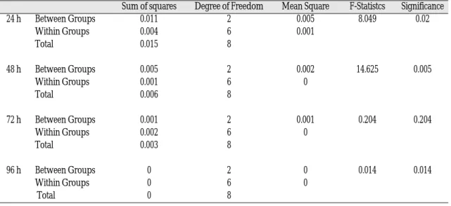
In ANOVA, the mean OD values from the XTT assays at 24, 48, and 96 h incubation were sig- nificantly different between and within all groups (p<0.05) (Table II). According to the data using the Ti discs with different surface topographies as the substrata, the results from the XTT assay were ver- ified to be significantly related between groups at Table I. Gene-Specific Primers used in RT-PCR
Target Sense Antisense Bp
Fibronectin 5’-CGAACATCCACACGGTAG-3’ 5’-ATCACATCCACACGGTAG-3’ 639 α5 integrin 5’-ACCAAGGCCCCAGCTCCATTAG-3’ 5’-GCCTCACACTGCAGGCTAAATG-3’ 375 CDK4 5’-CCAAAGTCAGCCAGCTTGACTGTT-3’ 5’-CATGTAGACCAGGACCTAAGGACA-3’ 193 p27kip 5’-AAACGTGCGAGTGTCTAACGGGA-3’ 5’-CGCTTCCTTATTCCTGCGCATTG-3’ 454
24, 48, and 96 h incubation. Multiple comparisons of the fibroblast viability/proliferation data from the XTT assay at 24 h incubation showed the mean OD value of E15 (0.174) to be significantly greater than the values of E30 (0.106) and NE0 (0.097) (p<0.05). At 48 and 96 h however, the mean OD values of E30 (0.134 and 0.092 at 48 and 96 h, respectively) were significantly greater than the values of E15 (0.097 and 0.082) and NE0 (0.079 and 0.080) (p<0.05). All other compar- isons between groups were not statistically sig- nificant (Fig. 1).
ANALYSIS ON GENE EXPRESSION
At 48 and 96 h incubation, as determined to be the optimal from the XTT assay, gene expres- sion of fibronectin, α5 integrin, CDK4, and p27kip were noted in all groups and there were no dif- ferences in the expression of transcripts at 96 h. At 48 h however, an unexpected increase in the expression of all the transcripts were noted in E15 compared with other two groups (Fig. 2).
Table II. Analysis of variance (p < 0.05)
Sum of squares Degree of Freedom Mean Square F-Statistcs Significance
24 h Between Groups 0.011 2 0.005 8.049 0.02
Within Groups 0.004 6 0.001
Total 0.015 8
48 h Between Groups 0.005 2 0.002 14.625 0.005
Within Groups 0.001 6 0
Total 0.006 8
72 h Between Groups 0.001 2 0.001 0.204 0.204
Within Groups 0.002 6 0
Total 0.003 8
96 h Between Groups 0 2 0 0.014 0.014
Within Groups 0 6 0
Total 0 8
Fig. 1. Time-line analysis on viability. Fig. 2. Gene expression Analysis.
SCANNING ELECTRON MICROSCOPY
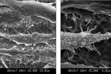
In SEM, orientation of fibroblasts parallel to the direction of the microgrooves in E15 and E30 were observed, whereas the cells in NE0 were observed to be oriented in random directions
(Fig. 3, 4 and 5). The majority of cells were found inside the microgrooves with increased formation of filopodia (Fig. 6 and 7). The width of a single fibroblast appeared to be identical to or slightly less than that of the microgrooves in E15.
Fig. 3. Human gingival fibroblasts adhered to smooth Ti substrata.
Fig. 4. Human gingival fibroblasts adhered to Ti substrata with microgrooves of 15 μm of spacing and 3.5 μm in depth and with further acid-etching.
Fig. 6. Human gingival fibroblasts inside the microgrooves of 15-μm spac- ing.
Fig. 7. Human gingival fibroblasts inside the microgrooves of 30-μm spac- ing.
Fig. 5. Human gingival fibroblasts adhered to Ti substrata with microgrooves of 30 μm spacing and 3.5 μm in depth and with further acid-etching.
DISCUSSION
In this study, the purpose of using XTT assay was twofold: 1) to assay the cell-substratum adhe- sion and the proliferation of fibroblasts plated on titanium substrata with different surface topogra- phies and 2) to determine, after plating, the opti- mal time of incubation at which fibroblasts on microgrooved/acid-etched Ti substrata would show significantly greater extent of the viability and proliferation compared with those on smooth Ti substrata. It was under the verifications of the abovementioned conditions that expression of genes related to cell-substratum adhesion and adhesion-dependent cell cycle progression of plated fibroblasts were analyzed in RT-PCR.
However, the data from the respective time-line of culture reveals the limitations of this pilot study because the mean OD values in all groups did not increase with time. Fibroblasts were not simultaneously plated on the prepared Ti substrata meaning that different cells in different envi- ronments were used in each group as well as in each time-line of culture. In addition, repeated uses of the fabricated Ti substrata may have lead to the result that the surfaces of the substrata became more and more unclean even after the thor- ough application of the ultrasonic device. Also, the surfaces of Ti discs were considered to be oxidized, that is, they underwent the process of corrosion after several usages. Taken all together, fibroblasts in this pilot study actually did not proliferate with time suggesting that the results of the time- line analysis from the XTT assay can only be translated in the differences in the viability or rather the ability to maintain, not the proliferation, of the cells between groups of substrata with differ- ent surface topographies. However, the results of our study from the XTT assay that E30 showed increased viability should not be underestimat- ed, for they clearly correspond with the results of
a previous study.7
The limitation of translation in the results from XTT assay could be considered one of the reasons for the unexpected results in RT-PCR. We expect- ed that the expression of genes involved in cell- substratum adhesions and adhesion-dependent G1 cell cycle progression would markedly increase in E30 compared with NE0 or E15 at 48 or 96 h incubations. Instead, all the genes in E15 showed increased expression compared with other groups suggesting that the increase in cellular length, which is elongation or polarity of the cell would increase the expression of genes involved in adhesion-dependent cell cycle progression. Indeed in SEM, a 15 μm-wide microgroove was observed to be capable of containing a single cell in itself.
The cells inside such grooves showed increases in elongation. This result corresponds with a previous study that 12.5 μm-wide microgrooves increased the expression of genes encoding various intra- cellular signaling proteins.4The effort to pro- vide various surface topographies on substrata such as our microgrooves is based on the dogma that the induction of changes in cell shape is tightly cou- pled to DNA synthesis and growth in adherent cells.8The changes in cell shape have mainly been induced by stimulating cell spreading in the field of mechanobiology where intracellular forces, also called cytoskeletal tension per se or cytoskeletal prestress, together with extracellular matrix (ECM) and cytoskeletal structure are con- sidered to play decisive roles in the control of var- ious biological activities, including cell prolif- eration and growth.9
A hypothesis of cellular mechanotransduction, where alterations in substrate surface topography may lead to changes in the probability of gene tran- scription,10provided us with the idea that surface topographies in combined microm and submicrom levels would also alter the gene expression of adhered cells.11A very recent study verified that
integrin and actin containing filopodia, the cellular organs involved in sensing the environment, formed the initial cell-matrix contacts and further generated mature focal adhesions.12 Another recent study suggested that actin and β1 inte- grin in cellular projections including filopodia at the leading edge of a migrating cell probe ligand and create sticky fingers.13Taken together, active formation of filopodia on acid-etched surfaces inside the microgrooves observed in this study puts an special emphasis on the use of such surface in the induction of periimplant soft tissue attachment.14 CONCLUSION
Human gingival fibroblasts were plated and incu- bated on three groups of titanium discs as NE0 (smooth Ti substrata), E15 (Ti substrata with microgrooves of 15 μm of spacing and 3.5 μm in depth and with further acid-etching), and E30 (Ti substrata with microgrooves of 30 μm spacing and 3.5 μm in depth and with further acid-etching).
From the results of the time-line viability/pro- liferation analysis using an XTT assay, gene expression analysis in RT-PCR, and on SEM observations, it can be concluded that Ti discs with surface topographies of combined microm and sub- microm levels such as microgrooved/acid-etched Ti substrata alter viability and gene expression of adhered human gingival fibroblasts.
REFERENCES
1. Brunette DM, Chehroudi B. The effects of the sur- face topography of micromachined titanium sub- strata on cell behavior in vitro and in vivo. J Biomech Eng 1999;121:49-57.
2. Jansen JA, den Braber ET, Walboomers XF, de Ruijter JE. Soft tissue and epithelial models. Adv Dent Res 1999;13:57-66.
3. Chou L, Firth JD, Uitto VJ, Brunette DM Substratum
surface topography alters cell shape and regu- lates fibronectin mRNA level, mRNA stability, secretion and assembly in human fibroblasts. J Cell Sci 1995;108:1563-1573.
4. Dalby MJ, Riehle MO, Yarwood SJ, Wilkinson CD, Curtis AS. Nucleus alignment and cell signaling in fibroblasts: response to a micro-grooved topog- raphy. Exp Cell Res 2003;284:274-282.
5. Bottaro DP, Liebmann-Vinson A, Heidaran MA.
Molecular signaling in bioengineered tissue mi- croenvironments. Ann N Y Acad Sci 2002;961:143- 153.
6. Roehm NW, Rodgers GH, Hatfield SM, Glasebrook AL. An improved colorimetric assay for cell pro- liferation and viability utilizing the tetrazolium salt XTT. J Immunol Methods 1991;142:257-265.
7. Sun F, Casse D, van Kan JA, Ge R, Watt F.
Geometric control of fibroblast growth on pro- ton beam-micromachined scaffolds. Tissue Eng 2004;10:267-272.
8. Folkman J, Moscona A. Role of cell shape in growth control. Nature 1978;273:345-349.
9. Ingber DE. Tensegrity II. How structural net- works influence cellular information processing net- works. J Cell Sci 2003;116:1397-1408.
10. Dalby MJ. Topographically induced direct cell mechanotransduction. Med Eng Phys 2005;27:730- 741.
11. Zinger O, Anselme K, Denzer A, Habersetzer P, Wieland M, Jeanfils J, Hardouin P, Landolt D.
Time-dependent morphology and adhesion of osteoblastic cells on titanium model surfaces fea- turing scale-resolved topography. Biomater 2004;25:2695-2711.
12. Partridge MA, Marcantonio EE. Initiation of at- tachment and generation of mature focal adhesions by integrin-containing filopodia in cell spread- ing. Mol Biol Cell 2006;17:4237-4248.
13. Galbraith CG, Yamada KM, Galbraith JA.
Polymerizing actin fibers position integrins primed to probe for adhesion sites. Science 2007;315:992- 995.
14. Kim H, Murakami H, Chehroudi B, Textor M, Brunette DM. Effects of surface topography on the connective tissue attachment to subcutaneous im- plants. Int J Oral Maxillofac Implants 2006;21:354- 365.
Reprint request to:
KEUN-WOOLEE, D.D.S., M.S.D., Ph.D.
DEPARTMENT OFPROSTHODONTICS,COLLEGE OFDENTISTRY, YONSEIUNIVERSITY
134, SHINCHON-DONG,SEODAEMUN-GU,SEOUL,120-095, KOREA [email protected]