서론
부분 혹은 완전 무치악 환자의 수복시 임플란트 고정성 보철 물은 예지성 있는 치료방법으로 선택될 수 있다. 고정성 임플란 트 보철물은 치아 삭제 과정이 필요하지 않고 가철성 국소의치 에 비해 높은 저작력을 얻을 수 있으며, 환자가 치아를 모두 가 지고 있다는 심리적 안정을 얻을 수 있다.1 하지만 임플란트의 장 기적인 안정을 위해서는 이상적인 위치와 각도의 임플란트 식립 이 동반 되어야 하며, 이를 위해서는 잔존 골의 양과 질 혹은 해 부학적 구조에 대한 정확한 평가가 필요하다. 하지만 Panorama radiograph 등의 방사선 사진은 환자의 자세와 악골의 만곡에 의 한 왜곡이 존재할 수 있고, 3차원적인 정보를 제공하지 않아 악 골의 폭경이나 신경과 같은 해부학적 구조에 대한 평가가 불가 능하다.2 이런 한계를 극복하기 위해 cone beam computerized
tomography (CBCT)가 소개되었고, 임플란트 진단 소프트웨어 와의 접목으로 보다 정교한 임플란트 수술 전 계획을 수립하는 것이 가능하게 되었다. 또한 computer-aided design / computer- aided manufacturing (CAD/CAM)의 발전을 통하여 계획했던 위치에 보다 근접하게 임플란트의 식립이 가능한 surgical guide 의 제작이 가능함으로써 guided surgery가 시도되고 있다.3,4
Guided surgery는 미래의 보철물에 대한 정보를 취합하여 가 상 계획 소프트웨어를 통해 stereolithographic template를 제작 하는 기술을 통해 술자가 계획한 최종 보철물에 적합한 위치에 임플란트 식립을 가능하게 한다. 이는 보다 재현성 있는 치료 계 획 설정이 가능하고, 하치조신경, 상악동 등의 해부학적 구조의 손상을 감소시키므로 안정적인 임플란트 식립을 시행할 수 있 다. 또한 임플란트 식립 전에 임시 보철물의 제작을 가능하게 하 고 필요 시 임플란트 식립 후 즉시 하중을 쉽게 가할 수 있어 술
Implant-Guided Surgery를 이용한 고정성 임플란트 보철물의 전악 수복 증례
김성모1 박진홍2 류재준1 신상완2 이정열2*
1
고려대학교 안암병원 치과 보철과,
2고려대학교 구로병원 임상치의학 연구소
Full mouth rehabilitation with Implant-Guided Surgery and Fixed prosthesis
Seong-Mo Kim
1, Jin-Hong Park
2, Jae-Jun Ryu
1, Sang Wan Shin
2, Jeong-Yol Lee
2*
1Department of Dental Prosthodontics, Anam Hospital, Korea University, Seoul, Republic of Korea
2Department of Prosthodontics, Institute for Clinical Dental Research, Korea University Medical Center, Korea University, Seoul, Republic of Korea
The development of cone beam computerized tomography (CBCT) allows three-dimensional analysis of the patient’s anatomy. The surgical guide is a combination of CBCT, computer-aided design/computer-aided manufacturing (CAD/CAM) and implant diagnostics software, which allows well planned prostheses design and ideal implant place- ment. Guided surgery minimizes possible anatomical damage and allows for more reproducible treatment planning. In this case, the operation time was shortened by using a surgical guide for multiple implants placement in a fully edentulous patient. Immediate loading were performed more easily using preliminary preparation of provisional pros- thesis. The patient was satisfied with improved esthetics and chewing function. (J Korean Acad Prosthodont 2018;56:126-33)
Keywords: Surgical stent; Guided surgery; Immediate loading; Implant supported fixed denture; Computer-aided design/computer-aided manufacturing (CAD/CAM)
*Corresponding Author: Jeong Yol Lee
Department of Prosthodontics, Institute for Clinical Dental Research, Korea University Medical Center, Korea University, 148 Gurodong-ro, Guro-gu, Seoul 08308, Republic of Korea +82 (0)2 2626 1922: e-mail, [email protected]
Article history: Received September 14, 2017 / Last Revision December 20, 2017 / Accepted December 21, 2017
2018 The Korean Academy of Prosthodontics
This is an Open Access article distributed under the terms of the Creative Commons Attribution Non-Commercial License (http://creativecommons.org/
licenses/by-nc/3.0) which permits unrestricted non-commercial use, distribution, and reproduction in any medium, provided the original work is properly cited.
c cc
자 및 환자의 불편감을 최소한으로 감소시킬 수 있다.
본 증례에서는 상, 하악 완전 무치악 환자를 대상으로 surgical stent를 이용하여 고정성 임플란트 보철물로 수복하였으며, 만족 할 만한 결과를 얻었기에 이를 보고하는 바이다.
증례
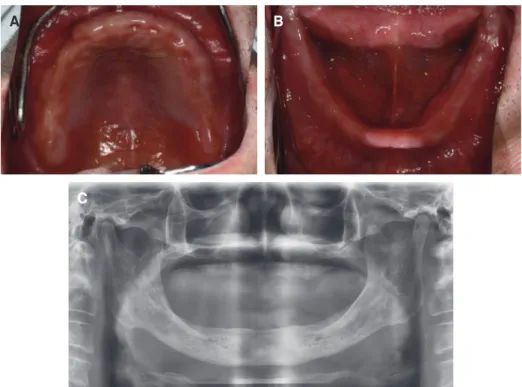
고려대학교 구로병원 치과 보철과에 59세의 남자 환자가 전악 보철 수복을 주소로 내원하였다. 상하 완전 무치악 상태로 3개 월 전 상, 하악의 잔존치아를 심한 치주염을 이유로 전악 발치하 였으며, 특별한 pre-medical history는 존재하지 않았다. 임상 및 방사선 검사 결과를 바탕으로 환자와 상의 후 임플란트를 이용 한 고정성 보철물 제작을 계획하였다 (Fig. 1).
수술 전 임시의치를 제작하였으며, 이를 복제하여 방사선적 진 단을 위한 stent로 활용하였다. Radiographic stent 착용 후 촬영 한 CBCT와 진단모형을 스캔한 자료를 중첩하여 임플란트 식 립을 계획하였다. 임플란트 진단 소프트웨어 (In2 guide on de- mand 3D software, Cybermed, Seoul, Korea)를 이용하였으며 치료 계획에 따라 surgical guide 및 provisional restoration을 제 작하였다 (Fig. 2).
국소마취 하 surgical guide를 이용하여 임플란트 식립을 시행 하였다. Surgical guide를 상악, 하악 각각 3개의 fixation pin으 로 고정하였으며, 상악에는 #14, #15, #16, #21, #23, #24, #26의 위치에 총 7개, 하악에는 #33, #34, #36, #43, #44, #46의 위치에
총 6개의 Neo CMI implant (NeoBiotech, Seoul, Korea) 를 식립 하였다. 단, drilling 후 surgical stent는 구강 내에서 제거 하였으 며, 이후 flap을 거상하여 최종 임플란트 위치를 결정하였다 (Fig.
3). 임플란트 식립 2일 후 낮은 초기 고정력을 보인 #21i, #23i,
#43i를 제외한 임플란트에 기성 abutment 체결 후 미리 제작해 놓은 PMMA provisional prosthesis을 relining 후 장착하여 즉시 하중을 가하였다 (Fig. 4).
수술 후 임시 보철물을 정기적으로 유지관리 및 평가하였으며 임플란트 식립 2개월 후 #21i, #23i 이차 수술을 시행하였다. 이 차 수술 1개월 후 primary impression 채득하여 individual tray 제작 시행하였으며, polyvinylsiloxane 인상재 (Aquasil XLV, Dentsply Sirona, York, PA, USA)와 Pick-up impression coping 을 사용해 보철물 제작을 위한 정밀인상채득을 시행하였다. 자 가중합 레진과 파라핀 왁스를 이용해 교합제를 제작한 뒤 교합 평면과 수직고경을 결정하였다. 악간관계 채득 및 안궁이전 시행 하여 교합기에 마운팅하였다. 교합기에 마운팅 된 주모형상에서 임시 보철물을 제작하였으며, 이를 구강내 장착하였다 (Fig. 5).
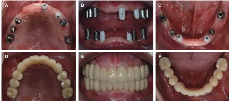
장착 3개월 후 provisional prosthesis를 scanning 하여 custom abutment와 최종 보철물을 CAD/CAM 방식으로 제작하였다.
제작된 보철물을 구내 시적하여 악간관계와 심미를 재평가하였 으며, 완성된 보철물을 구강 내에 장착 후 정기적인 유지관리 치 료 시행하였다. 관찰기간 동안 임상적, 방사선학적 검사 결과 이 상소견 발견되지 않았으며, 환자는 향상된 저작 능력과 심미적 인 안모에 만족한 모습을 보였다 (Fig. 6, Fig. 7).
Fig. 1. Preoperative view (A) upper, (B) lower intraoral photo, (C) panoramic view.
A B
C
Fig. 2. Fabrication of surgical stent. (A) Treatment planning of upper jaw, (B) Upper surgical stent, (C) Treatment planning of lower jaw, (D) Lower surgical stent.
A
C
B
D
Fig. 3. Implant placement. (A) Sitting of upper stent, (B) Upper jaw after initial drilling, (C) Upper jaw after surgery, (D) Sitting of lower stent, (E) Lower jaw after initial drilling, (F) Lower jaw after surgery.
D E F
A B C
Fig. 4. Delivery of provisional restoration. (A) Connection of ready-made abutment on upper jaw, (B) Upper jaw occlusal view after delivery of provisional res- toration, (C) Panoramic view, (D) Connection of ready-made abutment on lower jaw, (E) Lower jaw occlusal view after delivery of provisional restoration, (F) Follow up check after 2 month.
D E F
A B C
Fig. 5. Fabrication of second provisional prosthesis. (A) Master cast of upper jaw, (B) Master cast of lower jaw, (C) Jaw relation, (D) Face-bow trasfer, (E) De- sign of prosthesis, (F) Provisional restoration.
D E F
A B C
고찰
Guided surgery는 지지되는 조직에 따라 tooth, bone, mucosa supported guides로 분류될 수 있다.6 본 증례는 완전 무치악 환 자에서의 임플란트 수술로 mucosa supported guide를 제작하여 임플란트 식립수술을 진행하였다. Mucosa supported guide를 이용한 flapless surgery는 수술 후 환자가 느끼는 통증과 hemor- rhage를 줄일 수 있는 장점이 있으나,7 tooth supported guide에
비해 정확도가 부족하며, 임플란트 식립 위치의 오류가 나타날 수 있다.6 또한 bony interference에 의한 surgical guide의 불완 전한 seating이 발생할 수 있다.8 따라서 본 증례에서 drilling 후 flap을 거상하여 임플란트의 최종 식립 위치를 확인하였다.
Di Giacomo 등9은 deviation이 발생하는 이유로 guide tube 의 micromotion, guide drill 보다 0.2 mm 더 넓은 guide tube 의 직경, 낮은 해상도의 CBCT, surgical guide의 misplacement, osteotomy 후 freehand placement 등을 제시하였다. Schneider 등10은 systematic review를 통한 meta 분석 연구를 조사한 결과 수술전 계획단계에서 software 상의 임플란트와 실제 식립된 임 플란트 간의 평균 1.07 mm의 entry point deviation, 1.32 mm의 apex deviation, 0.43 mm height deviation, 5.26°의 angular de- viation을 보고하였다. 한편 surgical guide의 오차로 인한 해부 학적 구조의 침범을 방지하기 위해 최소한 2 mm의 safety zone 이 제시되었다.11
Raico Gallardo 등6은 supported tissue 간 오차를 측정하였으 며 그 결과 tooth supported guide가 가장 높은 정확도를 보였 고 mucosa supported guide, bone supported guide 순서로 낮은 정확도를 보였다. Bone supported guides는 많은 flap elevation 이 요구되므로 reflected tissue에 의한 오차가 발생할 수 있어 본 증례에서는 선택되지 않았으며 정확도를 높이기 위하여 3개의 fixation pin을 사용해 stent를 고정한 mucosa supported guide를 선택하였다. 본 증례에서 임플란트 식립시의 오차는 미리 제작 된 provisional prosthesis가 문제 없이 장착될 정도로 적은 수준 의 차이를 보였다. 이 보철물은 정밀인상채득 후 새롭게 제작된 Fig. 6. Delivery of final prosthesis. (A) Upper jaw occlusal view after abutment connection, (B) Frontal view after connection of abutment, (C) Lower jaw occlu- sal view after abutment connection, (D) Upper jaw occlusal view after delivery of final prosthesis, (E) Frontal view after delivery of final prosthesis, (F) Lower jaw occlusal view after delivery of final prosthesis.
D E F
A B C
Fig. 7. Analysis of smile line.
provisional prosthesis가 장착되기 전까지 문제 없이 사용되었다.
Guided surgery가 가지는 complication은 크게 surgical com- plication과 prosthetic complication으로 분류 할 수 있다.9 Sur- gical complication에는 surgical guide의 파절, drilling 시의 과 도한 열 발생, 구치부 식립시 limited interocclusal distance로 인 한 기구도달의 문제점 등이 존재한다.8,10-12 특히 surgical guide 장착 후 drilling을 시행할 경우 metal sleeve에 의한 irrigation의 방해가 발생할 수 있으므로 주의가 필요하며, 원활한 기구 조작 을 위해 술 전 개구량의 평가 또한 반드시 필요하다. Prosthetic complication에는 prosthesis misfit, 과도한 occlusal adjustment, incomplete seating 등의 문제점이 존재한다.9,10
본 증례에서는 임플란트 식립 2일 후 미리 제작된 provisional prosthesis를 연결하여 immediate loading을 시행하였다. 이전 의 immediate loading process는 기존에 사용하던 의치를 수정 하여 임플란트에 연결하거나 식립된 임플란트에 전통적인 인 상 과정을 통하여 provisional prosthesis를 제작하였다.1 그러 나 chair time이 증가하고 환자의 불편감을 야기할 수 있으며 제 작 과정이 번거롭다는 단점이 존재하였다.4 본 증례에서 수술 전 CAD/CAM으로 제작된 provisional restoration은 수립된 수 술 계획에 따라 제작되어 쉽게 식립된 임플란트에 장착할 수 있 었으며 chair time을 줄여 환자와 술자의 피로도를 줄일 수 있었 다. Immediate loading의 고려사항으로는 적절한 임플란트의 수 와 위치, 감소된 occlusal table, rigid splinting, 주기적인 occlu- sion check 등이 제시되며, 그 중 안정적인 초기고정이 매우 중요 한 요소로 여겨진다.13 따라서 본 증례에서 낮은 insertion torque value가 관찰된 #21i, #23i, #43i 임플란트를 제외하였으며, 1 Unit으로 제작된 provisional prosthesis로 rigid splinting을 시행 하고 주기적인 내원을 통해 교합조정을 시행하였다. Immediate loading을 시행함으로써 저작기능을 회복함과 함께 심미적인 안 모를 얻게 되었고 그로 인하여 환자가 치아 상실 전의 모습을 되 찾을 수 있었다.
본 증례에서는 provisional prosthesis의 형태를 스캐닝하여 지 대주와 중첩시키는 double scanning technique을 이용하여 최종 보철물을 제작하였다. 모델스캐너를 이용하여 임시 보철물을 스 캔하였으며, 최종보철물의 디자인 후 사용되었던 임시 보철물 이 미지 파일과 중첩하여 제작함으로써, 임시 보철물의 조절사항을 최종보철물에 반영하도록 하였다. 3개월 간 사용하였던 임시 보 철물과 유사한 형태로 제작되었기에 환자는 보다 쉽게 최종 보 철물에 적응할 수 있었으며, 최종 보철물 장착 후의 환자의 안모 에 대해서도 파악할 수 있었다.
결론
본 증례는 전악 무치악 환자에게 surgical guide를 이용하여 적 합한 수술 전 보철계획과 임플란트 식립을 시행하였으며 imme- diate loading을 통해 환자의 기능을 즉시 개선할 수 있었다. 이를 통하여 만족할만한 심미적, 기능적인 결과를 보였다.
ORCID
Seong-Mo Kim https://orcid.org/0000-0002-7225-0901 Jin-Hong Park https://orcid.org/0000-0002-3220-9912 Jae-Jun Ryu https://orcid.org/0000-0001-6903-5955 Sang Wan Shin https://orcid.org/0000-0002-3100-2020 Jeong-Yol Lee https://orcid.org/0000-0003-3079-0376
References
1. Misch CE. Contemporary implant dentistry. 3rd ed. St. Louis:
Mosby; 2008. p. 17-21, 814-8.
2. BouSerhal C, Jacobs R, Quirynen M, van Steenberghe D. Im- aging technique selection for the preoperative planning of oral implants: a review of the literature. Clin Implant Dent Relat Res 2002;4:156-72.
3. Widmann G, Bale RJ. Accuracy in computer-aided implant surgery-a review. Int J Oral Maxillofac Implants. 2006;21:
305-13.
4. Abboud M, Wahl G, Guirado JL, Orentlicher G. Application and success of two stereolithographic surgical guide systems for implant placement with immediate loading. Int J Oral Maxillofac Implants 2012;27:634-43.
5. Hultin M, Svensson KG, Trulsson M. Clinical advantages of computer-guided implant placement: a systematic review.
Clin Oral Implants Res 2012;23:124-35.
6. Raico Gallardo YN, da Silva-Olivio IRT, Mukai E, Morimoto S, Sesma N, Cordaro L. Accuracy comparison of guided sur- gery for dental implants according to the tissue of support: a systematic review and meta-analysis. Clin Oral Implants Res 2017;28:602-12.
7. Arisan V, Karabuda CZ, Ozdemir T. Implant surgery using bone- and mucosa-supported stereolithographic guides in totally edentulous jaws: surgical and post-operative outcomes of computer-aided vs. standard techniques. Clin Oral Implants Res 2010;21:980-8.
8. Yong LT, Moy PK. Complications of computer-aided-design/
computer-aided-machining-guided (NobelGuide) surgical implant placement: an evaluation of early clinical results. Clin Implant Dent Relat Res 2008;10:123-7.
9. Di Giacomo GA, da Silva JV, da Silva AM, Paschoal GH, Cury PR, Szarf G. Accuracy and complications of computer- designed selective laser sintering surgical guides for flapless dental implant placement and immediate definitive prosthesis installation. J Periodontol 2012;83:410-9.
10. Schneider D, Marquardt P, Zwahlen M, Jung RE. A sys- tematic review on the accuracy and the clinical outcome of computer-guided template-based implant dentistry. Clin Oral Implants Res 2009;20:73-86.
11. D’haese J, Van De Velde T, Komiyama A, Hultin M, De Bruyn H. Accuracy and complications using computer- designed stereolithographic surgical guides for oral rehabilita-
tion by means of dental implants: a review of the literature.
Clin Implant Dent Relat Res 2012;14:321-35.
12. Misir AF, Sumer M, Yenisey M, Ergioglu E. Effect of surgi- cal drill guide on heat generated from implant drilling. J Oral Maxillofac Surg 2009;67:2663-8.
13. Aparicio C, Rangert B, Sennerby L. Immediate/early loading of dental implants: a report from the Sociedad Española de Implantes World Congress consensus meeting in Barcelona, Spain, 2002. Clin Implant Dent Relat Res 2003;5:57-60.
Implant-Guided Surgery를 이용한 고정성 임플란트 보철물의 전악 수복 증례
김성모1 박진홍2 류재준1 신상완2 이정열2*
1
고려대학교 안암병원 치과 보철과,
2고려대학교 구로병원 임상치의학 연구소
Cone beam computerized tomography (CBCT)의 발전은 환자의 해부학적 구조를 3차원적으로 분석할 수 있게 하였다. Surgical guide는 CBCT와 CAD/CAM, 임플란트 진단 소프트웨어의 접목을 통해 미래의 보철물을 계획하고 적합한 위치에 임플란트를 식립할 수 있게 한다. Guided surgery
를 통해 해부학적 구조물에 대한 침범을 최소한으로 줄일 수 있고 보다 재현성 있는 치료계획의 설정이 가능하다. 본 증례는 전악 무치악 환자에
게 surgical guide를 이용하여 다수의 임플란트를 식립한 증례로 수술시간을 단축시킬 수 있었으며 임시 보철물을 미리 제작함으로써 보다 쉽게 immediate loading을 시행할 수 있었다. 환자는 개선된 안모와 저작기능에 만족하였다. (대한치과보철학회지 2018;56:126-33)
주요단어: Surgical stent; Guided surgery; 즉시하중; 고정성 임플란트 보철물; Computer-aided design/computer-aided manufacturing (CAD/CAM)
*교신저자: 이정열
08308 서울 구로구 구로동로 148 고려대학교 구로병원 임상치의학연구소 02 2626 1922: e-mail, [email protected]
원고접수일: 2017년 9월 14일 / 원고최종수정일: 2017년 12월 20일 / 원고채택일: 2017년 12월 21일
2018 대한치과보철학회
이 글은 크리에이티브 커먼즈 코리아 저작자표시-비영리 3.0 대한민국 라이선스에 따라 이용하실 수 있습니다.
c cc