2015, Vol. 59, No. 1
Printed in the Republic of Korea
http://dx.doi.org/10.5012/jkcs.2015.59.1.103 단신
(Notes)
Synthesis and Crystal Structures of Copper(II) Complexes with Schiff Base Ligands
Bon Kweon Koo
Department of Life Chemistry, Catholic University of Daegu, Gyeongbuk 712-702, Korea E-mail: [email protected]
(Received November 10, 2014; Accepted December 17, 2014) Key words: Crystal structure, Cu(II) complex, Schiff base, Benzilic acid
Schiff bases containing N, O/S donor atoms and their metal complexes have received considerable attention owing to their intriguing structural motifs1,2 and industrial, antifun- gal, and biological applications.3,4 Recently, dinuclear copper(II) complexes, [Cu2(L)2(CH3COO)](ClO4) (L = 2- benzoylpyridine S-methyldithiocarbazate) and [Cu2L2(SO4)]
(L = di-2-pyridyl ketone N(4),N(4)-(butane-1,4-diyl)thio- semicarbazone) in which sulfur atom from Schiff bases ligand together with acetate or sulfate oxygen atoms bridges the two copper(II) ions, respectively, were reported.5,6
Although many metal(II)-Schiff base complexes have been reported,7,8 dinuclear Cu(II) complexes consist of Schiff base and other the second ligand, especially, dinuclear complexes linked through the other second ligand except the solvent molecule or the counter anion of metal salt as starting material have been little published.9,10 In this context, we reported dinuclear Cu(II) complexes of an acetyl pyridine based dithiocarbamate or 4-phenyl-3-thiosemicarbazide with benzilic acid (H2BA) as second ligand.11 For metal ions, benzilic acid can provide a variety of chelating and/or bridging coordination modes displayed by the carboxylic or hydroxy groups.12 Many frameworks constructed by BA2− or HBA− with transition metal ions or rare earth ions have been reported, mainly using hydrothermal synthetic method.13−15 In this study, we extended these systems to a dinuclear compound of copper(II)-benzilate with 2-acetyl- pyridine/2-benzoylpyridine based benzhydrazide (Scheme 1) in order to study the crystal structure as part of our long- standing interest in synthesizing and extending the dimen- sionality of coordination compounds with mixed N,O/S coordination spheres.
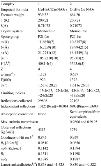
The molecular structures of 1 and 2 were determined using single crystal X-ray diffraction techniques. The crystallographic data and refinement parameters are listed in Table 1. Selected bond parameters are listed in Table 2. The molecular structure
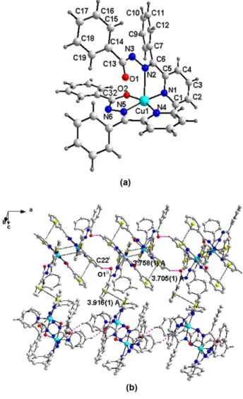
of complex 1 contains dinuclear [Cu2 (apb)2(HBA)(ClO4)] in which two enolate oxygen atoms and bidentate bridging HBA− anion bridge the two copper(II) centers and lattice water molecule (Fig. 1a). Each of the two copper atoms in [Cu2(apb)2(HBA)(ClO4)] has different coordination envi- ronments.
Cu1 adopts a five-coordinate square-pyramidal (τ = 0.27, the geometric parameter τ = |β − α|/60, where β and α are the two largest angles around the central atom; τ = 0 and 1 for the perfect square pyramidal and trigonal bipyramidal geometries, respectively.16) with a N2O3 donor. The pyridine nitrogen (N1), the azomethine nitrogen atom (N2) and the enolate oxygen atom (O1) together with the carboxyl oxygen atom (O(3)) from HBA− ligand comprise the basal plane of the square-pyramid whereas the enolate oxygen atom (O2) of another ligand occupies the apical position. The maximum displacement of them from the coordination plane is 0.005 (4) Å (N2). Cu1 atom displaces 0.022(1) Å out of the plane. The behavior of apb− results in the formation of two five-membered rings around Cu1 atom. Two planes [Cu1−N1−C5−C6−N2 and Cu1−N2−N3−C8−O1] are nearly planar with mean deviation of 0.048(5) and 0.018(4) Å, respectively, the dihedral angle between them being 2.3(1)°.
The environment around Cu2 atom can be best described as a distorted octahedral geometry in a N2O4 manner. One
Scheme 1. Chemical structures of Schiff bases and their abbre- viations.
104 Bon Kweon Koo
oxygen atom (O2), one azomethine nitrogen atom (N5) and one pyridine nitrogen atom (N4) from one apb− ligand and one oxygen atom (O4) from HBA− ligand occupy the basal positions, the two remaining positions in the octahedral geometry are the axial ones which are occupied by one enolate oxygen atom (O1) from the second ligand and one perchlorate oxygen atom (O6). The large difference between the two Cu−O distances (Cu1−O1 1.950(4)/Cu1−O3 1.900(3) Å in the basal plane and Cu1−O2 2.507(4) Å in the apical position) can be ascribed to a Jahn-Teller distortion.17 Copper(II) dimers with such similar a phenolato oxygen bridging have also been observed in other salicylaldehyde semicarbazone copper(II) complexes.18 The C−O bond length increases from the typical of ketonic linkage 1.23 Å8,19 to 1.303(6) for C8−O1 and 1.311(6) Å for C22−O2, respectively. Similarly, C8−N3 and C22−N6 suffer a significant decrease from the normal single bond of 1.52 Å20 to 1.333(7) and 1.318(6) Å, respectively.
These changes with enhanced single and double bond characters indicate the ligand in the present complex is coordinated in its deprotonated enolate form as observed in most complexes derived from carbamate/semicarbazone.21,22 The two apb− ligands have slightly different Cu−N(pyridine) bond distances and they are longer than the Cu−N(azomethine) dis- tances, this may be attributed to the fact that the azomethine nitro- gen is a stronger base compared with the pyridine nitrogen.23,24 The HBA− ligand together with each enolate oxygen atom bridges two Cu(II) centers. The Cu...Cu distance is 3.166(1) Å which is smaller than 3.245 Å25 of dimeric Cu(II) complex with μ-phenoxy bridge. Two planes [C31- C36 and C37−C42] in HBA− are nearly planar with mean deviation of 0.006 (6) Å, respectively, the dihedral angle between them being 76.1(2)o. The Cu1−O1−Cu2 bridg- ing angle is 84.3(1)o, which is much smaller than the value of 87.22o reported for the complex having the μ-pheno- lato and μ-acetate bridging groups as the smallest value found in this series of complexes, whereas the Cu1−O2−Cu2 angle is 89.2(1)o and is comparable to the value of 89.3(1)o for another bridging angle in the previous complex.26 The dinuclear units are further extended by the intermolecular hydrogen bonds (C39−H39...N6i, C40−H40...O6i) between the HBA− ligand and the oxygen atoms of the perchlorate Table 1. Crystal data and structure refinement for complexes1 and 2
Complex 1 2
Empirical formula C42H36ClCu2N6O9.5 C38H28 Cu N6O2
Formula weight 939.32 664.20
T (K) 200(2) 200(2)
λ(Å) 0.71073 0.71073
Crystal system Monoclinic Monoclinic
Space group P2(1)/n P2(1)/c
a (Å) 11.8658(7) 10.8357(8)
b (Å) 16.7559(10) 19.9942(15)
c (Å) 21.2741(12) 16.4349(13)
β (°) 105.2210(10) 95.603(2)
V (Å3) 4081.4(4) 3543.6(5)
Z 4 4
μ (mm−1) 1.173 0.657
F(000) 1920 1372
θ (°) 1.57 to 28.27 1.61 to 26.05 Limiting indices −15≤h≤15, −22≤k≤16,
−24≤l≤28 −13≤h≤13, −24≤k ≤22,
−13≤k≤20 Reflections collected 29808 22102
Independent reflections 10125 [R(int) = 0.0914] 6993 [R(int) = 0.0890]
Absorption correction None Semi-empirical from equivalents Max. and min. transmission 0.9806 and 0.9195 Observed reflections
[I≥2σ(I)] 4315 3759
Goodness-of-fit on F2 0.845 0.959
R1 [I≥2σ(I)] 0.0510 0.0656
wR2 [I≥2σ(I)] 0.1142 0.1594
R1 0.1511 0.1198
wR2 0.1749 0.1887
Largest peak and hole (e Å−3) 0.858 and −1.425 0.830 and −0.522
Table 2. Selected bond lengths (Å) and angles (˚) for complexes 1 and 2
Complex 1
Cu1–N2 1.893(5) Cu1–O3 1.900(3)
Cu1–O1 1.950(4) Cu1–N1 2.012(4)
Cu2–N5 1.927(4) Cu2–O4 1.949(3)
Cu2–O2 1.966(3) Cu2–N4 1.995(4)
N2–Cu1–O3 178.30(16) N2–Cu1–O1 80.52(18) O3–Cu1–O1 100.33(15) N2–Cu1–N1 81.65(19) O3–Cu1–N1 97.48(16) O1–Cu1–N1 162.14(16) N5–Cu2–O4 173.88(16) N5–Cu2–O2 79.96(16) O4–Cu2–O2 96.49(14) N5–Cu2–N4 80.67(18) O4–Cu2–N4 102.81(16) O2–Cu2–N4 160.63(16) Complex 2
Cu1–N5 1.922(4) Cu1–O2 1.979(3)
Cu1–N1 1.994(4) Cu1–N4 2.003(4)
Cu1–N2 2.396(4) C13–O1 1.214(5)
C32–O2 1.282(5) C13–N3 1.377(6)
C32–N6 1.327(6)
N5–Cu1–O2 79.77(15) N5–Cu1–N1 171.37(16) O2–Cu1–N1 97.21(14) N5–Cu1–N4 80.92(16) O2–Cu1–N4 160.14(15) N1–Cu1–N4 101.24(16) N5–Cu1–N2 113.03(15) O2–Cu1–N2 82.10(13) N1–Cu1–N2 74.27(14) N4–Cu1–N2 109.90(14)
anion or azomethine nitrogen atom to form 1D chain net- work along a-axis. The hydrogen bonds between perchlo- rate oxygen atom and water molecule (O8ii...O10wiii, O9...O10wiii) and π−π interactions between pyridine and phenyl rings of neighboring molecules with inter-ring dis- tance of 3.830(1) Å link the dimers into1D chain along c- axis, to form 2D network (Fig. 1b). In addition, the hydrogen
bonds [C3iv−H3...O10wv= 2.69 Å,C3iv...O10wv = 3.44(1) Å,
∠C3iv−H3...O10 wv= 136.3o andC12vi−H12...O10wv = 2.69 Å, C12vi...O10wv= 3.63(1) Å,∠C12vi−H12...O10wv= 167.1o; symmetry codes:(iv) 0.5+x, 0.5−y, 0.5+z. (v) 0.5+x, 1.5−y,
−0.5+z. vi) 0.5+x, 1.5−y, 0.5+z.] between apb− anion and water molecule finally bring the formation of 3D supra- molecular network.
The complex 2 consists of one Cu(II) ion and two coor- Figure 1. (a) Molecular structure of complex 1 with atomic
labeling (50% thermal ellipsoids). (b) The 2D layer framework of complex 1 formed by H-bond [C39−H39...N6i= 2.68 Å, C39...N6i = 3.49(1) Å, ∠C39−H39...N6i = 144.2o, C40−H40...O6i
= 2.47 Å, C40...O6i = 3.26(1) Å, ∠ C40−H40...O6i= 140.9o, O8ii...
O10wiii = 3.04(1) Å, O9...O10wiii = 3.05(1) Å] and π-π interac- tions [Cg1(N1C1−C5)...Cg2(C9−C14) = 3.830(1) Å]. Symmetry codes: i) 1+x, y, z; ii) 1−x, 1−y, 1−z; iii) 0.5−x, −0.5+y, 1.5−z.
Cg is centroids of the plane. H-bonds and π−π interactions have been shown as dashed and dotted lines, respectively. All H-atoms and water molecule have been omitted for clarity.
Figure 2. (a) Molecular structure of complex 2 with atomic labeling (50% thermal ellipsoids). (b) The 2D layer framework by the π−π interactions [Cg5(N1−C5)...Cg6(C14−C19) = 3.916(1) Å) between two 1D chains formed by H-bond [C22i−H22...O1ii
= 2.42 Å, C22i...O1ii = 3.22(1) Å, ∠ C22i-H22...1ii = 141.9o, Sym- metry codes: (i) −1+x, −1+y, z. (ii) 1−x, 1−y, 1−z.] and π−π inter- actions [Cg1(N1−C5)...Cg2(C26−C31) = 3.758(1), Cg3(N4−
C24)...Cg4(C33−C38) = 3.705(1) Å] in 2 along the a-axis. Cg is centroid of the plane. H-bonds and π−π interactions have been shown as dashed and dotted lines, respectively. All H-atoms, except hydrogen atoms for H-bobding in (b) have been omitted for clarity.
106 Bon Kweon Koo
dinated bpb− ligands (Fig. 2(a)). Cu(II) ion center adopts a five-coordinate square-pyramidal (τ = 0.19)16 with a N4O donor. The basal plane (N1N4N5O2) is nearly planar (mean deviation 0.016(3) Å) and the Cu1 is displaced by 0.122(1) Ǻ from the plane. The structural data (Table 2) are in agree- ment with those of the Cu(II) complexes which exhibit the similar geometry.27 Three planes [N1C1−C5, C7−C12 and C14−C19] in bpb− are nearly planar with the largest devi- ations of atoms from the mean planes: C5; 0.01(1), C8;
−0.006(5), and C17; 0.012(6) Å, respectively. The dihe- dral angles between two phenyl rings (N1C1−C5 and C7−
C12) and between two planes N1C1−C5 and C14−C19 are 69.24(16) and 14.03(17)°, respectively. In the structure, it is notable that oxygen atom from one of two bpb− is not coordinated to Cu(II) center. This result is supported by the x-ray data, that is, the C13−O1 is somewhat shorter than the C32−O2 as expected from oxygen O1 involved in un- coordination. However, the C32−O2 bond length increases from the typical of ketonic linkage 1.23 Å.8,19 Similarly, C32− N6 suffers a significant decrease from the normal single bond of 1.52 A to 1.327(6) Å.20 These changes with enhanced single and double bond characters indicate the ligand is coor- dinated in its deprotonated enolate form. The principal feature of the crystal packing is the formation of a two-dimen- sional network by C−H...O and π...π contacts. Two mono- meric molecules in the unit cell are linked by π...π stacking between pyridine and phenyl rings of neighboring mol- ecules with inter-ring distance of 3.705(1) (N4−C24...
C33−C38) and 3.758(1) Å (N1−C5...C26−C31) to give dimeric structure. The dimeric unit is further extended by inter- molecular H-bonds (C22−H22...O1) to give 1D chain net- work along a-axis. This chains also constructs 2D plane by the π...π stacking between pyridine and phenyl rings (C14−C19...N1−C5; centroid-to-centroid distance of 3.916 (1) Å) (Fig. 2(b)).
In conclusion, two copper(II) complexes with 2-acetylpyr- idine/2-benzoylpyridine based benzhydrazide have been synthesized from the methanolic solution of copper(II) perchlorate, Schiff base, and benzilic acid in the presence of triethylamine. Complex 1 is dinuclear structure of [Cu2(apb)2(HBA)(ClO4)] in which two copper(II) centers are bridged by bidentate HBA− and two enolate oxygen atoms of Schiff base. While, complex 2 is simple mono- meric 5-coordinate square pyramidal complex. Unfortu- nately, it has failed to obtain the dimeric complex of bpb− through benzilic acid in this work. The development of synthetic routes to the systems containing Schiff base and HBA− is still required for the rational design and synthe- sis.
EXPERIMENTAL Chemicals and Measurements
All chemicals are commercially available and were used as received without further purification. The ligands, Hapb and Hbpb were prepared as described in the literature.28,29 Elemental analyses (CHN) were performed on a Vario EL EA-Elementar Analyzer.
Preparation of [Cu2(apb)2(HBA)(ClO4)]·0.5H2O (1) To a methanolic solution (20 mL) of Hapb (0.239 g, 1 mmol) was added Cu(ClO4)2·6H2O (0.371 g, 1 mmol). To the result ing solution was added a methanolic solution (3 mL) of H2
BA (0.228 g, 1 mmol) and triethylamine (0.101 g, 1 mmol).
The solution turned to green and was refluxed for 3 h to yield green solid. The solid was isolated by filtration and airdried. The yellow filtrate was kept at room temperature to give green block crystals in good quality for X-ray crys- tallography. Yield: 63% (0.296 g) based on Cu. Anal. Calcd.
for C42H36N6O9.5ClCu2: C, 53.70; H, 3.86; N, 8.95. Found:
C, 53.83; H, 3.92; N,8.92%.
Preparation of [Cu(bpb)2] (2)
The compound was prepared similarly by the method described above for the preparation of 1, with use of Hbpb instead of Hapb ligand. The green solid was recrystallized from the CH3OH/(C2H5)2O = 1:2 solution to give black block crystals in good quality for X-ray crystallography. Yield:
72% (0.480 g) based on Cu. Anal. Calcd. For C38H28N6O2Cu:
C, 68.72; H, 4.25; N, 12.65. Found: C, 68.45; H, 4.41;
N,12.30%.
X-ray Structure Determination
Single crystals of 1 and 2 were obtained by the method described in the above procedure. Structural measurement for the complexes were performed on a Bruker SMART APEX CCD diffractometer using graphite monochroma- tized Mo-Kα radiation (λ = 0.71073 Å) at the Korea Basic Science Institute. The structure was solved by direct method and refined on F2 by full-matrix least-squares procedures using the SHELXTL programs.30 All non-hydrogen atoms were refined using anisotropic thermal parameters. The hydrogen atoms were included in the structure factor calcula- tion at idealized positions by using a riding model, but not refined. Images were created with the DIAMOND pro- gram.31
Acknowledgments. This research was supported by the Research Grants of Catholic University of Daegu in
2014. The author also acknowledges the Korea Basic Sci- ence Institute for providing the crystal structure results.
Supporting Information. Crystallographic data for the structure reported here have been deposited with CCDC (Deposition No. CCDC-1027686 (1) and -1031327 (2)).
The data can be obtained free of charge via http://www.ccdc.
cam.ac.uk/conts/retrieving.html or from CCDC, 12 Union Road, Cambridge CB2 1EZ, UK, E-mail: deposit@ccdc.
cam.ac.uk.
REFERENCES
1. Kumar, G.; Gupta, R. Chem. Soc. Rev. 2013, 42, 9403.
2. Champness, N. R. Dalton Trans. 2011, 40, 10311.
3. Sayin, K.; Kariper, S. E.; Sayin, T. A.; Karakas, D. Spec- trochim. Acta, Part A 2014, 133, 348.
4. Grivani, G.; Ghavami, A.; Kučeráková, M.; Dušek, M.;
Khalaji, A. D. J. Mol. Struct. 2014, 1076, 326.
5. Li, M. X.; Zhang, L. Z.; Chen, C. L.; Niu, J. Y.; Ji, B. S.
J. Inorg. Biochem. 2012, 106, 117.
6. Philip, V.; Suni, V.; Kurup, M. R. P.; Nethaji, M. Polyhedron 2006, 25, 1931.
7. Koo, B. K. J. Korean Chem. Soc. 2013, 57, 859.
8. Patole, J.; Sandbhor, U.; Padhye, S.; Deobagkar, D. N.;
Anson, C. E.; Powell, A. Bioorg. Med. Chem. Lett. 2003, 13, 51.
9. Massoud, S. S.; Guilbeau, A. E.; Luong, H. T.; Vicente, R.; Albering, J. H.; Fischer, R. C.; Mautner, F. A. Polyhedron 2013, 54, 26.
10. Sanchez, H.; Server-Carrio, J.; Escriva, E.; Soto, L.; Gar- cia-Lozano, J.; Sancho, A.; de Arellano, C. R. Polyhedron 2013, 50, 38.
11. Koo, B. K. Bull. Korean Chem. Soc. 2013, 34, 3233.
12. Qiu, Y.; Wang, K.; Liu, Y.; Deng, H.; Sun, F.; Cai, Y. Inorg.
Chim. Acta 2007, 360, 1819.
13. Li, C.-H.; Xie, H.-P.; Tan, X.-W.; Yang, Y.-Q.; Li, W. Jiegou Huaxue 2010, 29, 1317.
14. Halder, P.; Chakraborty, B.; Banerjee, P. R.; Zangrando, E.; Paine, T. K. Cryst. Eng. Commun. 2009, 11, 2650.
15. Cui, L.-F.; Li, D.-M.; Wu, J.-F.; Cui, X.-B.; Wang, T.-G.;
Xu, J.-Q. J. Mol. Struct. 2006, 797, 34.
16. Addison, A. W.; Rao, T. N.; Reedijk, J.; van Rijn, J.; Ver- schoor, G. C. J. Chem. Soc., Dalton Trans. 1984, 1349.
17. Baldini, M.; Belicchi-Ferrari, M.; Bisceglie, F.; Pelosi, G.;
Pinelli, S.; Tarasconi, P. Inorg. Chem. 2003, 42, 2049.
18. Vafazadeh, R.; Esteghamat-Panah, R.; Willis, A. C.; Hill, A. F. Polyhedron. 2012, 48, 51.
19. Mangalam, N. A.; Sivakumar, S.; Sheeja, S. R.; Kurup, M. R. P.; Tiekink, E. R. T. Inorg. Chim. Acta 2009, 362, 4191.
20. Dutta, S.; Basy, P.; Chakravorty, A. Inorg. Chem. 1991, 30, 4031.
21. Wilson, J. J.; Lippard, S. J. Inorg. Chem. 2011, 50, 3103.
22. Pal, I.; Dutta, S.; Basuli, F.; Goverdhan, S.; Peng, S.-M.;
Lee, G.-H.; Bhattacharya, S. Inorg. Chem. 2003, 42, 4338.
23. Sreekanth, A.; Kurup, M. R. P. Polyhedron 2004, 23, 969.
24. Li, M. X.; Zhang, L. Z.; Chen, C. L.; Niu, J. Y.; Ji, B. S.
J. Inorg. Biochem. 2012, 106, 117.
25. Patel, R. N.; Rawat, S. P.; Choudhary, M.; Sondhiya, V.
P.; Patel, D. K.; Shukla, K. K.; Patel, D. K.; Singh, Y.; Pandey, R. Inorg. Chim. Acta 2012, 392, 283.
26. Dutta, G.; Debnath, R. K.; Kalita, A.; Kumar, P.; Sarma, M.; Shankar, R. B.; Mondal, B. Polyhedron. 2011, 30, 293.
27. Anitha, C.; Sheela, C. D.; Tharmaraj, P. S.; Raja, J. Spec- trochim. Acta, Part A 2012, 98, 35.
28. Pelagatti, P.; Carcelli, M.; Pelizzi, C.; Costa, M. Inorg. Chim.
Acta 2003, 342, 323.
29. Mangalam, N. A.; Sivakumar, S.; Sheeja, S. R.; Kurup, M. R. P.; Tiekink, E. R. T. Inorg. Chim. Acta 2009, 362, 4191.
30. Sheldrick, G. M. SHELXTL, Version 6; Bruker AXS Inc.:
Madison, Wisconsin, USA, 2001.
31. Brandenburg, K. DIAMOND, Version 2.1; Crystal Impact GbR: Bonn, Germany, 1998.