Q Tan, et al
476 Ann Dermatol
Received August 19, 2016, Revised September 26, 2016, Accepted for publication October 19, 2016
Corresponding author: Hua Wang, Department of Dermatology, Children’s Hospital of Chongqing Medical University, No 136 2nd ZhongShan Rd, Yu Zhong District, Chongqing 400014, China. Tel: 86-23-63631445, Fax:
86-23-63622874, E-mail: huawangpfk@163.com
This is an Open Access article distributed under the terms of the Creative Commons Attribution Non-Commercial License (http://creativecommons.
org/licenses/by-nc/4.0) which permits unrestricted non-commercial use, distribution, and reproduction in any medium, provided the original work is properly cited.
Copyright © The Korean Dermatological Association and The Korean Society for Investigative Dermatology
pISSN 1013-9087ㆍeISSN 2005-3894 Ann Dermatol Vol. 29, No. 4, 2017 https://doi.org/10.5021/ad.2017.29.4.476
CASE REPORT
Pyoderma Gangrenosum in a Patient with X-Linked Agammaglobulinemia
Qi Tan, Fa-liang Ren1, Hua Wang2
Ministry of Education Key Laboratory of Child Development and Disorders, Key Laboratory of Pediatrics in Chongqing, 1Chongqing International Science and Technology Cooperation Center for Child Development and Disorders, 2Department of Dermatology, Children’s Hospital of Chongqing Medical University, Chongqing, China
X-linked agammaglobulinemia (XLA) is a primary im- munodeficiency disorder caused by germline mutations of B-cell tyrosine kinase (BTK) gene. It is characterized by de- creased serum immunoglobulins levels and circulating ma- ture B cells. This defect in humoral immunity leads to in- creased susceptibility to infection. Pyoderma gangrenosum (PG) is an uncommon, ulcerating, neutrophilic dermatosis.
Here we report PG in an 8-year-old patient with XLA. The pa- tient received intravenous immunoglobulin treatment in conjunction with prednisone and topical application of 0.03% tacrolimus ointment and the ulcer was almost com- pletely healed in the 2 weeks of follow-up. The coexistence has been rarely reported. XLA may be a possible cofactor in the pathogenesis of PG. (Ann Dermatol 29(4) 476∼478, 2017)
-Keywords-
Agammaglobulinemia, Immunity, Pyoderma gangrenosum, X chromosome
INTRODUCTION
X-linked agammaglobulinemia (XLA) is a primary im- munodeficiency first characterized by Bruton1 in 1952.
With an incidence of 1 in 100,000 to 1 in 200,000 of the population2, these individuals carry a mutation in the B-cell tyrosine kinase (BTK) gene encoding for a tyrosine kinase critical for B-cell maturation3. In myeloid and den- dritic cells, BTK has been found to be a component of Toll-like receptor (TLR) signaling, important for recog- nition of foreign pathogens4. The coexistence of pyoderma gangrenosum (PG) in a patient with XLA has been rarely reported. PG is a neutrophilic dermatosis that occurs both as a primary disorder as well as secondary to an under- lying disease. To date, only five previous reports have documented the association of PG with XLA5-9. We report herein a case of PG in a patient with XLA.
CASE REPORT
An 8-year-old boy was transferred to our hospital for man- agement of multiple, painful ulcers on the left lower extremity. The lesion began as a few small plaques on the left shin that gradually coalesced and broke down into an ulcer. Intravenous antibiotics were initiated but the ulcers continued to increase in size and number, which was as- sociated with increased pain. There was no history of trau- ma to the lower extremities. He had been diagnosed with XLA at two years of age when he presented with recurrent pneumonia, pleural effusion and otitis media. He receives intravenous immunoglobulin replacement of every four weeks.
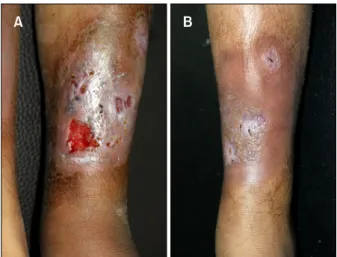
On clinical examination, the patient was febrile and had multiple ulcers with irregular, undermined, violaceous margins and indurated bases on his left shin (Fig. 1A). The
PG Related to XLA
Vol. 29, No. 4, 2017 477 Fig. 1. (A) The left shin ulcer with surrounding irregular,
undermined, violaceous margins and induration. (B) Nearly healed leg ulcer 2 weeks after treatment with intravenous immunoglobulin in conjunction with prednisone and topical application of 0.03% tacrolimus ointment.
Fig. 2. A mixed neutrophilc and lymphocytic infiltrate and hemorrhage in the upper and lower dermis (H&E; A: ×40, B: ×200).
right knee joint was swollen, warm, and tender with a de- tectable effusion, range of motion was limited by pain.
There were no skin lesions on the trunk or genitalia. There was no associated lymphadenopathy.
Laboratory investigations showed that the patient had a normal white cell count, but a mild anemia (hemoglobin, 83 g/L; red blood cell, 4.8×10⁹). Further laboratory data included an increased erythrocyte sedimentation rate (60 mm/h; normal range, 0∼15 mm/h) and C-reactive protein level (46 mg/L; normal range, 0∼8 mg/L). Immunological investigations were negative for antineutrophil cytoplas- mic antibodies and antinuclear antibody. Blood and bone marrow cultures and virology screens gave negative results. Flow cytometric evaluation of peripheral blood
lymphocytes showed markedly decreased numbers of CD19pos cells (0% of lymphocyte count), while other lym- phocyte subsets were normal. The immunoglobulin (Ig)A and IgM levels were below normal (IgA <0.068 g/L [normal range, 0.51∼2.59 g/L], IgM <0.054 g/L [normal range, 0.48∼2.26 g/L]), whereas the IgG level was in the lower range of normal (normal range, 5.28∼21.9 g/L).
The rest of the routine laboratory test results were within normal limits. During his hospitalization, the synovial flu- id was negative for infectious organisms. Magnetic reso- nance imaging (MRI) demonstrated soft-tissue inflamma- tion, and MRI revealed no evidence of osteomyelitis. The edge of the ulcer was biopsied and sent for histopatho- logic and microbiological cultures. The histopathological examination revealed a mixed neutrophilc and lympho- cytic infiltrate and hemorrhage in the upper and lower dermis (Fig. 2). Special staining (Gram stain, periodic acid-Schiff stain) did not reveal any microorganisms. The culture of a biopsy specimen taken from the edge of the ulcer was negative for bacteria, fungi, and atypical mycobacteria. Based on distinctive clinical features, ex- clusion of infectious origin, and histopathologic findings, the diagnosis of PG was established. The patient received intravenous immunoglobulin treatment in conjunction with prednisone (1 mg/kg/d) and topical application of 0.03% tacrolimus ointment. On follow-up 2 weeks later, the ulcer was almost completely healed (Fig. 1B) and swelling of knee joint was relieved and no recurrence has appeared in the 2 months of follow-up.
DISCUSSION
We encountered a case of PG in a Bruton’s XLA patient, in which defect in the BTK gene resulted in a decrease serum
Q Tan, et al
478 Ann Dermatol
levels of immunoglobulin subtypes. However, there have been no reports that demonstrate an association between XLA and PG. Although the pathophysiology of PG is poor- ly understood, it is thought to be immune-mediated9, spe- cially neutrophil chemotaxis and cytokine levels thought to be involved10. PG lesions either as a purely cutaneous manifestation or be associated with a systemic disease, in- cluding ulcerative colitis and Crohns disease, polyarthritis, IgA-gammopathy, malignancies and also with further con- ditions like chronic active hepatitis and Behcet syn- drome11. To date, most patients have been described in single case reports, the association between PG and XLA needs larger sample sized studies supported obviously.
Clinically PG starts with a sterile pustule, nodule or bulla that rapidly progresses and turns into painful ulceration with raised, undermined borders and necrotic eschar.
PG-like ulcer caused by Helicobacter cinaedi and Campylobacter species have been reported in a patient with X-linked agammaglobulinaemia2. There was no clear difference about the morphologic characteristics of the mucocutaneous lesions between those and this case. In our patient, Tissue cultures were negative for infectious organisms.
The treatment of PG is still a therapeutic challenge. Initial therapy for PG consists of corticosteroids, dapsone, mino- cycline, methotrexate, cyclosporine, mycophenolate and intravenous immunoglobulin12. The majority of reported cases of XLA-associated PG required the use of im- munosuppressive agents and immunoglobulin substitution for treatment. As BTK appears to be a component of TLR signaling, TLR activation leads to production of cytokines, notably tumor necrosis factor alpha (TNF-α), which pro- mote the inflammatory cascade9. Therefore, anti-TNF agents, such as etanercept, have also been anecdotally re- ported to be successful in the treatment of PG13. Several reports has been demonstrated an improvement of cuta- neous lesions of PG following topical treatment14. The combination use of intravenous immunoglobulin and pre- dnisone and topical tacrolimus ointment showed great im- provement in our patient. These cases suggest that treat- ment for PG should be individualized depending on the severity of the symptoms and underlying associated disease.
CONFLICTS OF INTEREST
The authors have nothing to disclose.
REFERENCES
1. Bruton OC. Agammaglobulinemia. Pediatrics 1952;9:722-728.
2. Dua J, Elliot E, Bright P, Grigoriadou S, Bull R, Millar M, et al. Pyoderma gangrenosum-like ulcer caused by Helicobacter cinaedi in a patient with x-linked agammaglobulinaemia.
Clin Exp Dermatol 2012;37:642-645.
3. Conley ME, Rohrer J, Minegishi Y. X-linked agammaglo- bulinemia. Clin Rev Allergy Immunol 2000;19:183-204.
4. Jefferies CA, Doyle S, Brunner C, Dunne A, Brint E, Wietek C, et al. Bruton's tyrosine kinase is a Toll/interleukin-1 receptor domain-binding protein that participates in nuclear factor kappaB activation by Toll-like receptor 4. J Biol Chem 2003;278:26258-26264.
5. Van der Hilst JC, Smits BW, van der Meer JW. Hypogam- maglobulinaemia: cumulative experience in 49 patients in a tertiary care institution. Neth J Med 2002;60:140-147.
6. Barrière H, Litoux P, Stalder JF, Berger M, Delahaye M.
Pyoderma gangrenosum associated with congenital hy- pogammaglobulinemia. Ann Dermatol Venereol 1979;106:
695-696.
7. Bloom D, Fisher D, Dannenberg M. Pyoderma gan- grenosum associated with hypogammaglobulinemia; report of two cases. AMA Arch Derm 1958;77:412-421.
8. Marcussen PV. Hypogammaglobulinemia in pyoderma gangrenosum. J Invest Dermatol 1955;24:275-280.
9. Schwartzfarb EM, Weir D, Conlan WA, Romanelli P, Kirsner RS. Pyoderma gangrenosum in a patient with Bruton's X-linked agammaglobulinemia: shared pathogenesis of altered tumor necrosis factor alpha? J Clin Aesthet Dermatol 2008;1:26-29.
10. Adachi Y, Kindzelskii AL, Cookingham G, Shaya S, Moore EC, Todd RF 3rd, et al. Aberrant neutrophil trafficking and metabolic oscillations in severe pyoderma gangrenosum. J Invest Dermatol 1998;111:259-268.
11. Necas M, Semrádova V, Vaskù V. Pyoderma gangraenosum associated with autoimmune thyreopathy and hyperandrogenic syndrome. Acta Dermatovenerol Alp Pannonica Adriat 2005;14:57-60.
12. Cafardi J, Sami N. Intravenous immunoglobulin as salvage therapy in refractory pyoderma gangrenosum: report of a case and review of the literature. Case Rep Dermatol 2014;6:239-244.
13. Charles CA, Leon A, Banta MR, Kirsner RS. Etanercept for the treatment of refractory pyoderma gangrenosum: a brief series. Int J Dermatol 2007;46:1095-1099.
14. Deckers-Kocken JM, Pasmans SG. Successful tacrolimus (FK506) therapy in a child with pyoderma gangrenosum.
Arch Dis Child 2005;90:531.