Role of heat shock protein 90
in angiotensin II-induced
myocardial hypertrophy
Kyung Hye Lee
Department of Medical Science
The Graduate School, Yonsei University
Role of heat shock protein 90
in angiotensin II-induced
myocardial hypertrophy
Directed by Professor Yangsoo Jang
Doctoral Dissertation
submitted to the Department of Medical Science,
the Graduate School of Yonsei University in
partial fulfillment of the requirements for the
degree of Doctor of Philosophy
Kyung Hye Lee
December 2006
This certifies that the Doctoral Dissertation
of Kyung Hye Lee is approved
.
Thesis Supervisor : Yangsoo Jang
Thesis Committee Member
Thesis Committee Member
Thesis Committee Member
Thesis Committee Member
The Graduate School
Yonsei University
Acknowledgments
Many people contributed to complete this thesis, and it would not be possible if their support did not exist. For that, I would like to thank many professors, colleagues and my family for their invaluable help and guidance during the research.
I wish to express my sincere gratitude to Dr. Yangsoo Jang, Dr. Jeon Han Park , Dr. Duck Sun Ahn, and Dr. Sang-Kyou Lee for in-depth discussion and the writing of this manuscript. I would like to thank Dr. Ji Hyung Chung for his contribution to the development of data processing and his technical advice and encouragement.
I would like to appreciate Dr. Ki-Chul Hwang and Dr. Kwang-Hoe Chung for their consideration and concern.
I wish to acknowledge to my colleagues, Hye-Jin Shin, Seung Won Yang, Su Hyouck Kim, Kang Won Chang, Hyun Joo Choi for their thoughtful help and encouragement. I also thank Eun Kyung Im, Jun Hye Kwon, Su Jung Kang, So Yeon Im, Bo Hee Shin, Sae A Sun, Hee Jin Sung, Bong Jun Cho and Eric J.C. Oh for their concern and support.
Finally, I would like to give my gratitude to my family for their sacrifice and understanding. This thesis would not exist without their concern and assistance. I wish to express my respect to my father and mother for their endless love and care and thank my husband for his constant encouragement. I also thank my precious son.
December 2006
TABLE OF CONTENTS
ABSTRACT
··· 1I. INTRODUCTION
··· 3II. MATERIALS AND METHODS
··· 91. Materials ··· 9
2. Cell culture ··· 10
3. [3H]Leucine incorporation ··· 11
4. Immunocytochemistry assay ··· 11
5. NF-κB Luciferase activity assay ··· 12
6. Nuclear Protein Extraction ···13
7. SDS–PAGE and Immunoblot analysis ···13
8. Immunoprecipitation ···14
9. In vitro cleavage assay ···15
10. IKK kinase assay ···15
12. JC-1 staining assay ···16
13. Measurement of caspase-3 and -8 activity ···17
14. RT-PCR Analysis···18
II. RESULTS
···191. AngII induces hypertrophy and p65 NF-κB nuclear translocation ··· 19
in cardiac cells 2. AngII induces activation of NF-κB in cardiac cells ···21
3. Hsp90 inhibitor suppresses AngII-induced hypertrophy and ··· 24
NF-κB activation in cardiac cells 4. Hsp90 inhibitor cleaves IκBα through caspase-8 activation in cardiac cells ··· 28
1) IκBα cleavage by Hsp90 inhibitor ··· 28
2) Effects of calpain inhibitors on the IκBα cleavage ··· 31
3) Caspase-8 induces IκBα cleavage in GA-treated cardiac cells ···33
5. Hsp70 is not involved in IκBα cleavage ···36
6. Hsp90 inhibitor induces apoptosis in cardiac cells ···38
7. Caspase-8 directly cleaves IκBα protein···46
1) Hsp90 inhibitor inhibits AngII-induced activation of IKKα/βactivation···48
2) Hsp90 inhibitor inhibits AngII-induced ubiquitination of IκBα and ···52
maintains its interaction with p65 NF-κB 9.Recovery of NF-κB activation by inhibition of caspase-8 activity ···55
10.Hsp90 inhibitor increases FasL expression and decreases c-FLIPS expression ···57
IV. DISCUSSION
··· 59V. CONCLUSION
··· 65REFERENCES
···66LIST OF FIGURES
Fig.1. Rennin-angiotensin system and angiotensin peptides
Fig.2. AngII induces hypertrophy in cardiac cells
Fig.3. AngII induces nuclear translocation of p65 NF-κB in cardiac cells Fig.4. AngII activates NF-κB in cardiac cells
Fig.5. Blockage of Hsp90 function inhibits AngII-induced cardiac hypertrophy
Fig.6. Blockage of Hsp90 function inhibits AngII-induced NF-κB activation in cardiac cells
Fig.7. Hsp90 inhibitor induces IκBα cleavage in cardiac cells Fig.8. Effects of calpain inhibitors on the IκBα cleavage Fig.9. IκBα cleavage by caspase-8 in GA-treated cardiac cells Fig.10. Hsp70 is not involved in IκBα cleavage
Fig.11. GA induces cell death in cardiac cells
Fig.12. GA induces apoptosis in cardiac cells
Fig.13. Caspase-8 activation by GA in cardiac cells
Fig.15. Caspase-8 directly cleaves IκBα protein Fig.16. GA inhibits AngII-induced IKKα/β activation Fig.17. Phosphorylation of IκBα fragment by IKKα/β
Fig.18. GA inhibits AngII-induced ubiquitination of IκBα and maintains its interaction with p65 NF-κB
Fig.19. Recovery of NF-κB activation by inhibition of caspase-8 activity Fig.20.Effect of GA on the expression of FasL and c-FLIPS
Abstract
Role of heat shock protein 90
in angiotensin II-induced myocardial hypertrophy
Kyung Hye Lee
Department of Medical Science The Graduate School, Yonsei University
(Directed by Professor Yangsoo Jang)
Angiotensin II (AngII), vasoactive octapeptide, has a modulatory role on hypertrophic effect in cardiac cells. Hypertrophy is well characterized by an increased cell size and protein synthesis without cell division. Numerous studies have evidenced that AngII induces nuclear factor-κB (NF-κB) activation in cardiac cells and that activation of NF-κB is required for hypertrophic process. Interestingly, recent reports proposed that Hsp90 function was required for NF-κB activation. In this study, we investigated the role of Hsp90 on AngII-induced cardiac hypertrophy and molecular mechanism NF-κB pathway. Hsp90 inhibitors inhibited AngII-induced cellular hypertrophy and NF-κB luciferase activity in cardiac cells. Hsp90 inhibitor also inhibited the phosphorylation of IκBα and level of IKKα/β induced by
AngII. Interestingly, accumulation of IκBα cleavage fragment was found in Hsp90 inhibitor-treated group. This IκBα cleavage, when pretreated with z-IETD-fmk, an inhibitor of caspase-8, was recovered. IκBα is directly cleaved by caspase-8 in vitro. Cleaved IκBα fragment does not contain the amino terminus baring two lysine residues which can be ubiquitinated by proteosome complex. So, even if IκBα is phosphorylated by any kinases, it can not go through proteosomal degradation, resulting in the maintenance of IκBα interaction with NF-κB. Hsp90 inhibitor induces cardiac cell apoptosis with caspase-8 activation and increase of FasL expression. However, the level of c-Flips was decreased by Hsp90 inhibitor. When
caspase-8 inhibitor was added into Hsp90 inhibitor-treated cells, NF-κB activation was recovered. These results suggest that Hsp90 sustains NF-κB activity by blocking the caspase-8 activation, thereby representing a potential molecular switch where apoptosis is inhibited without affecting hypertrophy in cardiac cells.
Role of heat shock protein 90
in angiotensin II-induced myocardial hypertrophy
Kyung Hye Lee
Department of Medical Science The Graduate School, Yonsei University
(Directed by Professor Yangsoo Jang)
I. INTRODUCTION
Cardiac hypertrophy is an increase in the size of cardiac myocytes to generate increased muscle mass, usually driven by increased workload for the heart. Although it is an adaptive response to physical exercise, excessive hypertrophy can result in arrhythmia, dilated cardiomyopathy, fibrotic disease, heart failure and sudden death.1 Hypertrophy is well characterized by an increased cell size and protein synthesis without cell division. Cardiac hypertrophy is induced by various growth factors,
vasoactive peptides, cytokines and several neurohormones such as epinephrine, norepinephrine, aldosterone and cathecholamines.2
Angiotensin II (AngII), vasoactive octapeptide, is the effector peptide of the renin-angiotensin system (RAS) and is involved in electrolyte homeostasis regulating cardiac growth. The system was well represented by Zaman MA et al and the diagram was quoted in Fig. 1. Renin released from the juxtaglomerular cells of the kidney cleaves the liver-derived angiotensinogen to produce the inactive decapeptide angiotensin I (AngI), which is then converted to the active octapeptide AngII by angiotensin-converting enzyme (ACE). The RAS can be considered as a system in which circulating AngII is delivered to target tissues or cells and can be mediated several biological actions.3
Nat Rev Drug Discov 2002;1:621-636
Action of AngII are mediated by angiotensin type 1 (AT1) and angiotensin type 2 (AT2) receptors, which are G-protein coupled seven transmembrane glycoproteins. AT1 receptor is present in many tissues and organs, including the heart, blood vessels, kidney, and adipocytes, whereas the AT2 receptor is expressed mainly in the fetus and has low levels of expression after birth. AT1 receptor is responsible for most of the pathophysiological actions of AngII.4 Among other pathological factors, AngII is an important humoral factor responsible for cardiac hypertrophy, and is associated with the induction of immediate-early genes such as c-fos, c-jun and
Egr-1 which is followed by activation of late genes such as β-myosin heavy chain,
α-skeletal and α-smooth muscle actin and atrial natriuretic factor (ANF).2 Ang II-induced cellular hypertrophy is mainly mediated via stimulation of AT1 receptor. So, pharmacological agents that antagonize the RAS at the level of ACE or the AT1 receptor have come into prominence in the treatment of hypertension, cardiac hypertrophy, heart failure and cardiac remodeling. Practically, administration of either an ACE inhibitor or an AT1 receptor antagonist not only significantly induces the regression, but also prevents the development of cardiac hypertrophy in experimental animal models. Previous studies have shown that AngII also induces myocyte apoptosis.
Numerous studies have evidenced that AngII induces nuclear factor-κB (NF-κB) activation in cultured cardiac cells5,6, and reported that activation of NF-κB is required for hypertrophic growth of primary rat neonatal ventricular
cardiomyocytes.7,8 NF-kB is a ubiquitiously-expressed cytoplasmic transcription factor whose primary mode of regulation is through nuclear translocation. Most abundant forms of NF-κB transcription factor is heterodimer of p65 and p50 in five mammalian family members : RelA/p65, cRel, RelB, NF-κB1 (p50/p100), NF-κB2 (p52/p100).9 In un-stimulated cells, NF-κB is sequestered in the cytoplasm by interaction with inhibitor of κB (IκB) which masks nuclear localization signal in Rel homology domain (RHD) of NF-κB . Phosphorylation on ser-32 and ser-36 of IκBα leads to its ubiquitination and degradation, permitting NF-κB to translocate to the nucleus and stimulates transcription of specific target genes. One of the best known pathway on IκBα phosphorylation is activation of IκB kinase (IKK) complex, which is composed of catalytic IKKα and IKKβ and regulartory IKKγ/NEMO, and these possess essential kinase activity to phosphorylate IκBα. IKK complex is regulated by NF-κB-inducing kinase (NIK) or other kinases. NF-kB also regulates expression of many genes that are involved in anti-apoptotic pathway, cell cycle progression and cell survival. Activation of NF-κB can promote cell survival by inducing the expression of genes encoding anti-apoptotic proteins such as Bcl-2 family. It was also reported that the cellular FADD-like IL-1β-converting enzyme (FLICE)-inhibitory protein (c-FLIP) which can interfere with procaspase-8 activationwas induced by NF-κB transcriptional activation.10,11 Caspase-8 is a cysteine protease that initiates activation of other caspases. Procasepase-8, an inactive precursor, is present in cytosol as monomer, and proximity of inactive caspase-8 enhances
auto-processing itself resulting heterotetrameric active caspase-8. This proximity was provided by death receptor ligation offering death domain recruitment including death effector domain of caspase-8. As an important modulator of caspase-8, c-FLIP appears to be an important regulator determining the life and death in various types of cells.12
Heat shock proteins (Hsps) has been previously shown to regulate the NF-κB cascade. Hsps have been implicated in protective cardiac responses to ischemia, hypoxia and other kinds of stressful or injurious stimuli. Several groups have reported that Hsps regulates NF-κB activation. Recent reports proposed that Hsp90 function was required for IKK activation and its homeostasis. After TNF-α stimulates the cells, Hsp90 recruits IKK complex to the TNF-α receptor and cytokines are expressed by NF-κB activation. Interestingly, it is found that Hsp70 regulates development of cardiac hypertrophy.13 When cells are subjected to heat shock or other stresses, heat shock proteins helps to maintain protein stability and to re-nature of target for degradation unfolded proteins. The cardiotrophin-1 (CT-1), an hypertrophic stimuli, treatment causes an increase in the Hsp70 and Hsp90 in cardiac cells.14 It was also reported that Hsp70 was induced in acute hypertension through increased blood pressure,15 and Hsps were up-regulated by hypertrophic stimuli, AngII, in vascular smooth muscle cells.16 Especially, recent studies demonstrated that heat shock treatment suppresses AngII-induced NF-κB activation,17 and revealed liposomal delivered Hsp70 blocks NF-κB activation in
renal tubular cells.18 However, disruption of Hsp90 function resulted in blockage of the NF-κB activation.19 Other studies also suggest that Hsp90 regulates the stability and function of receptor-interaction proteins (RIP) and IKKβ, the key components of the NF-κB activation pathway. Despite these evidences in our understanding of the important roles of Hsps in NF-κB cascade, no information is currently available regarding the crosstalk between Hsp90 and NF-κB cascade in the modulation of cardiac hypertrophy and cell death induced by AngII. In this study, we investigated the role of Hsp90 on AngII-induced cardiac hypertrophy, as well as the associated signaling mechanisms. In particular, this work demonstrated how Hsp90 regulates NF-κB signal and caspase cascade in regulating the cardiac hypertrophy or death induced by AngII.
II. MATERIALS AND METHODS
1. Materials
Angiotensin II was purchased from Sigma (St. Louis, Mo, USA). Geldanamycin, 17-AAG (17-(Allylamino)-17-dimethoxygeldanamycin), MG-132 (Z-Leu-leu-leu-aldehyde) and cycloheximide were obtained form A.G. Scientific (San Diego, CA, USA). Heat shock protein inhibitor I (3,4-Methylenendioxy-benzylidine- γ-butyrolactam) was purchased from Calbiochem (San Diego, CA, USA). Recombinant rat tumor necrosis factor-alpha (TNF-α) was purchased from Biosource (Camarillo, CA, USA). As calpain inhibitors, PD150606 (3-(4-Iodo phenyl)-2-mercapto-(Z)-2-propenoicAcid), calpeptin (Benzyloxycarbonyl dipeptidyl aldehyde), calpastatin were also purchased from Calbiochem (San Diego, CA, USA). Caspase-3 inhibitor (z-DEVD-fluoromethylketone, z-DEVD-fmk) was purchased from A.G. Scientific. Caspase-8 inhibitor (z-IETD-fmk) was purchased from R&D systems (Minneapolis, MN, USA). Other caspase inhibitors (VAD-fmk, z-VDVAD-fmk, z-LEHD-fmk) were purchased from Tocris (Ellisville, MO, USA). Polyclonal heat shock protein antibodies (anti-Hsp70, anti-Hsc70 and anti-Hsp90) were purchased from Santa Cruz Biotechnology (Santa Cruz, CA, USA). NF-κB p65, IκB-α, IKK-α/β and IKK-γ antibodies were also obtained from Santa Cruz Biotechnology. Phospho-IκB-α (ser32), phospho-IKK-α (ser180)/IKKβ (Ser181) and FLIP antibodies were purchased from Cell Signaling (Beverly, MA, USA).
Horseradish peroxidase-conjugated secondary antibodies and enhanced chemiluminescence (ECL) western blotting detection system were obtained from Santa Cruz Biotechnology and Amersham Biosciences (Uppsala, Sweden), respectively. L-[4,5-3H]Leucine and γ-32P-labeled dATP were also obtained from Amersham Biosciences.
2. Cell culture
Rat neonatal ventricular cardiac myocytes were prepared as described previously with minor modifications.21 All animal procedures were carried out according to a protocol approved by the Yonsei University Animal Care Committee. Heart from neonatal (1-2 days old) Sprague-Dawley rats were excised, and washed with phosphate-buffered saline (PBS) in order to deplete red blood cells and minced. The minced myocardial tissues were incubated with 0.1% collagenase type II (Gibco BRL, Grand Island, NY, USA) with PBS at 37°C repeatedly. Ten 10 min digestions were carried out, the cells were collected by centrifugation and resuspended in plating media, minimum Essential Medium Alpha Medium (α-MEM, Gibco BRL) containing 10% fetal bovine serum (FBS) and 100 U/ml penicillin/streptomycin (Gibco BRL). Cells were pre-plated for 1 h at 37°C in 5% CO2 incubator to allow
removal of fibroblasts. Unattached cardiac myocytes were plated in gelatin coated dishes and incubated for 24 h in plating media and then spontaneously contracting confluent cells could be seen. In order to reduce fibroblast contamination, 0.1 mM
5-bromo-2’deoxyuridine (Brd-U) (Sigma, St. Louis, Mo, USA) was added in the media. Rat heart-derived embryonic myoblast H9c2 cells were purchased from the American Type Culture Collection (ATCC, Manassas, VA, USA) and cultured in Dulbecco’s modified Eagle’s medium (DMEM) containing 10% FBS and 100 U/ml penicillin/streptomycin (Gibco BRL).
3. [3H]Leucine incorporation
[3H]Leucine incorporation was measured as described previously.20 Cells were grown in 24-well plates and made quiescent in serum-free medium for 24 h. Cells were then treated with AngII for 24 h after pretreatment with or without inhibitors for 1 h. The stimulated cells were pulsed with 1 µCi/ml [3H]leucine during the last 4 h before harvest. After being washed with ice-cold PBS, cells were treated with 5% trichloroacetic acid for 30 min and then washed twice with ice-cold PBS. Finally, cells were solubilized in 500 µl of 1 N NaOH for 30 min. After neutralization with 0.5 N HCl andfollowing add of aquazol (PerkinElmer, Wellesley, MA), an aliquot was taken to determine the incorporated radioactivity by liquid scintillation counting. Radioactivity was measured in a liquid scintillation counter, Beckman LS 3801 (Beckman, Fullerton, CA, USA) or 1450 microbeta Trilux (Wallac, Turku, Finland).
4. Immunocytochemistry assay
Rochester, NY, USA) and grown with serum free α-MEM for 24 h. After 48 h treatment of 100 nM of AngII or 1% of FBS, cells were fixed with 4% paraformaldehyde. And then after the washing with PBS containing 0.2% NP-40, cells were blocked with 10% goat serum-containing PBS and incubated with rabbit polyclonal ANF antibody for overnight. After three times washing with PBS, cells were incubated with FITC-conjugated goat anti-rabbit antibody as the secondary antibody for 1 h. Photographs of cells were taken under fluorescence by immunofluorescence microscopy (Olympus, Melville, NY, USA).
5. NF-κB luciferase activity Assay
Cells were plated at 6 × 10⁴ in 24-well plate 24 h prior to transfection. Cells were transfected with 100 ng of pNF-κB/LUC luciferase vector, and pcDNA3.1/lacZ was co-transfected to cells as an internal control of transfection efficiency detection. All transfections were performed using Lipofectamine plusTM reagent (Invitrogen, Carlsbad, CA, USA). The media were changed 3 h after transfection, and the cells were grown overnight in fresh complete media. After recovery, cells were starved and exposed to AngII or several inhibitors. Luciferase activity was determined using Luciferase Reporter Assay Kit (BD Biosciences, Franklin Lakes, NJ, USA). The luciferase activity was measured using luminometer Victor 3 (Perkin Elmer, Wellesley, MA, USA). The activity of the NF-κB reporter luciferase was standardized to that of β-galactosidase activity. It was determined using ONPG for
its substrate by measuring absorbance at 420 nm.
6. Nuclear protein extraction
Nuclear and cytosolic extracts were prepared as described previously21. Briefly, AngII-treated cells were washed, and then scraped into 1 ml of ice-cold PBS, and pellet at 3,000 rpm at 4°C for 5 min. The pellets were suspended in 10 mM Tris (pH 8.0), with 1.5 mM MgCl2, 1 mM DTT, 0.1% NP-40 and incubated on ice for 15 min.
Nuclei were separated from cytosol by centrifugation at 12,000 rpm at 4°C for 15 min. The cytosolic supernatants were removed, and the precipitated pellets were suspended in 10 mM Tris (pH 8.0) containing 50 mM KCl, 100 mM NaCl and incubated on ice for 30 min. After agitation for 30 min at 4°C, the lysate was centrifuged at 12,000 rpm for 5 min at 4°C, and the supernatant was collected. The nuclear protein concentration was determined using Bradford protein assay kit (Bio-Rad).
7. SDS–PAGE and immunoblot analysis
Cells were treated with AngII in the presence or absence of appropriate inhibitors for the indicated time periods at 37°C. The medium was aspirated, and the cells were rinsed with cold PBS and immediately scraped into 1.5 ml tubes at -80°C. Harvested cells were solubilized in a lysis buffer (20 mM Tris, pH 7.5, 150 mM NaCl, 1 mM Na2EDTA, 1 mM EGTA, 1% Triton, 2.5 mM sodium pyrophosphate,
1 mM β-glycerophosphate, 1 mM Na3VO4, 1 mM phenylmethylsulfonyl fluoride,
1 µg/ml aprotinin, and 1 µg/ml leupeptin) and centrifuged at 12,000 rpm for 20 min at 4°C. The protein concentrations of the supernatants were determined using a Bradford protein assay kit (Bio-Rad). Cell lysates containing equal amounts of protein were subjected to SDS–polyacrylamide gel electrophoresis, and the proteins were transferred to polyvinylidene difluoride (PVDF) membrane (Millipore, Bedford, MA, USA). After blocking in 10 mM Tris–HCl buffer (pH 8.0) containing 150 mM sodium chloride, 0.1% Tween 20, and 5% (w/v) nonfat dry milk, the membrane was treated with appropriate primary antibodies, followed by incubation with appropriate horseradish peroxidase-conjugated secondary antibodies. The bands were detected using an enhanced chemiluminescence reagent kit (Santa Cruz Biotechnololgy, and Amersham Biosciences) and quantified using densitometry.All of Western blot analyses were repeated at least three times in independent experiments.
8. Immunoprecipitation
Cells were washed once in PBS and lysed in a lysis buffer (Cell signaling) containing 20 mM Tris (pH7.5), 150 mM NaCl, 1 mM Na2EDTA, 1 mM EGTA, 1%
triton, 2.5 mM sodium pyrophosphate, 1 mM β-glycerophosphate, 1 mM Na3VO4, 1
mg/ml leupeptin and 1 mM PMSF. Protein concentrations were determined using Bradford protein assay kit (Bio Rad). Cell extract (200 µg) was pre-cleared with
protein A/G agarose plus (Santa Cruz Biotechnology) and incubated with 2 µg of antibody on a rotating platform plate at 4°C for 2 h. Immunoprecipitation was facilitated by the addition of 15 µl of protein A/G agarose at 4°C for 1 h on a rotating platform plate. After serial washes, it was subjected to SDS-PAGE and detected using immunoblot anlaysis.
9. In vitro cleavage assay
Caspase reaction was performed with 1 µg of GST-IκBα and 1 µg of caspase-8 or 1 uint of caspase-1, 2, 3, 4, 5, 6, 7, 9, or 10 in 20 mM HEPES (pH 7.4), 100 mM NaCl, 0.5% NP-40, 10 mM DTT, 1 mM PMSF for 1 h at 37°C. Samples were subjected to 10% SDS-PAGE and analyzed by immunoblot assay with anti-IκBα antibody.
10. IKK kinase assay
Immunoprecipitation was performed using 2 µg of rabbit anti-IKKα/β antibodies and protein A/G agarose plus. Precipitate sample were incubated with GST-IκBα (1
µg) and purified active caspase-8 (0.5 µg) in kinase buffer containing 20 mM
HEPES (pH 7.4), 10 mM MgCl2, 300 µM sodium ortho-vanadate, 10 mM β-glycero
phosphatate, 1 mM NAF, 1 mM DTT, 1 mM PMSF, 1 mM benzamidine, 10 µg/ml aprotinin, 1 µg/ml leupeptin. Total volume of reaction mixture was 50 µl as follows: 1 µg GST-IκBα, 1 µl γ-[32P]-ATP (>3000 Ci/mmol, 250 Ci/25µl), Enzyme reaction was performed at 30°C for 40 min and then reaction mixtures were loaded on 12.5%
SDS-PAGE. After electrophoresis, the gel was vacuum-dried and performed autoradiography by exposure to X-ray film at -70°C for 1 d.
11. Cell viability assay
Cells were plated in 48-well plates at a density of 1 × 10⁴ per well. After starving with serum-free DMEM for 24 h, cells were exposed to the appropriate treatment. Cell viability was determined by the MTT assay. The 3-[4,5-dimethylthiazol-2-yl]-2,5-diphenyltetrazolium bromide (MTT) solution (Sigma) was added to each well to a final concentration of 0.5 mg/ml and was incubated at 37°C for 1 h. The formazan crystal was dissolved by adding dimethylsulfoxide (DMSO) and absorbance was measured at the 570 nm with a spectrophotometer.
12. JC-1 staining assay
Cells were plated in 4-well chamber slide at a density of 1 × 10⁴ per well. After starving with serum-free DMEM, cells were treated with z-IETD-fmk (100 µM) for 30 min, and geldanamycin (1 µM) was treated for 12 h. Samples were incubated with 5 µg/ml of 5,5',6,6'-tetrachloro-1,1'3,3'-teterathylbenzimicazolocarbocyanine iodide (JC-1) (Molecular Probe, Eugene, OR, USA) for 10 min and washed with PBS. JC-1 is a cationic, lipophilic dye for the detection of changes in mitochondrial inner membrane electrochemical potential in living cells. Electrochemical potential gradient causes concentrated dye in mitochondria emit red fluorescence, while break
down of the gradient makes dye dispersed in the cytosol, leading to green fluorescence. Mitochondrial JC-1 aggregates were visualized with LSM 510 Meta (Carl Zeiss, Thornwood, NY, USA) confocal microscope for the excitation at 488 nm and the emission at 530 nm and 590 nm.
13. Measurement of caspases activity
The activity of caspases in the cells were determined with ApoAlertTM Caspase-3 and ApoAlertTM Caspase-8 Fluorescent assay kits (BD Biosciences, Palo Alto, CA, USA) by fluorescent detection (at 505 nm) of 7-amino-4-trifluoromethyl coumarin (AFC) after cleavage from the peptide substrate DEVD-AFC and IETD-AFC, respectively. Cells were incubated in DMEM without FBS and treated with geldanamycin for 15 h. After then, cells were harvested, resuspended in chilled cell lysis buffer and incubated on ice for 10 min. Cells were centrifuged and supernatants were transferred to a fresh tube. Protein concentration was determined by the Bradford assay (Bio-Rad). The 50 µl of 2× reaction buffer containing 10 mM DTT was added and each sample was incubated with 5 µl of DEVD-AFC (1 mM) or IETD-AFC (1 mM) at 37°C for 1 h. The AFC-substrate conjugate usually emits blue light, however, cleavage of the substrate by the appropriate caspase liberates AFC, which fluorescence yellow-green (λ max=505 nm). Caspase-3 and caspase-8 activity were read in a Luminescence Spectrometer LS50B (Perkin Elmer Life and Analytical Sciences, Boston, MA, USA) with a 400 nm excitation filter and 505 nm
emission filter.
14. RT-PCR Analysis
RT-PCR was used to measure the expression of FasL. Cells were plated on 100 mm tissue culture dishes, and starved with serum deprived DMEM for 24 h. After geldanamycin treatment for 15 h, cells were harvested and total RNA was purified by 1 ml of TRIZOL® Reagent (Invitrogen, Carlsbad, CA, USA). 1 µg of total RNA was reverse transcribed with 200 units of Molony Murine Leukemia (M-MLV) Reverse Transcriptase (Invitrogen, Carlsbad, CA, USA) for 50 min using oligo(dT)12-18 in a 20 µl reaction. The first-strand cDNA (2 µl) was amplified by
ExTaq (TaKaRa, Shiga, Japan) with 10 pmol of primer sets and 0.25 mM dNTP.
PCR condition was as follows: 30 cycles of denaturation at 94°C for 30 s, annealing at 54°C for 30 s, and extension at 72°C for 30 s. The primer sequences were as follows: 5'-AAGGCGGCCTTGTGATCA-5' (forward), 5'-TGCAGGCATTAAGG ACCA-5' (reverse).
III. RESULTS
1. AngII induces hypertrophy and p65 NF-κB nuclear translocation in cardiac cells
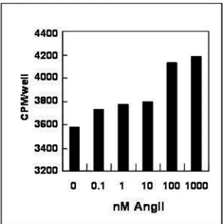
AngII stimulation induces long-term changes in cardiovascular tissues including cell growth and hypertrophy. Cellular hypertrophy is an adaptive process for synthesis of proteins without DNA replication, leading to protein accumulation. To confirm the hypertropic effect of AngII in cardiac cells, [3H] leucine incorporation assay was performed in rat neonatal cardiac myocytes. At the cellular level, the hypertrophic response is characterized by an increase in protein synthesis which can be analyzed by measurement of [3H] leucine incorporation into cells. Cardio myocytes cultured in serum-free condition were treated with AngII (0.1 ~ 1000 nM) for 48 h. AngII increased [3H] leucine incorporation in a concentration-dependent manner in cardiac myocytes (Fig. 2A). The specific marker of hypertrorphy, artrial natriuretic factor (ANF) was also visualized by immuno cytochemical staining assay using specific anti-ANF antibody. ANF was detected around nucleus in AngII-treated group compared with unAngII-treated cells (Fig. 2B). We showed the same result in rat heart-derived myoblast H9c2 cells, and no significant difference was observed between primary cardiac cells and H9c2 cells (data not shown). These findings clearly demonstrate that in agreement with the previous reports, AngII treatment of cardiac cells resulted in a significant increase in cellular hypertrophy.
Fig. 2. AngII induces hypertrophy in cardiac cells. A. Cells were treated with
AngII and assessed for hypertrophy by [3H]leucine incorporation assay in three independent experiments. Incorporated radioactivity was determined by liquid scintillation counting. B. Cells were incubated with 100 nM AngII or 1% FBS for 48 h. Cells were stained for ANF protein using polyclonal anti-ANF antibody and FITC-conjugated secondary antibody and then visualized by fluorescence microscopy.
A.
A.
A.
A.
B
B
B
B....
2. AngII induces activation of NF-κB in cardiac cells
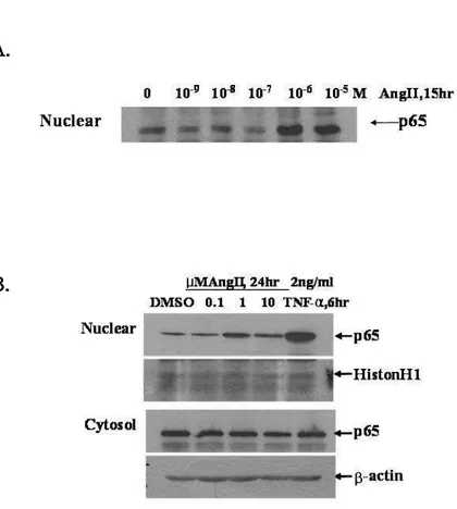
It has been well reported that NF-κB is activated during hypertrophy in cardiac cells. To validate the NF-κB activation on AngII-induced hypertrophy, NF-κB p65 subunit of nuclear fraction were visualized by immunoblot analysis. AngII stimulated the nuclear translocation of p65 NF-κB in neonatal cardiac myocytes (Fig. 3A). As shown in Fig. 3B, p65 NF-κB subunit was translocated to nucleus at 1 µM AngII in H9c2 cells in the same manner with primary cardiac myocytes. TNF-α, is a well known cytokine that activates NF-κB, also induced p65 nuclear translocation.
To confirm the transcriptional activity of AngII-induced nuclear translocation of p65, NF-κB luciferase activity assay was examined. Because of low efficiency of transfection in rat neonatal cardiac myocytes, H9c2 was used to perform luciferase activity assay. After cells were transiently transfected with a luciferase reporter construct (pNF-κB/LUC luciferase vector) possessing consensus pNF-κB binding sites, AngII was treated. NF-κB luciferase activity was enhanced by the addition of AngII in a concentration-dependent manner (Fig. 4A). To confirm whether the translocation of p65 is due to AngII-induced phosphorylation of IκBα, immunoblot assay was performed using phospho-specific IκBα antibody. IκBα was decreased in AngII- or TNF-α-treated group and its reduction is a result of proteosomal degradation (Fig. 4B). It was shown that pretreatment of MG132, a proteosomal inhibitor, blocked IκBα degradation. Theses are evidences that AngII activates
Fig. 3. AngII induces nuclear translocation of p65 NF-κB in cardiac cells.
Neonatal rat cardiac myocytes (A) or H9c2 cells (B) were cultured in presence or absence of AngII for the indicated times. Cells harvested and extracted the nuclear protein. The p65 nuclear translocation was visualized by immunoblot assay using anti-p65 NF-κB antibody. Equal amounts of nuclear and cytosolic protein were confirmed using Histon H1 and β-actin antibody, respectively.
A.
A.
A.
A.
B
B
B
B....
Fig. 4. AngII activates NF-κB in cardiac cells. A. Cells transfected with 100 ng of
pNF-κB/LUC luciferase vector were treated with AngII as indicated concentration for 24 h. Luciferase activity was determined using Luciferase Reporter Assay Kit and measured using luminometer. B. Cells were incubated with 1 µM AngII for 30 min or 2 ng/ml TNF-α for 10 min, either in the presence or absence of 20 µM MG132. Immunoblot assay was performed with anti-phospho-IκBα antibody.
A
A
A
A....
B
B
B
B....
3. Hsp90 inhibitor suppresses AngII-induced hypertrophy and NF-κB activation in cardiac cells
Heat shock proteins (Hsps) are a group of chaperone proteins that helps to maintain protein stability and to re-nature or target for degradation of unfolded proteins when cells are subjected to heat shock or other stresses. Since cardiac hypertrophy is an adaptive response of the heart for increasing workload, it is supposed that Hsps are induced and have a role as chaperone during hypertrophic stress. To investigate the role of Hsp90 in AngII-induced hypertrophy in cardiac cells, specific inhibitors of Hsp90 (geldanamycin;GA or 17-AAG) were treated prior to AngII treatment. These compounds are specific inhibitors of Hsp90 by blocking the binding of ATP to Hsp90. GA reduced AngII-induced [3H]leucine incorporation in a concentration-dependent manner (Fig. 5A). The pretreatment of 17-AAG also significantly inhibited AngII-induced [3H]leucine incorporation in similar pattern with that of GA (Fig. 5B). These findings clearly demonstrate that blockage of Hsp90 function inhibits AngII-induced cardiac hypertrophy. Recent reports illustrated that Hsp90 modulated NF-κB activity in TNFα-induced activation of
NF-κB. To understand the role of Hsp90 on activity of NF-κB in AngII-induced
hypertrophy, NF-κB luciferase assay was performed using GA. GA has an effect on lowering the activity of NF-κB in AngII-induced activation in cardiac cells (Fig. 6A). NF-κB activity is controlled by translocation of NF-κB through IκBα degradation or modification of p65 such as phosphorylation after nuclear localization. To assess
whether Hsp90 regulates the nuclear localization of NF-κB, cells were fractionated into nuclear and cytosolic extract after treatment of 17-AAG in cardiac cells. As shown in Fig. 6B, 17-AAG inhibited the AngII-induced p65 nuclear translocation. However, the inhibitor of Hsp70 did not affect the p65 nuclear translocation. These data represent that blockage of Hsp90 activity inhibits the activation of NF-κB, and Hsp90 might be an important regulator of NF-κB pathway in AngII-induced hypertrophy in cardiac cells.
Fig. 5. Blockage of Hsp90 function inhibits AngII-induced cardiac hypertrophy.
Cells were treated with indicated concentration of geldanamycin (A) or 17-AAG (B) prior to incubation with 1 µM AngII for 24 h. The [3H]leucine incorporation assay was performed and radioactivity measured with liquid scintillation counter
Fig. 6. Blockage of Hsp90 function inhibits AngII-induced NF-κB activation in cardiac cells. A. After transfection with pNF-κB Luc vector for 24 h, cells were
starved then exposed to various concentrations of GA for 1 h prior to AngII treatment. B. Cells were treated with 1 µM 17-AAG or 100 µM Hsp70 inhibitor (HII) for 1 h prior to addition of 1 µM AngII. Cells were harvested and fractionated into nuclear and cytosolic protein. The NF-κB p65 subunit level was determined by
A
A
A
A
B
B
B
B
immunoblot assay using anti-p65 specific antibody.
4. Hsp90 inhibitor cleaves IκBα through caspase-8 activation in cardiac cells 1) IκBα cleavage by Hsp90 inhibitor
Hsp90 inhibitors suppressed AngII-induced hypertrophy and nuclear translocation of NF-κB activation in cardiac cells (Fig. 6). These results suggest that Hsp90 inhibitor can modulate some signal proteins in the NF-κB activation pathway. In order to determine if Hsp90 inhibitor affects the p65 NF-κB subunit, the protein level of p65 was measured by immunoblot analysis. The p65 level exhibited little or no change after treatment with GA (data not shown). However, immunoblot analysis of the whole extract revealed that the smaller band of IκBα began to appear in GA-treated H9c2 cells (Fig. 7A, B). Suppression of Hsp90 activity also strongly affected the time-course of the cleavage of IκBα (Fig. 7A) The smaller band of IκBα became to appear by 1 µM GA treatment, and 10 µM GA induced an increase of a significant amount of the smaller band of IκBα (Fig. 7B). GA acts as a specific inhibitor of Hsp90 by blocking the binding of ATP to Hsp90. In this experiment, Hsp90 showed no differences in the GA-treated cells compared to the control cells (Fig. 7A, B). To determine if GA also affects IKKα/β stability as known, the protein level of IKKα/β was also examined with specific antibody to IKKα/β. IKKα/β was significantly decreased in a time-dependent and concentration-dependent manner (Fig. 7A, B). To observe whether the smaller band of IκBα is the product of proteosomal degradation or de novo protein synthesis, a proteosome inhibitor, MG132 (20 µM) and a protein
treatment withGA. These inhibitors did not exhibit a decrease of the smaller band of IκBα, but MG132 generated a large amount of the smaller band of IκBα, interestingly (Fig. 7C). These results suggest that the smaller band might be a cleavage product of IκBα due to treatment with GA.
Fig. 7. Hsp90 inhibitorHsp90 inhibitorHsp90 inhibitorHsp90 inhibitor induces IκBα cleavage in cardiac cells. H9c2 cells were
exposed to GA for indicated time (A) or with various concentrations (B). Cells were harvested and subjected to immunoblot analysis. H9c2 cells were incubated with 1
µM GA for 15 h in the presence or absence of 20 µM MG132 or 100 ng/ml of
cycloheximide (CHX) (C). IκBα was determined by immunoblot analysis using anti-IκBα antibody.
A
A
A
A....
C
C
C
C....
B
B
B
B....
2) Effects of calpain inhibitors on the IκBα cleavage
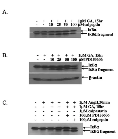
In previous study by Schaecher et al, a calcium-activated neutral proteinase, calpain, exhibited cleavage of IκBα in human peripheral blood mononuclear cells (PBMC) under activation with anti-CD3/CD28 (HIT3A/CD28.2), and 150 µM calpeptin, a calpain inhibitor, blocked cleavage of IκBα.22 To investigate whether calpain could mediate cleavage of IκBα, various calpain inhibitors were added 1 h prior to treatment with GA. Calpain inhibitors (calpeptin and PD150606) were treated as the indicated concentrations and cleavage of IκBα was analyzed by the immunoblot assay using IκBα antibody. Contrary to previous reports, cleavage of IκBα was not decreased by calpain inhibitors (Fig. 8A, B). When another calpain inhibitor (1 µM calpastatin) was treated, same result was shown (Fig. 8C). These results suggest that GA-induced IκBα cleavage is not mediated by calpain in H9c2 cells.
- + + + + + 1μμμμM GA, 15hr - - 10 25 50 100 μμμμM calpeptin IκκκκBαααα IκκκκBααααfragment - + + + + + 1μμμμM GA, 15hr - - 10 25 50 100 μμμμM calpeptin IκκκκBαααα IκκκκBααααfragment - + + + + + 1μμμμM GA, 15hr - - 10 25 50 100 μμμμM PD150606 β ββ β-actin IκκκκBαααα IκκκκBααααfragment - + + + + + 1μμμμM GA, 15hr - - 10 25 50 100 μμμμM PD150606 β ββ β-actin IκκκκBαααα IκκκκBααααfragment
-
+ + + + +
1μμμμM AngII,30min-
-
+ + + +
1μμμμM GA, 15hr-
-
-
+ -
-
1μμμμM calpastatin-
-
-
-
+ -
100μμμμM PD150606-
-
-
-
- +
100μμμμM calpeptin IκκκκBαααα IκκκκBααααfragment-
+ + + + +
1μμμμM AngII,30min-
-
+ + + +
1μμμμM GA, 15hr-
-
-
+ -
-
1μμμμM calpastatin-
-
-
-
+ -
100μμμμM PD150606-
-
-
-
- +
100μμμμM calpeptin IκκκκBαααα IκκκκBααααfragmentFig. 8. Effects of calpain inhibitors on the IκBα cleavage. Cells were treated with
calpeptin (A) or PD150606 (B) with various concentrations prior to the incubation of 1 µM GA for 15 h. Cells were pretreated with 1 µM calpastatin or 100 µM PD150606 or 100 µM calpeptin for 30 min and further incubation was carried with 1
µM GA for 15 h prior to the addition of 1 µM of AngII for 30 min (C). Samples were
harvested and IκBα level was determined by immunoblot analysis.
A
A
A
A....
B
B
B
B....
C
C
C
C....
3) Caspase-8 induces IκBα cleavage in GA-treated cardiac cells
Barkett et al. have already revealed that caspase-3 specifically cleaved IκBα at
a conserved Asp-Ser sequence in vitro.23 In order to determine whether caspases are responsible for the cleavage of IκBα, various membrane permeable caspase inhibitors were incubated with GA-treated H9c2 cells. z-VAD-fmk, z-VDVAD-fmk, z-DEVD-fmk, z-IETD-fmk and z-LEHD-fmk were used for inhibition of a broad spectrum of caspase, caspase-2, caspase-3, caspase-8 and caspase-9, respectively. GA-induced cleavage of IκBα was significantly inhibited by exposure of 100 µM of a pan-caspase inhibitor (z-VAD-fmk) (Fig. 9A). Whereas pre-treatment of a caspase-8 inhibitor (z-IETD-fmk) reduced IκBα cleavage, cells treated with z-VDVAD-fmk, z-DEVD-fmk, and z-LEHD-fmk did not exhibit inhibition of IκBα cleavage (Fig. 9A). It was confirmed that IκBα cleavage was decreased by the addition of z-IETD-fmk, in a concentration-dependent fashion (Fig. 9B). And, on the contrary, caspase-3 inhibitor slightly increased GA-mediated cleavage of IκBα in H9c2 cells (Fig. 9C).
Caspases has been known to recognize and cleave specific substrates that contain a DXXD-G/S/A consensus peptide motif and cleave after the second aspartic acid of DXXD. Because a putative sequence for recognition by caspases is present in the amino-terminus as DRHD31-S32, the cleavage product of IκBα should be 3-5 kD less compared to intact IκBα in immunoblot analysis by using the antibody against the carboxyl-terminus of IκBα. To further investigate the effect of caspase inhibitors on the GA-induced decrease of IKKα/β, immunoblot analysis with anti-IKKα/β antibody was performed. A GA-mediated decrease of IKKα/β was not recovered by
caspase inhibitors, suggesting that caspase activity is not required for a decrease of IKKα/β levels (Fig. 9A, B). Taken together, IκBα was a target of caspase-8 during the GA-induced inhibition of hypertrophy.
IKKαααα/ββββ - - 100μμμμM - + + + + + + 1μμμμM GA VA D VD VA D DE VD IE TD LE HD IκκκBακ ααα IκκκκBαααα fragment IKKαααα/ββββ - - 100μμμμM - + + + + + + 1μμμμM GA VA D VD VA D DE VD IE TD LE HD IκκκBακ ααα IκκκκBαααα fragment - + + + + + 1μμμμM GA - - 10 20 50 100 μμμμM z-IETD-fmk β β β β-actin IKKαααα/ββββ IκκκκBαααα IκκκBακ ααα fragment - + + + + + 1μμμμM GA - - 10 20 50 100 μμμμM z-DEVD-fmk IκκκκBαααα IκκκκBααααfragment
Fig. 9. IκBα cleavage by caspase-8 in GA-treated cardiac cells. A. Cells were
incubated with 100 µM z-VAD-fmk, z-VDVAD-fmk, z-DEVD-fmk, z-IETD-fmk and z-LEHD-fmk for 30 min prior to 1 µM GA for 15 h. Cells were pretreated with indicated doses of z-IETD-fmk (B) or z-DEVD-fmk (C) for 30 min, and then exposed with 1 µM GA for 15 h. Cells were harvested and immunoblot analysis was performed using anti-IκBα or IKKα/β antibodies.
A
A
A
A....
B
B
B
B....
C
C
C
C....
5. Hsp70 is not involved in IκBα cleavage
Hsp90 inhibitors are well-known inducers of Hsp70. Hsp70 induction was
appeared at approximately the same time as that of IκBα cleavage (data not shown). Because of this result, the following experiment was carried out to reveal whether Hsp70 causes IκBα cleavage or not. The pcDNA3.1/Hsp70 was delivered by lipofectamine plus into H9c2 cells, and the level of IκBα was detected by an immunoblot analysis (Fig. 10A). Hsp70 was over-expressed by 2 µg of pcDNA3.1/Hsp70 successfully. Enhanced Hsp70 expression had an effect on increasing the level of IκBα and Hsp90. Similar to this result, the effect of Hsp70 on IκBα accumulation has been well illustrated. However, IκBα cleavage was not determined at the Hsp70-overexpressing cells. Thus, Hsp70 is not involved in the cleavage of IκBα during the GA-induced increase of Hsp70 in H9c2 cells. As expected, Hsp70 induction reduced NF-κB activity in accordance with IκBα accumulation (Fig. 10B).
0 200 400 600 800 1000 1200 1400 10 50 L u c. A c ti v it y ( lu c /o n p g ) -1μμμμM AngII - + + + ng Hsp70 -0 200 400 600 800 1000 1200 1400 10 50 L u c. A c ti v it y ( lu c /o n p g ) -1μμμμM AngII - + + + ng Hsp70
-Fig. 10. Hsp70 is not involved in IκBα cleavage. A. H9c2 cells were transiently
transfected with indicated amount of pcDNA3.1/Hsp70. After 48 h, the cells were harvested and the protein levels of Hsp70, Hsp90 and IκBα were evaluated with immunoblot analysis. B. H9c2 cells were transfected with 100 ng of p-NF-κB/LUC luciferase vector and pcDNA3.1/lacZ, and simultaneously co-transfected with pcDNA3.1/Hsp70. After 24 h, cells were starved for 24 h and treated with 1 µM of AngII for 30 min. The NF-κB luciferase activity was performed with Luciferase Reporter Assay Kit.
B
B
B
B....
A
A
A
A....
6. Hsp90 inhibitor induces apoptosis in cardiac cells
We previously showed that GA induced caspase-8-mediated IκBα cleavage. Caspases have been known to be activated during apoptosis, a form of regulated programmed cell death. Cell death was observed in rat cardiac myoblast cell line H9c2 cells with a phase contrast microscope (Fig. 11A). After exposure to GA, the cells developed a rounded shape and detached from the bottom of the culture dish. To investigate the effect of GA on cell death, the cells were treated with various doses of GA, and cell viability was determined using the MTT assay (Fig. 11B). The viability was decreased in a concentration-dependent manner, and about 73% of the cells were viable in cell treated with 1 µM GA compared with the DMSO-treated cells. When the cells were treated with 1 µM GA for 15 h and then stained using an ApopTag®Plus Peroxiadase In Situ Apoptosis Detection Kit, the cells were stained
with TdT enzyme bounded HRP-conjugated at the breaks of DNA (Fig. 12A). JC-1 (5,5',6,6'-tetrachloro-1,1',3,3'-teterathylbenzimicazolo carbocyanine iodide) is up-taken into the mitochondria, and can be used as an indicator of mitochondrial potential, as a distinction between normal and apoptotic cells. Photographs demonstrated that treatment with GA results in green fluorescence shift from red as compared with the DMSO-treated cells (Fig. 12B). GA did not give the mitochondrial potential disruption under the treat of 100 µM z-IETD-fmk in H9c2 cells. This showed that GA induces mitochondrial damage, a part of which was mediated by caspase-8, indicating that Hsp90 inhibition could activate mitochondrial
Fig. 11.GA induces cell death in cardiac cells. A. Morphological changes of cells
were monitored with phase contrast microscope and photographed after treatment with GA. B. H9c2 cells were incubated with various concentrations of GA. After 24 h, viable cells were quantified by an MTT assay.
B
B
B
B....
A
A
A
A....
Fig. 12.GA induces apoptosis in cardiac cells... .A. H9c2 cells were treated with 1
µM GA for 15 h and incubated with TdT enzyme bounded HRP-conjugated
antibody. B. H9c2 cells were incubated with 1 µM GA in the presence or absence of 100 µM z-IETD-fmk. The cells were treated with 5 µg/ml of JC-1 for 10 min. The disruption of mitochodrial membrane potentials was measured using fluorescence microscopy on the excitation at 488 nm and the emission at 530 nm and 590 nm.
B
B
B
B....
A
A
A
A....
In order to know whether GA activated caspases, caspase activity assay and immunoblot analysis were performed. Caspase-8 activity increased in a dose-dependent manner and began to increase at 1 µM GA (Fig. 13A). A fragment of active caspase-3 was detected after treatment with 5 and 10 µM GA (Fig. 13B). The protein level of 55 kD pro-caspase-8 decreased at 10 µM GA (Fig. 13B). These results show that GA induces caspase-3- and caspase-8-dependent apoptosis in H9c2 cells. Recent reports have suggested that caspase activation was not the only mechanism of Hsp90 inhibition-induced apoptosis in Hodgkin’s lymphoma cell lines. Cytochrome C, which normally resides exclusively in the intermembrane space of mitochondria, is released into the cytosol during apoptosis. Release of cytochrome C from the mitochondria inactivates the electron transfer chain and triggers activation of caspase-3 through Apaf-1. GA induced the cytosolic release of cytochrome C in a concentration-dependent manner in H9c2 cells (Fig. 13B).
To verify whether caspase activation is necessary in GA-induced apoptosis, an MTT assay was carried out after pre-treating with caspase inhibitors in GA-exposed H9c2 cells. GA-inhibited cell viability was recovered by caspase inhibitors, z-VAD-fmk, z-VDVAD-z-VAD-fmk, z-DEVD-z-VAD-fmk, z-IETD-fmk and z-LEHD-fmk (Fig. 14A), and various doses of z-IETD-fmk, an inhibitor of caspase-8 (Fig. 14B). However, caspase is not a necessary mediator to induce apoptosis, because of its partial recovery. This implies that GA might mediate caspase-independent apoptosis in part. Mitochondrial proteins, EndoG and AIF were increased at the 1 µM GA-treated
nuclear fraction (data not shown). EndoG and AIF are released from the mitochondria into the cytosol, and translocated to the nucleus to induce apoptosis unrelated to caspases. These results demonstrate that GA induced not only caspase-dependent apoptosis but also caspase-incaspase-dependent apoptosis in H9c2 cells.
procaspase-8(55kD) 0 1 2 5 10 GA 39 26 19 13 32kD procaspase -3 17kD μ μ μ μM GA 39 26 19 13 32kD procaspase -3 17kD active caspase-3 Cyto.C Cyt. Total.
Fig. 13. Caspase-8 activation by GA in cardiac cells. A. H9c2 cells were treated
with GA for 15 h. Total cell lysate was incubated with 5 µl of IETD-AFC (1 mM) for 1 h. Caspase-8 activity was quantified by fluorescence spectrometer at 400 nm excitation filter and 505 nm emission filter. B. H9c2 cells were treated with GA for 15 h. Total cell lysate was subjected to SDS-PAGE and analyzed using immunoblot assay. The level of cytosolic cytochrome C was also analyzed by immunoblot assay.
A
A
A
A....
B
B
B
B....
Fig. 14. Caspase-independent cell death by GA in cardiac cells. A. H9c2 cells
were incubated with 100 µM of VAD-fmk, VDVAD-fmk, DEVD-fmk, z-IETD-fmk, and z-LEHD-fmk for 1 h prior to treatment with 10 µM GA. After 24 h, cell viability was determined by an MTT assay.
.
A
A
A
A....
B
B
B
B....
7. Caspase-8 directly cleaves IκBα protein
In order to confirm that caspase-8 is directly responsible for IκBα cleavage, purified GST-IκBα and active caspases were incubated in test tube, then visualized by an immunoblot assay using an IκBα antibody. Because GST is tagged at the NH2
-terminus of IκBα, the cleavage product has a mass of the same thing compared to the cleavage fragment of IκBα without GST in an immunoblot assay using a carboxyl-terminus-specific IκBα antibody. IκBα was cleaved by caspase-3 and caspase-8 directly, and the cleavage was inhibited by z-DEVD-fmk and z-IETD-fmk, respectively (Fig. 15). The other caspases had no effect on the cleavage of IκBα. Because z-DEVD-fmk showed no change in the GA-induced IκBα cleavage in the cells (Fig. 9C) and GA activated caspase-3 and caspase-8 in H9c2 cells (Fig. 13), GA activated both caspase-3 and caspase-8, but GA-mediated caspase-3 activation could not cleave IκBα, which suggests that GA-mediated caspase-3 is not sufficient to cleave IκBα in vivo. These data show that the amino-terminus of IκBα was cleaved by caspase-3 and caspase-8 in vitro, but the GA-induced active caspase-8
only had the ability to cleave IκBα.
Fig. 15. Caspase-8 directly cleaves IκBα protein. A. Purified GST- IκBα (100 ng)
was treated with purified active caspases for 1 h. Samples were subjected to SDS-PAGE and cleaved IκBα was analyzed by immunoblot assay. B. Purified GST- IκBα (100 ng) was incubated in the absence or presence of 20 µM IETD-CHO or z-DEVD-CHO for 30 min. After the mixture was treated with purified active caspase-8 or caspase-3 for 1 h, cleaved IκBα was analyzed by immunoblot analysis.
A
A
A
A....
B
B
B
B....
8. Hsp90 inhibitor inhibits IκBα phophorylation and degradation in cardiac cells
1) Hsp90 inhibitor inhibits AngII-induced IKKα/ββββ activation
The expression level of IKKα/β was decreased and IκBα cleavage was increased after GA exposure (Fig. 7). To investigate whether AngII-induced IKK activity changes along with IKKα/β, an immunoblot assay was examined using specific IKKα/β and phospho-IKKα/β (Ser181/182) antibodies. Compared to phosphorylated IKKα/β, the protein level of IKKα/β was dramatically decreased (Fig. 16A upper two panels). To elucidate whether the GA-induced IKK dephosphorylation has an effect on IκBα phosphorylation, a phospho-IκBα antibody (Ser32) was used for an immunoblot assay. IκBα was phosphorylated by treatment of 10 ng/ml of TNF-α, and the expression level was reduced without cleavage, which indicated that IκBα might be reduced by proteosomal degradation through ubiquitination. However, GA cleaved IκBα and reduced its phosphorylation, suggesting that inactive IKKα/β could not phosphorylate IκBα (Fig. 16 lower two panels). The fact that the phospho-IκBα level did not completely decrease although most of the IKKα/β level was decreased indicates that other kinases are present to phosphorylate IκBα.
Interestingly, AngII failed to phosphorylate cleaved IκBα, indicating that the N-terminal deleted fragment of IκBα could not be phosphorylated, at least on Ser32. To confirm whether the IκBα fragment has a possibility of being phosphorylated by AngII, an IKK activity assay was performed (Fig. 17). First, an in vitro cleavage
assay was performed with 1 µg of purified GST-IκBα and 1 µg of purified active caspase-8. The cells treated with 1 µM AngII for 30 min were immunoprecipitated with a specific antibody for IKKα/β, and the precipitates were incubated with the a mixture of the in vitro cleavage reaction. Immunoprecipitates with the anti-IKKα/β
antibody phosphorylated not only GST-IκBα but also cleaved IκBα, and its phosphorylation was enhanced by the addition of AngII (Fig. 17), which demonstrated that the caspase-8-mediated IκBα fragment can phosphorylated by active IKKα/β. Although the fragment of IκBα possessed the phosphorylation residues, ser32 and ser36, because capase-8 cleaved at the front of ser32 of IκBα, only ser36 was assumed to be a target of phosphorylation. Taken together, these results suggest that inhibition of IKK activity could be responsible for the blocking of IκBα phosphorylation by AngII, and IκBα proteolysis by caspase-8 repressed phosphorylation of cleaved IκBα on ser32.
Fig. 16. GA inhibits AngII-induced IKKα/β activation. H9c2 cells were treated
with GA and AngII. The cells were harvested and IKKα/β was determined by immunoblot analysis.
Fig. 17. Phosphorylation of IκBα fragment by IKKα/β. H9c2 cells were treated
with 1 µM AngII. The cells were harvested and immunoprecipitated with against IKKα/β antibody. Mixture of GST-IκBα (1µg) incubated with active caspase-8 (1µg) was incubated with the immunoprecipitates and kinase assay was performed as described in materials and methods.
2) Hsp90 inhibitor inhibits AngII-induced ubiquitination of IκBα and maintains its interaction with p65 NF-κB
Since IκBα has lysine residues in the amino-terminus modulated by ubiquitin ligase at Lys21 and Lys22, it might be deleted when the proteolytic cleavage of IκBα is processed by caspase-8. To determine whether ubiquitination of IκBα is disrupted by treatment with GA, an immunoblot assay with ubiquitin-specific antibody was performed, and followed by immunoprecipitation with a phospho-IκBα antibody. AngII induced poly-ubiquitination of phosphorylated IκBα. Even though IκBα was phosphorylated and ubiquitinated by AngII, it was not degraded in proteosome because of the addition of 20 µM MG-132 prior to treatment with AngII. The ubiquitination of IκBα was disrupted by treatment with GA (Fig. 18A). This result indicates that cleavage of N-terminus of IκBα by caspase-8 in GA-treated cells, allowing the release of ubiquitination residue from intact IκBα, decreased ubiquitination chance of phospho-IκBα.
The 26S proteosomal degradation of IκBα is followed by ubiquitination of IκBα. As a result of these processes, IκBα can not interact with NF-κB. Interaction of the IκBα protein with the p65 NF-κB subunit usually results in retention of the NF-κB in the cytoplasm and inhibition of the transcriptional activity of NF-κB.9 In order to confirm that GA enhanced the interaction between IκBα and NF-κB, an immunoprecipitation assay was examined. The interaction between IκBα and the p65 NF-κB subunit was slightly increased in the presence of 1 µM and 2 µM GA
(Fig. 18B). Therefore, GA-mediated diminishment of poly-ubiquitination of phospho-IκBα resulted in increased interaction between IκBα and NF-κB. These results show that GA has an effect on the inactivation of NF-κB through the reduction of ubiquitination and maintenance of interaction of IκBα with the p65
Fig. 18. GA inhibits AngII-induced ubiquitination of IκBα and maintains its interaction with p65 NF-κB. A. After treatment with GA for 15 h, H9c2 cells were
exposed to 20 µM MG132 for 30 min prior to incubation of 1 µM AngII for 30 min. Cells were lysed and immunoprecipitated with antibody against phospho-IκBα, and followed by immunoblot analysis with anti-ubiquitin antibody. B. Cell lysate was immunoprecipitated with IκBα antibody and immunoblot analysis was performed using anti-p65 antibody.
A
A
A
A....
B
B
B
B....
9. Recovery of NF-κB activation by inhibition of caspase-8 activity
As shown in Fig. 18, GA-induced caspase-8 activation exerts cleavage of IκBα and subsequent inhibition of ubiquitination. To confirm that these effects were mediated by caspase-8, a caspase-8 inhibitor, z-IETD-fmk was pretreated for 30 min, and the level of ubiquitination of phospho-IκBα was checked (Fig. 19A, upper panel). When caspase-8 was inactivated by z-IETD-fmk, ubiquitinated phospho-IκBα was increased compared with the GA treatment group in the absence of z-IETD-fmk. To confirm that this effect was not a result of an increase of IκBα phosphorylation, the same lysate was visualized by immunoblotting using a phospho-IκBα antibody (Fig. 19A, lower panel). As expected, pretreatment of z-IETD-fmk did not induce phosphorylation of IκBα, indicating that ubiquitination of IκBα was increased by inhibition of IκBα cleavage without phosphorylation of IκBα.
To determine whether recovery of IκBα ubiquitination has an effect on the
κB activity, κB luciferase activity was measured (Fig. 19B). GA-mediated NF-κB inactivation was partially recovered by the addition of a caspase-8 inhibitor.
Because recovered ubiquitination of phospho-IκBα occurred in z-IETD-fmk-pretreated cells compared with GA-treated H9c2 cells, and correlated with increased NF-κB transcriptional activity (Fig. 19), GA-mediated NF-κB inactivation was due to the blocking of ubiquitination through caspase-8 mediated IκBα cleavage.
Fig. 19. Recovery of NF-κB activation by inhibition of caspase-8 activity. A.
H9c2 cells were pre-incubated with 20 µM z-IETD-fmk for 30 min prior to addition of 1 µM GA for 15 h and cells were exposed to MG132. After 30 min treatment with 1 µM AngII, cells were collected and immunoprecipitation analysis was carried out using phospho-IκBα antibody, and followed by immunoblot assay with anti-ubiquitin antibody. B. H9c2 cells were transfected with p-NF-κB/LUC luciferase vector. After 24 h, H9c2 cells were treated with z-IETD-fmk and GA and incubated with 1 µM of AngII for 30 min. NF-κB activity was determined by Luciferase Reporter Assay Kit.