INTRODUCTION
Primary hypothyroidism is a common clinical condition, complicated by ascites in less than 4% of cases. Myxedema ascites, caused by hypothyroidism, is also a rare cause of ascites in less than 1% of new onsets (1). There is often a delay in diagnosis, and patients may go undiagnosed for a long time (2). In such cases, the use of thyroid hormone replacement usually leads to a progressive decrease in ascites, which ulti- mately disappear (3). Therefore, when any patient presents with ascites of uncertain etiology, hypothyroidism should be considered as a differential diagnosis.
Here, we report a 71-yr-old man with myxedema ascites that resolved completely with thyroid hormone replacement therapy. This may be the first case reported in Korea.
CASE REPORT
A 71-yr-old man presented with abdominal distension and anorexia that had lasted for 10 days. His past medical histo- ry was unremarkable except for prostate surgery for benign prostate hyperplasia six years previously. He did not smoke or drink alcoholic beverages. His blood pressure was 140/90 mmHg, the pulse rate 70 beats/min, and he weighed 55 kg.
The patient was alert and oriented. No jugular vein disten- sion was found and a cardiopulmonary examination was unre- markable. The abdomen was markedly distended, with taut skin and a circumference of 85 cm. There was shifting dull- ness and fluid waves evident on palpation. There was no pre- tibial edema. His white blood cell count was 5,500 cells/ L, hemoglobin was 10.5 g/dL and platelet count was 165,000/
L. The prothrombin time was 11.2 sec (international nor- malized ratio, INR, 1.0) and the activated partial thrombo- plastin time was 28 sec. The serum alkaline phosphatase level was 109 U/L, total bilirubin 1.0 mg/dL, aspartate aminotrans- ferase (AST) 70 U/L, alanine aminotransferase (ALT) 18 U/L, amylase 60 mg/dL, and total cholesterol 185 mg/dL. The total protein level was 5.8 g/dL and albumin 3.3 g/dL. Uri- nalysis results were normal. Hepatitis B and C serologies were negative. His initial chest radiograph showed normal with- out findings of cardiomegaly or pulmonary edema.
Samples of ascitic fluid showed elevated total protein (3.5 g/dL) and a low serum-to-ascites albumin gradient (SAAG;
0.8 g/dL). These were not characteristics of portal hyperten- sion. The white blood cell count in the fluid was 81/ L, and 84% of the cells were lymphocytes. Gram staining and cytol- ogy were negative. Bacterial, fungal and mycobacterial cul- tures were also negative.
Given these negative findings, we performed imaging stud- Jeong-Seon Ji, Hiun-Suk Chae,
Young-Seok Cho, Hyung-Keun Kim, Sung-Soo Kim, Chang-Wook Kim, Chang-Don Lee, Bo-In Lee, Hwang Choi, Kang-Moon Lee, Hye-Kyung Lee*, Kyu-Yong Choi
Departments of Internal Medicine and Pathology*, College of Medicine, The Catholic University of Korea, Seoul, Korea
Address for correspondence Hiun-Suk Chae, M.D.
Division of Gastroenterology, Department of Internal Medicine, Uijeongbu St. Mary’s Hospital, The Catholic University of Korea, 65-1 Gumo-dong, Uijeongbu 480-130, Korea
Tel : +82.31-820-3046, Fax : +82.31-847-2719 E-mail : [email protected]
761 J Korean Med Sci 2006; 21: 761-4
ISSN 1011-8934
Copyright � The Korean Academy of Medical Sciences
Myxedema Ascites
: Case Report and Literature Review
Myxedema ascites caused by hypothyroidism is rare, so its diagnosis is often delayed and patients frequently receive unnecessary procedures such as liver biopsies and exploratory laparotomies. We report a 71-yr-old man with clinical ascites that was the first manifestation of hypothyroidism, and which resolved completely in response to thyroid hormone replacement therapy. To our knowledge, this is the first report of myxedema ascites in Korea. A review of the literature revealed 51 well-document- ed cases of myxedema ascites. Analyses of ascites from patients in this condition usually show high protein (>2.5 g/dL) and low white blood cell counts, with a high proportion of lymphocytes. A consistent feature is the good response to thyroid hor- mone replacement therapy, which has always led to resolution of the ascites. Myxe- dema ascites is thus rare but easy to treat; it should be borne in mind, especially if the ascites fluid has a high protein content.
Key Words : Myxedema; Ascites; Hypothyroidism
Received : 6 April 2005 Accepted : 27 June 2005
762 J.-S. Ji, H.-S. Chae, Y.-S. Cho, et al.
ies to evaluate the possible cause of the ascites. Ultrasonogra- phy and computed tomography (CT) of the abdomen revealed massive ascites and a normal-sized liver and spleen (Fig. 1).
Esophagogastroduodenoscopy (EGD) disclosed no evidence of portal hypertension such as esophageal varices or gastropa- thy. A laparoscopic biopsy of the peritoneum and liver was performed to rule out any common cause of high protein, low SAAG ascites such as peritoneal malignancies, tuberculosis or infections. Laparoscopy showed that the surface of the liver was slightly irregular and the peritoneum was normal in app- earance. A microscopic examination of the liver biopsy revealed accumulations of yellow bile pigment in the hepatocytes, suggesting intrahepatic cholestasis without findings of liver cirrhosis (Fig. 2). Histology of the peritoneum yielded non- specific findings.
We then evaluated the possibility of a cardiogenic origin.
Echocardiography showed normal sized cardiac chambers, and the patient’s left ventricular ejection fraction was 54%.
However, there was no evidence of congestive heart failure as the cause of the ascites.
To prevent activation of the renin-angiotensin-aldosterone system and sodium retention from a vicious cycle of ascites, we prescribed an aldosterone antagonist (spironolactone 50 mg daily) and an inhibitor of proximal renal tubule sodium
Fig. 1.CT of the abdomen showing massive ascites and normal- sized liver and spleen.
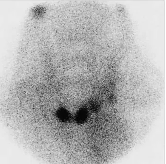
Fig. 4.Technetium-99m thyroid scan showing only two small nodu- lar foci in the thyroid bed.
Fig. 2.Microscopic finding of the liver showing accumulation of yellow bile pigment in the hepatocytes (H&E, ×400).
Fig. 3.Thyroid ultrasonograph revealing atrophic change of both thyroid lobes (arrowheads).
Myxedema Ascites 763
absorption (furosemide 20 mg daily). However, the patient’s response to diuretics was poor; his body weight decreased by only 1 kg and the abdominal circumference by only 2 cm after diuretic therapy for 10 days.
The patient was discharged with a prescription for 20 mg furosemide and 50 mg spironolactone daily, because of his poor financial status. He did not attend the outpatient clinic for another month, when he was readmitted because of his increased abdominal girth. Compared with discharge, his body weight had increased by 3 kg and the abdominal cir- cumference by 5 cm. The ascitic fluid showed elevated total protein and a low SAAG value, as found previously. We decid- ed to evaluate some relatively unusual causes of ascites. We were interested in the high protein component of the ascites fluid, so we performed thyroid function testing. These showed the following values: T30.14 nM/L (normal 0.92-2.78 nM/L);
T46.0 nM/L (normal 58-140 nM/L); free T40.13 pM/L (nor- mal 10.3-35 pM/L); and thyroid-stimulating hormone level (TSH) 70.3 mU/L (normal 0.4-5 mU/L). These findings neces- sitated further thyroid tests. The antimicrosomal antibody titer was 1:102,400 (normal, <1:100) and the antithyroglob- ulin antibody titer was negative. Ultrasonography revealed atrophic change to both thyroid lobes (Fig. 3), and a techne- tium-99m thyroid scan revealed only two small nodular foci in the thyroid bed (Fig. 4).
Thyroid hormone replacement therapy was started with gradually increasing doses of levothyroxine, from 0.05 mg to 0.15 mg daily to treat the patient’s hypothyroidism. Over the following three months, he became euthyroid with com- plete resolution of his ascites, and the liver function tests re- turned to normal. His body weight decreased by 9 kg and the abdominal circumference was reduced by 28 cm. There has been no recurrence of ascites and his euthyroid condition was maintained over three years of follow-up with levothy- roxine 0.15 mg daily. The latest follow-up thyroid function studies showed a free T4level of 19.3 pM/L and a TSH of 2.7 mU/L.
DISCUSSION
Hypothyroidism is a relatively rare cause of ascites. How- ever, the importance of its diagnosis is that use of thyroid hormone replacement results in complete resolution. If there is new onset ascites, diagnostic workup should begin with the analysis of ascitic fluid. Usually total protein in the ascitic fluid and the SAAG value give a useful framework for anal- ysis of whether the ascitic fluid is a transudate or an exudate.
The total protein in the ascitic fluid was >2.5 g/dL in the exudate and <2.5 g/dL in the transudate. Of the various caus- es, peritoneal malignancies, tuberculous peritonitis, pyogenic peritonitis and pancreatic ascites can all lead to high-protein ascites. Patients with liver cirrhosis and congestive heart fail- ure show low protein ascites. The SAAG correlates directly
with portal pressure (4). Ascites fluid associated with portal hypertension shows a low total albumin level, and the SAAG is greater than 1.1 g/dL (high gradient) (5, 6). SAAG is usu- ally high in patients with liver cirrhosis and congestive heart failure. A gradient of <1.1 g/dL (low) usually suggests that the ascites is not caused by portal hypertension. The SAAG is low in patients with peritoneal malignancies, tuberculous peritonitis, pyogenic peritonitis and pancreatic ascites. There- fore, a low gradient ascites should initiate an evaluation for primary peritoneal process, most importantly infections and malignancies.
There has been a suggestion that the SAAG may exceed 1.1 in patients with myxedema ascites, based on a review of eight patients (7). Because so few cases have been studied and portal hypertension or heart failure do not seem to be the me- chanisms causing ascites in patients with myxedema, we can- not conclude that a high SAAG is a typical feature in this disease (8). Moreover, the patient reported here showed a low SAAG.
Portal hypertension secondary to liver cirrhosis is the lead- ing cause of ascites (more than 80% of cases) and peritoneal involvement in patients with malignant diseases is the sec- ond at about 10% (9). Therefore, if the composition of ascitic fluid and ultrasonography are not consistent with portal hyper- tension or other specific diseases, the physician should con- sider peritoneal malignancy and perform a peritoneal biop- sy. If this is negative and the ascitic fluid shows a high pro- tein content, then hypothyroidism should be considered as a differential diagnosis. In this patient, because the SAAG was <1.1 g/dL and there was a lack of esophageal varices or gastropathy on EGD and characteristic findings on ultrasono- graphy, there was no evidence of portal hypertension. More- over, a peritoneal biopsy showed nonspecific findings. As the ascitic fluid analysis revealed a high protein content, we per-
Number of patients
Mean Ranges Remarks
Ascites protein 49 3.9 1.8-5.1 Forty-eight patients
(g/dL) (98%) showed
ascites protein levels >2.5 g/dL SAAG (g/dL) 11 1.5 0.8-2.3 Because of the small
number of patients, the characteristics
were unclear Ascites WBC count 48 60 10-400 Predominance of
(per L) lymphocytes
(mean 81%) Duration of ascites 51 8 1 month
months to 8 yr
Response to treatment 51 Regression of ascites Table 1.Characteristics of reported patients with myxedema as- cites
SAAG, serum-to-ascites albumin gradient; WBC, white blood cell.
formed thyroid function testing, which proved decisive.
A review of the literature turned up 51 well-documented cases of myxedema ascites (Table 1) (2, 3, 7, 8, 10-20). A very consistent finding was the high total protein level (>2.5 g/
dL) (7). Total protein levels exceeded 2.5 g/dL in almost all cases, with a mean of 3.9 g/dL. The mean SAAG was 1.5 g/
dL with a range of 0.8-2.3 g/dL. White blood cell counts were rather low, usually with a predominance of lymphocytes; the mean white blood cell count was 60/ L with a mean of 81%
lymphocytes. In our patient, white blood cell count was 81/
L and lymphocyte proportion, 84%. There was usually a sig- nificant delay in the diagnosis, with a mean of eight months.
Prompt recognition of myxedema ascites prevents the inap- propriate use of diuretics and unnecessary procedures, includ- ing repeated paracenteses, liver biopsies and exploratory laparo- tomies (10). A constant feature was the good response to thy- roid hormone replacement therapy, which led to elimination of the ascites in every instance.
The mechanism of ascites fluid formation in patients with myxedema is unclear. There are two main hypotheses. The first is that low levels of circulating thyroid hormones cause increased extravasation of plasma proteins because of abnor- mal capillary permeability and the lack of a compensatory increase in lymph flow and protein return rate (21). The sec- ond hypothesis is that hyaluronic acid accumulates in the skin and produces edema by a direct hygroscopic effect. However, hyaluronic acid has only been found in minute quantities in patients with myxedema ascites: not large enough to exert a direct hygroscopic effect. However, it could interact with albu- min to form complexes that prevent the lymphatic drainage of extravasated albumin (22).
To our knowledge, this is the first report of myxedema as- cites in Korea. Although the diagnosis was delayed for about one and half months, treatment by thyroid hormone replace- ment medication led to complete regression of the ascites and normalization of liver function in this patient.
In conclusion, myxedema ascites is rare but easy to treat.
Treatment with thyroid hormone replacement therapy leads to complete regression of the ascites. A very prominent fea- ture in such cases is the high total protein level (>2.5 g/dL).
Once routine evaluation of ascites excludes common causes such as liver cirrhosis, peritoneal malignancies and infections, congestive heart failure and pancreatic ascites, thyroid func- tion tests should be performed on patients with high protein levels in the ascites fluid.
REFERENCES
1. Watanakunakorn C, Hodges RE, Evans TC. Myxedema: a study of 400 cases. Arch Intern Med 1965; 116: 183-90.
2. McDonough CH, Lee L, de Beur SJ, Arai S, Vogelsang GB. Myxede-
ma ascites in the posttransplant setting: case report. Am J Hematol 2002; 71: 216-8.
3. De Feudis L, Scudieri M, Orlando D, Traisci G. Ascites as preeminent manifestation of primary hypothyroidism: clinical case. Ann Ital Med Int 1999; 14: 294-7.
4. Hoefs JC. Serum protein concentration and portal pressure determine the ascitic fluid protein concentration in patient with chronic liver disease. J Lab Clin Med 1983; 102: 260-73.
5. Rector WG Jr, Reynolds TB. Superiority of the serum-ascites albumin difference over the ascites total protein concentration in separation of ‘‘transudative’’ and ‘‘exudative’’ ascites. Am J Med 1984; 77: 83-5.
6. Hoefs JC. Diagnostic paracentesis: a potent clinical tool. Gastroen- terology 1990; 98: 230-6.
7. de Castro F, Bonacini M, Walden JM, Schubert TT. Myxedema as- cites: report of two cases and review of the literature. J Clin Gastro- enterol 1991; 13: 411-4.
8. Otero Bedoya J, Landeira G, Corino M, Tamashiro A, Fassio E. Ascites due to hypothyroidism in a patient with alcoholic cirrhosis. Acta Gastroenterol Latinoam 2001; 31: 77-81.
9. Runyon BA. Care of patients with ascites. N Engl J Med 1994; 330:
337-42.
10. Chiprut RO, Knudsen KB, Liebermann TR, Dyck WP. Myxedema ascites. Am J Dig Dis 1976; 21: 807-8.
11. Hazard J, Merot J, Klotz A, Potel M. Ascites revealing thyroid defi- ciency. Ann Med Interne 1969; 120: 755-9.
12. Kinney EL. Myxedema ascites. Am Fam Physician 1987; 36: 134.
13. Kocen RS, Atkinson M. Ascites in hypothyroidism. Lancet 1963; 1:
527-30.
14. Leung FW, Nortman DF, Shinaberger JH. Myxedema ascites in a patient undergoing chronic hemodyalisis. Dyalisis Transplant 1982;
11: 708-9.
15. Madenberg F, Byfield GV, Baker LA. Occurrence of ascites in myx- edema. Arch Intern Med 1954; 93: 787-95.
16. Desrame J, Mathurin P, Rozov R, Sabate JM, Poynard T, Opolon P, Denis J. Isolated ascites revealing a hypothyroidism: study of 2 cases.
Gastroenterol Clin Biol 1998; 22: 732-5.
17. Skinner J. Mixedema with thyroid antibodies presenting with ascites.
Proc R Soc Med 1962; 55: 997.
18. Turner JA, Rapoport J. Mixedema ascites. Postgrad Med J 1977; 53:
343-4.
19. Von Knorring J, Friman C. Mechanism of myxedema formation. N Engl J Med 1980; 302: 469.
20. Alberti LE, Lopez-Gomez A, Alberti-Flor JJ. Spontaneous bacterial peritonitis in a patient with myxedema ascites. Digestion 2003; 68:
91-3.
21. Parving HH, Hansen JM, Nielsen SL, Rossing N, Munck O, Lassen NA. Mechanisms of edema formation in myxedema increased pro- tein extravasation and relatively slow lymphatic drainage. N Engl J Med 1979; 301: 460-5.
22. Bonvalet JP, David R, Baglin A, Hatt PY. Myxedema with inappro- priate antidiuresis and hyperaldosteronism. Ann Med Interne 1970;
121: 949-55.
764 J.-S. Ji, H.-S. Chae, Y.-S. Cho, et al.