INTRODUCTION
It has been reported that a high dose chemotherapy with autologous stem cell transplantation (ASCT) enhances pa- tient’s survival rate and suppresses the recurrence rate in chemo-sensitive primary tumors such as non-Hodgkin’s lymphoma (1). However, the treatment also produces delayed toxicities including treatment-related myelodysplastic syn- drome (t-MDS) and acute myelogenous leukemia (t-AML).
Among the chemotherapeutic agents, etoposide, a topoiso- merase II inhibitor is well-known to cause t-AML with a chrac- teristic chromosomal abnormality of 11q23 rearrangement (2). We report here a case of a 28-yr-old Korean woman with t-MDS and t-AML with progressively evolving cytogenetic abnomalities including 11q23 rearrangement after BCNU/
etoposide/Ara-C/cytoxan high-dose chemotherapy and ASCT for non-Hodgkin’s lymphoma.
CASE REPORT
A 28-yr-old, four months pregnant woman was admitted with complaints of an icteric skin change and right upper quadrant abdominal mass. Her blood pressure was 120/80 mmHg, pulse rate 100/min, respiration rate 18/min, and body temperature 36.0℃. Her skin and sclera were icteric.
Respiratory and heart sounds were normal. A huge non-ten- der, 10×10 cm sized hard mass was palpable on the right upper quadrant of the abdomen.
The initial laboratory findings were as follows: white blood cell count 7,800/ L (seg 91%, lymphocyte 6%, and mono- cyte 3%), hemoglobin 8.3 g/dL, hematocrit 24.5%, and platelet count 296,000/ L on complete blood cell count (CBC), blood urea nitrogen 5 mg/dL, creatinine 0.5 mg/dL, protein 6.4 g/dL, albumin 4.0 g/dL, aspartate aminotrans- ferase 38 IU/L, alanine aminotransferase 37 IU/L, total biliru- bin 11.1 mg/dL, and direct bilirubin 5.6 mg/dL on blood
Geundoo Jang, Sang-We Kim, Cheolwon Suh, Eun-Kyoung Kim, Hyeseung Bahng, Young Hoon Jeong, Il Gwon Park, Woo-Kun Kim, Sang-Hee Kim, Eul-Ju Suh*, Chan-Jeoung Park*, Hyun-Sook Ji*, Jung-Shin Lee
Department of Medicine, Department of Clinical Pathology*, University of Ulsan, College of Medicine, Asan Medical Center, Seoul, Korea
Address for correspondence Sang-We Kim, M.D.
Department of Medicine, University of Ulsan, College of Medicine, Asan Medical Center, 388-1 Poongnap-dong, Songpa-gu, Seoul 138-736, Korea
Tel : +82.02-3010-3215, Fax : +82.02-3010-6961 E-mail : [email protected]
555
A Case of Treatment-Related Myelodysplastic syndrome and Acute Myelogenous Leukemia Following High-Dose Chemotherapy with Autologous Stem Cell Transplantation for Non-Hodgkin's Lymphoma
Treatment-related myelodysplastic syndrome (t-MDS) and acute myelogenous leukemia (t-AML) are now well established as complications of cytotoxic che- motherapy. We experienced a 28-yr-old female patient who developed t-MDS/t- AML with characteristic chromosomal abnormalities including 11q23 chromoso- mal rearrangement following high-dose chemotherapy with autologous stem cell transplantation (ASCT) for non-Hodgkin’s lymphoma. The patient was admitted with bulky abdominal masses of B cell lineage non-Hodgkin’s lymphoma. After 2 cycles of systemic chemotherapy of the Vanderbilt regimen, the patient under- went ASCT with high dose chemotherapy of the BEAC regimen. She also received radiation of 48 Gy for the residual periportal lymphadenopathy. The initial cyto- genetic analysis of the infused mononuclear cells revealed a normal karyotype.
Twenty two months after the ASCT, pancytopenia was noted and her bone mar- row aspirate showed dysplastic hemopoiesis with myeloblasts up to 12% of non- erythroid nucleated cells. The patient was diagnosed as t-MDS (refractory ane- mia with an excess of blasts). Cytogenetic analysis showed complex chromoso- mal abnormalities including 11q23 rearrangement, which is frequently found in topoisomerase II inhibitor-related hematologic malignancies. Four months later, it was noted that the t-MDS had evolved into an overt t-AML. Cytogenetic analysis showed an evolving pattern with more complex abnormalities. The patient was treated with combination che-motherapy, but her leukemic cells were resistant to the therapy.
Key Words : Myelodysplastic Syndromes; Leukemia, Myelocytic, Acute; Transplantation, Autologous;
Gene Rearrangement
Received : 27 June 2001 Accepted : 20 August 2001
A B
Fig. 1.(A) A bulky mass is noted by abdominopelvic CT scan at the initial diagnosis; (B) The mass size decreases after 2 cycles of chemotherapy of the Vanderbilt regimen.
chemistry. Bilirubin was +++, urobilinogen +, WBC ±on routine urinalysis. No abnormality was found on chest radio- graphy and computed axial tomography (CT) scan of the chest. A bulky mass compressing the common bile duct (Fig.
1A) with an involvement of porta caval space and another mass near the left common and internal iliac arteries were detected on CT scan of the abdomen and pelvis. The mass
proved to be a malignant lymphoma of B cell lineage by posi- tive immunohistochemical staining against pan B (L-26) (Fig.
2) with percutaneous needle biopsy. There was no evidence of bone marrow involvement by lymphoma and the result of chromosomal study was normal.
The patient was diagnosed as malignant B cell lymphoma, and treated with percutaneous transhepatic biliary drainage and artificial termination. Then she was given 2 cycles of chemotherapy of the Vanderbilt regimen (cycle 1; cytoxan 1,500 mg/m2day 1, 2, etoposide 400 mg/m2day 1-3, vin- cristine 1.4 mg/m2day 8, 22, bleomycin 10 U/m2day 8, 22, methotrexate 200 mg/m2day 15, leucovorin 15 mg/m2q 6 hr for 6 doses 24 hr after methotrexate, prednisolone 60 mg/m2 day 1-7, cycle 2; cytoxan 1,500 mg/m2day 29, etoposide 100 mg/m2day 29-31, doxorubicin 45 mg/m2day 1, 2, Bleomycin 10 U/m2day 36, 50, methotrexate 200 mg/m2day 43, leu- covorin 15 mg/m2q 6 hr for 6 doses 24 hr after methotrex- ate, prednisolone 60 mg/m2day 29-35) chemotherapy (3).
A partial remission was observed on a follow-up CT scan (Fig. 1B). Cytoxan (4 g/m2) and granulocyte-colony stimu- lating factor (G-CSF, 10 g/kg) were adminstered for stem cell mobilization, and the BEAC regimen (BCNU 300 mg/
m2, etoposide 800 mg/m2, cytarabine 800 mg/m2, and cytox- an 140 mg/kg) was employed for the conditioning of ASCT (4).
On the 27th day after ASCT, her CBC (WBC 3,200/ L, Hb 10.9 g/dL, and platelet 119,000/ L) suggested a suc- cessful engraftment. Follow-up CT scan revealed much more decreased abdominal mass. She received radiotherapy of 48 Gy on periportal area for the residual mass. Afterward, she was followed-up on an outpatient basis in a near complete
Fig. 2.Light microscopic finding (×200) of B cell lineage non- Hodgkin’s lymphoma showing positive immunohistochemical staining against a pan B antigen (L-26).
remission state.
Twenty two months after the ASCT, her CBC showed a progressive pancytopenia. Her bone marrow examination revealed slightly hypogranular granulocytes with an increase of myeloblasts (12% of nonerythroid nucleated cells, Fig.
3A). She was diagnosed with refractory anemia with an excess of blasts according to the FAB classification. Chromosomal study showed complex abnormalities including trisomy 6 and t(11;11)(q23;q25) (Fig. 4A). Because an HLA-compati- ble related donor was not available, she had to wait for an HLA-matched unrelated donor from the bone marrow reg- istry receiving a supportive care. The chromosomal study of the patient’s frozen mononucler cells that had been stored
after mobilization did not show any cytogenetic abnormali- ties.
Four months after the diagnosis of t-MDS, she was read- mitted due to cough and purulent sputum. A consolidated lesion was found in the right lower lobe of her lung by chest radiography. Peripheral blood smear revealed blasts up to 27%
and bone marrow aspiration and biopsy showed large clusters of myeloblasts (50% of nonerythroid nucleated cells) (Fig.
3B). Chromosomal study showed more complex abnormali- ties than before (Fig. 4B).
After alleviation of pneumonic consolidation, the patient was treated with cytarabine and idarubicine chemotherapy for t-AML. However her bone marrow on day 14 harbored
A B
Fig. 3.Light microscopic findings of bone marrow. (A) t-MDS with hypogranular granulocytes and increased myeloblasts (12% of nonerythroid nucleated cells, ×1,000). (B) t-AML with a large cluster of leukemic blasts (50% of nonerythroid nucleated cells, ×400).
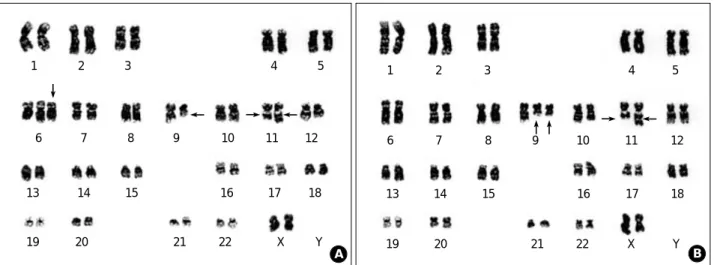
A B
Fig. 4.Cytogenetic analysis of bone marrow. (A) Complex chromosomal abnormalities including +6, del(9)(q12q32), and t(11;11)(q23;q25) at the diagnosis of t-MDS. (B) Karyotypic evolution including -6, del(X)(q24), +del(9)(q12q32) at the diagnosis of t-AML.
1
6
13
19 20 21 22 X Y
14 15 16 17 18
7 8 9 10 11 12
2 3 4 5 1
6 7 8 9 10 11 12
18 17 16 15
14 13
19 20 21 22 X Y
2 3 4 5
persistent leukemic cells. Pneumonic consolidation reap- peared along with severe neutropenia. Despite aggressive treatment with broad-spectrum antibiotics, amphotericin B, and G-CSF, the patient eventually succumbed to her dis- ease.
DISCUSSION
The employment of high-dose chemotherapy has improved the survival of patients with malignancies such as non-Hodg- kin’s lymphoma (1). However its long-term effects are of increasing concern. One of the most serious adverse effects is the development of secondary malignancies including treatment-related leukemia (5).
MDS and acute leukemia are clonal neoplastic hemato- logic disorders from various causes such as familial factor, ionizing radiation, benzene, and chemotherapeutic agents.
It is well known that chemotherapy or radiotherapy of pri- mary cancer can induce t-MDS and t-AML especially in pa- tients with Hodgkin’s and non-Hodgkin’s lymphomas who received high-dose chemotherapy with or without total body irradiation for ASCT (24). These patients represent the high- est risk group to develop t-MDS and/or t-AML between 2- 8 yr following therapy, and most have chromosomal abnor- malities that were not present at the time of diagnosis of the primary cancer (6).
Recently, it has become evident that there are at least two different forms of chemotherapy-related leukemia: alkylat- ing agent-related and topoisomerase II inhibitor-related leukemias. Alkylating agents such as cyclophosphamide, melphalan, busulfan, semustine, and carmustine have been implicated in t-MDS and t-AML. These agents form covalent bonds between the carbon of an alkyl moiety and nucleophilic DNA bases. These induce both intra- and interstrand DNA cross-linkings and breaks and the DNA cross-linking causes a DNA misleading. Although the leukemogenicity of the alkylating agents is not fully known, they may induce leuke- mia through such mechanisms as point mutations, deletions, and inappropriate recombinations, because of the enhanced and aberrant DNA repair (7, 8). There are evidences that the leukemogenic risk is dose dependent (9, 10) and most t- AML present initially as myelodysplasia and develop within 1 yr from the diagnosis of MDS (11). Cytogenetic studies have shown unbalanced chromosomal aberrations, most commonly the loss of whole chromosomes 5 or 7, or various parts of the long arms of these chromosomes (11, 12). Mor- phologically, the French-American-British (FAB) subtypes M1, M2, M6, M7 and even undetermined phenotypes have been reported (13).
It is thought that topoisomerase II inhibitors such as etopo- side (VP-16) and teniposide induce secondary leukemias by inhibition of the topoisomerase II, which regulates the super- helical rearrangement of the DNA. The enzyme cleaves a
single strand of the DNA duplexes at a specific sites by a covalent binding between its tyrosine residue and 5′-phos- phate residue of the DNA, and produces a transient double strand break, which enables the knotted or interlinked DNA to pass (14-16). Thus topoisomerase II inhibitor produces secondary DNA damages such as a complex conversion. In the process of repair, an illegitimate and non-homologous recombination can occur and this may be responsible for leukemogenic potential. Topoisomerase II inhibitor-related leukemia has a relatively short incubation period, median 2 to 3 yr following treatment without a prodromal myelodys- plastic phase compared to alkylating agent-related leukemia (13). The chromosomal study may show the characteristic balanced translocations involving chromosome band 11q23 or less often, 21q22 (17-20). Common partner chromosomes for the 11q23 translocations are the chromosomes 1, 2, 4, 6, 9, 10, 16, 17, 19, 22, and X (13). These chromosomal aber- rations are more frequently associated with the development of AML M4 or M5 according to the FAB classification (16, 19). The mixed lineage leukemia (MLL) gene is located at chromosomal band 11q23 and is frequently involved in t- AML following topoisomerase II inhibitor treatment (21).
Recently, Krishnan et al. reported 22 patients with morpho- logic evidence of t-MDS and t-AML among 612 patients who had been treated with high-dose chemotherapy with ASCT for Hodgkin’s disease and non-Hodgkin’s lymphoma (22).
A multivariate analysis of the entire cohort revealed that the stem cell priming with etoposide (relative risk 7.7, p value=
0.002) was independently associated with an increased risk of developing a secondary leukemia with the 11q23/21q22 abnormalities.
A cumulative dose of etoposide higher than 2-3 g/m2is known to be associated with the development of t-AML, although this association has not been found in all studies (23). In this case, a total of 2.3 g/m2cumulative dose of etopo- side was given as well as alkylating agents and irradiation.
The patient was diagnosed with t-MDS 22 months after ASCT, which progressed to t-AML after 4 months. The cyto- genetic studies revealved a normal karyotype before ASCT, the 11q23 rearrangement within chrmosome 11 at the diag- nosis of t-MDS, and more complex chromosomal abnormal- ities at t-AML.
In conclusion, our patient suffered from t-MDS/t-AML following an aggressive treatment for non-Hodgkin’s lym- phoma. Her characteristic 11q23 rearrangement might be attributed to the use of a high cumulative dose of topoiso- merase II inhibitor, etoposide.
REFERENCES
1. Philip T, Guglielmi C, Hagenbeek A, Somers R, Van der Lelie H, Bron D, Sonneveld P, Gisselbrecht C, Cahn JY, Harousseau JL, Coiffier B, Biron P, Mandelli F, Chauvin F. Autologous bone mar-
row transplantation as compared with salvage chemotherapy in relapses of chemotherapy-sensitive non-Hodgkin's lymphoma. N Engl J Med 1995; 333: 1540-5.
2. Super HJ, McCabe NR, Thirman MJ, Larson RA, Le Beau MM, Pedersen-Bjergaard J, Philip P, Diaz MO, Rowley JD. Rearrange- ments of the MLL gene in therapy-related acute myeloid leukemia in patients previously treated with agents targeting DNA-topoiso- merase II. Blood 1993; 82: 3705-11.
3. Waits TM, Greco FA, Greer JP, Johnson DH, Wolff SN, Stein RS, McMaster ML, Hainsworth JD. Effective therapy for poor-progno- sis non-Hodgkin's lymphoma with 8 weeks of high-dose intensity combination chemotherapy. J Clin Oncol 1993; 11: 943-9.
4. Van Besien K, Tabocoff J, Rodriguez M, Andersson B, Mehra R, Przepiorka D, Dimopoulos M, Giralt S, Suki S, Khouri I, Spitzer G, Jagannath S, Dicke K, Le Maistre CF, Deisseroth A, Cabanillas F, Champlin RE. High-dose chemotherapy with BEAC regimen and autologous bone marrow transplantation for intermediate grade and immunoblastic lymphoma: durable complete remissions, but a high rate of regimen-related toxicity. Bone Marrow Transplant 1995; 15: 549-55.
5. Darrington DL, Vose JM, Anderson JR, Bierman PJ, Bishop MR, Chan WC, Morris ME, Reed EC, Sanger WG, Tarantolo SR. Inci- dence and characterization of secondary myelodysplastic syndrome and acute myelogenous leukemia following high-dose chemoradio- therapy and autologous stem-cell transplantation for lymphoid malignancies. J Clin Oncol 1994; 12: 2527-34.
6. Pedersen-Bjergaard J, Philip P, Larsen SO, Andersson M, Daugaard G, Ersboll J, Hansen SW, Hou-Jensen K, Nielsen D, Sigsgaard TC, Specht L, Osterlind K. Therapy-related myelodysplasia and acute myeloid leukemia. Cytogenetic characteristics of 115 consecutive cases and risk in seven cohorts of patients treated intensively for malignant diseases in the Copenhagen series. Leukemia 1993; 7(12):
1975-86.
7. Kaina B. Critical steps in alkylation-induced aberration formation.
Mutat Res 1998; 404: 119-24.
8. Sanderson BJ, Shield AJ. Mutagenic damage to mammalian cells by therapeutic alkylating agents. Mutat Res 1996; 355: 41-57.
9. Curtis RE, Boice JD, Jr., Stovall M, Bernstein L, Greenberg RS, Flannery JT, Schwartz AG, Weyer P, Moloney WC, Hoover RN.
Risk of leukemia after chemotherapy and radiation treatment for breast cancer. N Engl J Med 1992; 326: 1745-51.
10. Tucker MA, Meadows AT, Boice JD, Jr., Stovall M, Oberlin O, Stone BJ, Birch J, Voute PA, Hoover RN, Fraumeni JF Jr. Leukemia after therapy with alkylating agents for childhood cancer. J Natl Cancer Inst 1987; 78: 459-64.
11. Michels SD, McKenna RW, Arthur DC, Brunning RD. Therapy- related acute myeloid leukemia and myelodysplastic syndrome: a clinical and morphologic study of 65 cases. Blood 1985; 65: 1364-
72.
12. Pedersen-Bjergaard J, Philip P, Larsen SO, Jensen G, Byrsting K.
Chromosome aberrations and prognostic factors in therapy-related myelodysplasia and acute nonlymphocytic leukemia. Blood 1990;
76: 1083-91.
13. Ng A, Taylor GM, Eden OB. Treatment-related leukaemia: a clini- cal and scientific challenge. Cancer Treat Rev 2000; 26: 377-91.
14. Felix CA. Secondary leukemias induced by topoisomerase-targeted drugs. Biochim Biophys Acta 1998; 1400: 233-55.
15. Liu LF, Liu CC, Alberts BM. Type II DNA topoisomerases: enzymes that can unknot a topologically knotted DNA molecule via a reversible double-strand break. Cell 1980; 19: 697-707.
16. Smith MA, Rubinstein L, Ungerleider RS. Therapy-related acute myeloid leukemia following treatment with epipodophyllotoxins:
estimating the risks. Med Pediatr Oncol 1994; 23: 86-98.
17. Pedersen-Bjergaard J, Philip P. Balanced translocations involving chromosome bands 11q23 and 21q22 are highly characteristic of myelodysplasia and leukemia following therapy with cytostatic agents targeting at DNA-topoisomerase II. Blood 1991; 78: 1147-8.
18. Bernard OA, Berger R. Molecular basis of 11q23 rearrangements in hematopoietic malignant proliferations. Genes Chromosomes Cancer 1995; 13: 75-85.
19. Prieto F, Palau F, Badia L, Beneyto M, Perez-Sirvent ML, Orts A, Castel V. 11q23 abnormalities in children with acute nonlympho- cytic leukemia (M4-M5). Association with previous chemotherapy.
Cancer Genet Cytogenet 1990; 45: 1-11.
20. Rubnitz JE, Behm FG, Downing JR. 11q23 rearrangements in acute leukemia. Leukemia 1996; 10: 74-82.
21. Thirman MJ, Gill HJ, Burnett RC, Mbangkollo D, McCabe NR, Kobayashi H, Ziemin-van der Poel S, Kaneko Y, Morgan R, Sand- berg AA, Chaganti RSK, Larson RA, Le Beau MM, Diaz MO, Rowley JD. Rearrangement of the MLL gene in acute lymphoblas- tic and acute myeloid leukemias with 11q23 chromosomal translo- cations. N Engl J Med 1993; 329: 909-14.
22. Krishnan A, Bhatia S, Slovak ML, Arber DA, Niland JC, Nadema- nee A, Fung H, Bhatia R, Kashyap A, Molina A, O'Donnell MR, Parker PA, Sniecinski I, Snyder DS, Spielberger R, Stein A, For- man SJ. Predictors of therapy-related leukemia and myelodysplasia following autologous transplantation for lymphoma: an assessment of risk factors. Blood 2000; 95: 1588-93.
23. Stewart C, Ratain M. Topoisomerase interactive agents. In: Devita VT Jr., Hellman S, Rosenberg S, editors. Cancer Principles & Prac- tice of Oncology. 6th ed: Lippincott Williams & Wilkins; 2001: 415- 31.
24. Pedersen-Bjergaard J, Andersen MK, Christiansen DH. Therapy- related acute myeloid leukemia and myelodysplasia after high-dose chemotherapy and autologous stem cell transplantation. Blood 2000; 95: 3273-9.