ISSN 0378-6471 (Print)⋅ISSN 2092-9374 (Online)
https://doi.org/10.3341/jkos.2017.58.8.1003
Case Report
스펙트럼영역 빛간섭단층촬영에서 발생한 중복 중심와 오류 1예
A Case of Double Fovea Artifact Detected with Spectral-domain Optical Coherence Tomography
박효송1,2⋅정현교1,2⋅신우범1,2⋅양종윤1
Hyo Song Park, MD1,2, Hyun Kyo Jeong, MD1,2, Woo Beom Shin, MD1,2, Jong Yun Yang, MD1
실로암안과병원1, 연세대학교 의과대학 안과학교실 시과학연구소2 Siloam Eye Hospital1, Seoul, Korea
The Institute of Vision Research, Department of Ophthalmology, Yonsei University College of Medicine2, Seoul, Korea
Purpose: In the present study, a case of double fovea artifact on spectral-domain optical coherence tomography (SD-OCT) was reported.
Case summary: A nine-year-old male presented with blurred vision of both eyes. His best corrected visual acuity (BCVA) was 20/20 in both eyes, and complete ophthalmologic evaluation including fundus examination and fundus photography revealed no abnormality in both eyes. He underwent SD-OCT imaging with the Cirrus HD-OCT. The Macular Cube 512 × 128 protocol of his right eye revealed an unusual pseudo-duplication of the fovea in the vertical meridian. The same protocol in his left eye also ren- dered a pseudo-duplication of two foveas in the vertical and horizontal meridians on the retinal thickness map. Re-examination with the same OCT system and protocol was performed two weeks later after the patient received counseling on fixation during the examination, and it revealed normal contour of the fovea in both eyes.
Conclusions: Double fovea artifact seen in SD-OCT is a rare artifact that can possibly lead to misdiagnosis and inappropriate clinical treatment. Since the artifact was resolved with better fixation of the patient, repeating the scan with better patient com- pliance is necessary when such an artifact is encountered.
J Korean Ophthalmol Soc 2017;58(8):1003-1007
Keywords: Artifacts, Spectral-domain optical coherence tomography
■Received: 2017. 5. 11. ■ Revised: 2017. 7. 13.
■Accepted: 2017. 7. 28.
■Address reprint requests to Jong Yun Yang, MD
Siloam Eye Hospital, #181 Deungchon-ro, Gangseo-gu, Seoul 07668, Korea
Tel: 82-2-2650-0880, Fax: 82-2-2650-0895 E-mail: [email protected]
*Conflicts of Interest: The authors have no conflicts to disclose.
ⓒ2017 The Korean Ophthalmological Society
This is an Open Access article distributed under the terms of the Creative Commons Attribution Non-Commercial License (http://creativecommons.org/licenses/by-nc/3.0/) which permits unrestricted non-commercial use, distribution, and reproduction in any medium, provided the original work is properly cited.
빛간섭단층촬영(optical coherence tomography, OCT)은 망막의 단층을 비침습적으로 촬영하여 망막, 그중에서도 특히 황반 질환의 진단, 경과 관찰, 치료에 매우 중요한 검 사이다.1 최근 개발된 spectral domain OCT (SD-OCT)는 이
전에 사용되었던 time domain OCT (TD-OCT)에 비해 그 속 도와 해상도가 개선되었으나, 소프트웨어에 의한 오류(seg- mentation software error), 검사자에 의한 오류(operator-in- duced acquisition error), 환자에 의한 오류(patient motion or eccentric fixation) 등이 발생할 수 있음이 알려져 있다.2,3 Ray et al4은 TD-OCT에서 발생하는 오류를 내망막 경계설 정 오류(inner retina misidentification), 외망막 경계설정 오 류(outer retina misidentification), 이미지등록 오류(out of register), 중심부이탈 오류(off center), 부적합 영상획득 오 류(degraded), 주변부 영상절단 오류(cut edge) 등 6가지로 분류하였다. Han and Jaffe2는 SD-OCT를 대상으로 하여, 앞서 TD-OCT에서 나타난 6가지 오류에 추가적으로 불완
A
B
C
D
Figure 2. Cirrus HD-OCT of the right eye. (A) Retinal thickness map from the Macular Cube 512 × 128 volume scan. Two con- tiguous foveal depressions are present. (B) Horizontal, transfoveal B-scan of the right eye that demonstrates a normal foveal contour of one of two foveal depressions. (C) Vertical scan of the same eye. Note two foveal depressions. (D) Optical coherence tomography fundus image overlay (blue asterisk) on infrared photomicrograph (red asterisk) of the right eye. The abrupt transition is noted by white arrows. Note gray shadow distinguishable from other areas in the background.
A
B
Figure 1. Fundus photography and autofluorescent photog- raphy of both eyes. Fundus photography (A), autofluorescent fundus photography (B) of a nine-years-old patient with dou- ble fovea artifact on HD-OCT scan.
전 분할 오류(incomplete segmentation error), 완전 분할 실 패(complete segmentation failure), 도상 혹은 거울상 오류 (inverted or mirror image artifact), 정지 혹은 고정상(static or fixed image), 완전 도상(complete inversion), 그리고 분 할 이동(segmentation shift)의 6가지 오류를 추가하였다. 이 러한 빛간섭단층촬영 오류들 이외에도 드물게 SD-OCT 상 에서 중심와가 두 번 촬영되어 나타나는 중복 중심와 오류 (double fovea artifact)가 나타날 수 있음이 보고된 바 있 다.5,6 이러한 오류는 국내에서 아직 보고된 바 없어 본원에
서 확인된 중복 중심와 오류 1예를 보고하고자 하는 바이다.
증례보고
9세 남자 환아가 양안의 시력저하를 주소로 내원하였다.
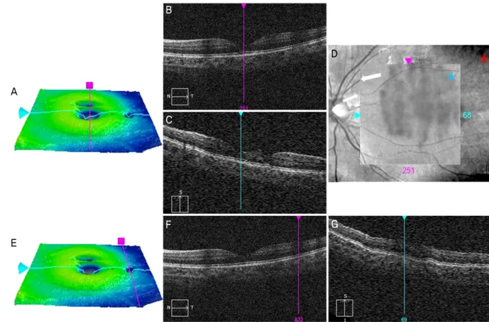
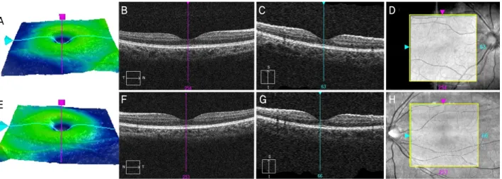
초진 시 우안 나안시력 0.25, 좌안 나안시력 0.2로 측정되었 으며, 최대교정시력은 양안 1.0이었다. 양안 안압은 각각 18 mmHg, 17 mmHg로 측정되었다. 세극등 검사에서 특이 소견은 없었으며, 안저검사와 안저촬영에서 양안 모두 하 나의 중심와를 확인할 수 있었다(Fig. 1). 추가적으로 Cirrus HD-OCT (Carl Zeiss-Meditec, Dublin, CA, USA)를 이용한 황반 검사를 시행하였고, 그 결과 우안의 Macular Cube 512 × 128 프로토콜의 retinal thickness map에서 인접한 두 개 의 중심와를 확인할 수 있었다(Fig. 2). 또한 좌안의 Macular Cube 512 × 128 프로토콜의 retinal thickness map에서는 중앙의 중심와에서 수직선상으로 분리된 중심와 소견이 관 찰되었고(Fig. 3), 수평선상으로도 분리된 중심와 소견이 관 찰되었다(Fig. 3). 관찰된 중심와를 지나는 수직 B-scan 상 분리된 두 개의 중심와 위치에서 각각 중심와 모양이 관찰 되었다(Fig. 3). 그러나 수평선상으로 위치한 중심와를 지나 는 수평 B-scan 상 가쪽 황반의 중심와 위치에서 전형적인 중심와 모양이 관찰되지 않았다(Fig. 3). 내원 2주 후 같은 기계와 프로토콜을 사용하여 재검사가 시행되었고, 이때 검사 중 시선 고정이 잘 이루어지도록 검사 전 설명을 시행 하였다. 그 결과 양안의 Macular Cube 512 × 128 프로토콜 에서 하나의 중심와를 확인할 수 있었다(Fig. 4).
A
E
B
C
F
D
G
Figure 3. Cirrus HD-OCT of the left eye. (A) Retinal thickness map from the Macular Cube 512 × 128 volume scan on Cirrus HD-OCT. Three foveal depressions are noted. (B) Horizontal, transfoveal B-scan of the left eye that demonstrates a normal foveal contour. Foveal depression of the lateral side visible on retinal thickness map’s horizontal meridian is presented not as a foveal pit, but as a normal retinal contour. (C) Vertical B-scan of the same eye presenting two foveal depressions. (D) Optical coherence tomog- raphy fundus image overlay (blue asterisk) on infrared photomicrograph (red asterisk) of the left eye. Some abrupt discontinuations of vessels are noted by white arrows. Note gray shadow distinguishable from other areas in the background. (E-G) Foveal duplication at the horizontal meridian to the anatomical fovea shown in retinal thickness map is not clearly presented in both horizontal and verti- cal scans. However, signal intensity at the site of foveal duplication seems to be significantly lower than the other part of the scans.
고 찰
해부학적으로 인간에게서 중복 중심와가 보고된 예는 지 금까지 단 한 건뿐이다.7 그런데 정상적인 중심와를 가지고 있음에도 SD-OCT 상에서 둘 이상의 중심와로 나타나는 오 류를 보이는 증례들이 보고되고 있다.4,5 Baskin et al5은 이와 같은 오류를 처음으로 보고하면서 이것이 Cirrus HD-OCT의 스캔 변환 알고리즘의 기계적인 오류(hardware error)인지 환자의 위쪽으로의 미세 도약 안구 운동(microsaccade) 이 후 정면 주시로 인한 환자에 의한 오류인지를 결론짓지 못 하였다. 이후 Kalliath and Shukla6는 안진을 가진 환자 한 명을 포함한 네 명의 환자에게서 나타난 중복 중심와 오류 를 보고하였다. 이 보고에서 환자들에게 정면을 주시할 것 을 재교육시킨 후 같은 기계와 같은 프로토콜을 사용하여 재검을 시행하였을 때 안진이 있는 환자를 제외한 나머지 세 명의 환자에게서 오류가 사라졌고, 중복 중심와 오류가
환자의 주시 실패로 인한 것일 가능성을 시사하였다. 본 증례에서 중복 중심와 오류는 환자에게 검사 중의 주 시에 대한 적절한 교육 이후 재검을 거쳤을 때 사라지는 것 을 확인할 수 있었다. 이는 이전의 연구들에서 제시한 미세 도약 안구 운동으로 인한 오류의 발생 가능성을 시사한다. 또한 본 증례의 양안 모두에서 중복 중심와 오류는 이전 보 고들에서와 마찬가지로 수직 B-scan 상에, 그리고 해부학 적 중심와를 중심으로 위쪽 황반에 위치하는 것을 확인할 수 있었다. 이렇게 중복 중심와 오류가 반복적으로 해부학 적 중심와 위쪽에 나타나는 것은 검사 과정에서 검사에 대 한 충분한 교육을 받지 못한 환자가 위에서 아래로 내려오 는 보이는 광원을 눈으로 자연스럽게 좇아가면서 발생한 현상으로 설명될 수 있다. 만일 이러한 가정이 옳다면, 이 에 대한 대책으로 눈에 보이지 않는 광원을 이용한 기계 등 의 사용이 하나의 대안이 될 수 있을 것이다.
본 증례 환자의 좌안 retinal thickness map에서 실제의
A
E
B C D
F G H
Figure 4. Cirrus HD-OCT of the both eyes two weeks after the first visit. (A-D) Macular Cube 512 × 128 volume scan on Cirrus HD-OCT of the right eye. (E-H) Macular Cube 512 × 128 volume scan on Cirrus HD-OCT of the left eye. No duplication of fovea was noted. No abrupt discontinuation or shadow was noted.
중심와의 수평선상에 위치한 추가적인 중심와 오류가 관찰 되었으나 수평 B-scan 상에서는 명확한 중심와 오류가 나 타나지 않았다(Fig. 3). 대신에 그 위치의 수평 B-scan 상 신호감도가 다른 부분에 의해 현저하게 떨어지는 부분으로 관찰되었다(Fig. 3). 일반적으로 중복 중심와 오류가 관찰되 는 수직선상이 아닌 다른 위치에서 발생한 중복 중심와 오 류라는 점과 B-scan 상에서 신호 강도가 떨어지는 부분에 서 이러한 오류가 발생했다는 점은 중복 중심와 오류의 발 생에 환자에 의한 오류 이외에도 소프트웨어에 의한 오류 에 의한 가능성이 있음을 시사한다.
본 증례의 중복 중심와 오류를 보이는 양안 모두에서 Cirrus HD-OCT의 Macular Cube 512 × 128 프로토콜에서 제공하는 fundus overlay image 상 그림자 현상이 보였다 (Fig. 2, 3). 이러한 그림자 현상이 대부분 수직선 형태로 생 기는 것을 미루어 볼 때, OCT 촬영 시 환자가 광원을 위에 서 아래로 추종하면서 생긴 미세 도약 안구 운동으로 인한 오류로 유추할 수 있다. 이것은 광원의 추종으로 발생하는 영상의 위상차로 인해 그림자 현상이 발생한 것으로 사료 된다. 그림자 현상에서 수평선 형태로 생기는 것도 발견할 수 있는데, 마찬가지의 원리로 안구의 순간적인 좌우 운동 으로 인해 발생할 것으로 생각된다(Fig. 2 white arrows). 2 주 후 같은 프로토콜의 fundus overlay image 상에서는 이 러한 그림자 현상은 관찰되지 않았고, 중복 중심와 오류 역 시 없었다(Fig. 4). Fundus overlay image 상에서의 그림자 현상과 중복 중심와 오류의 직접적인 연관성을 확인하기 위해서는 다른 추가적인 연구들이 시행되어야 하겠으나, 빛간섭단층촬영 영상에 이러한 그림자 현상이 확인될 경우
에는 획득한 영상 전체의 질에 문제가 있는지 재확인이 필 요할 것이며 그 해석에 주의해야 할 것이다.
이번 증례에서 중복 중심와 오류는 다른 검사 결과와의 불일치로 인해 쉽게 발견되고 그 오류를 바로잡을 수 있었 다. 그러나 만일 이 같은 오류가 제대로 발견되지 못할 경 우, 진단 실패와 잘못된 처치를 야기할 수 있다. 따라서 중 복 중심와 오류가 발견되었을 경우 반드시 환자에게 검사 전 반과 주시에 대한 재교육을 거친 후 환자의 협조하에 재검사 를 시행하여 오류 여부를 분명히 확인하여야 할 것이다.
REFERENCES
1) Kim US. Optical coherence tomography. Clin Neuroophthalmol 2014;4:17-21.
2) Han IC, Jaffe GJ. Evaluation of artifacts associated with macular spectral-domain optical coherence tomography. Ophthalmology 2010;117:1177-89.e4.
3) Ho J, Sull AC, Vuong LN, et al. Assessment of artifacts and re- producibility across spectral- and time-domain optical coherence tomography devices. Ophthalmology 2009;116:1960-70.
4) Ray R, Stinnett SS, Jaffe GJ. Evaluation of image artifact produced by optical coherence tomography of retinal pathology. Am J Ophthalmol 2005;139:18-29.
5) Baskin DE, Gault JA, Vander JF, Dugan JD Jr. Double fovea artifact. Ophthalmology 2011;118:429.e1.
6) Kalliath J, Shukla D. Foveal duplication artifact with spectral-do- main optical coherence tomography. Ophthalmic Surg Lasers Imaging Retina 2013;44:94-6.
7) Behera UC, Shukla D, Kim R. Pseudoduplication of fovea in a hu- man eye. Arch Ophthalmol 2007;125:1428-30.
= 국문초록 =
스펙트럼영역 빛간섭단층촬영에서 발생한 중복 중심와 오류 1예
목적: 스펙트럼영역 빛간섭단층촬영에서 발생한 중복 중심와 오류를 보고하고자 한다.
증례요약: 9세 남자가 시력저하를 주소로 내원하였다. 최대교정 시력은 양안 1.0이었으며, 안압은 정상이었다. 안저검사와 안저촬영, 스펙트럼영역 빛간섭단층촬영을 시행하였고, 안저검사와 안저촬영상 정상 중심와가 확인되었다. 스펙트럼영역 빛간섭단층촬영상 우 안에는 수직상으로 위치한 한 개의 중심와 오류가 발견되었다. 좌안에는 각각 수직상 및 수평상으로 위치한 두 개의 중심와 오류가 관찰되었다. 내원 2주 후 같은 기계와 프로토콜을 사용하여 재검사가 시행되었고, 이때 검사 중 시선 고정이 잘 이루어지도록 검사 전 설명을 시행하였다. 그 결과 양안에서 모두 하나의 중심와를 확인할 수 있었다.
결론: 해부학적 중심와 위쪽에 발생한 중복 중심와 오류는 주로 환자의 시선 고정 실패에 의해 발생한 환자 오류일 가능성이 있으므로 중복 중심와 오류가 발견되었을 경우 반드시 환자에게 검사 전반과 주시에 대한 재교육을 거친 후 환자의 협조하에 같은 기계와 프로 토콜로 재검사를 시행하여 오류 여부를 분명히 확인하여야 할 것으로 생각된다.
<대한안과학회지 2017;58(8):1003-1007>