pISSN: 0378-6471 eISSN: 2092-9374 DOI : 10.3341/jkos.2010.51.11.1471
= 증례보고 =
야간혈압하강이 녹내장의 진행에 미치는 영향
서홍융 류원열 노세현 동아대학교 의과대학 안과학교실
목적: 야간혈압하강이 시야 결손의 진행에 미치는 영향을 알아보고자 하였다.
대상과 방법: NTG 140명(280안)과 POAG 84명(168안)을 대상으로 24시간 활동 혈압 검사와 시야검사를 시행하였다. 야간혈압하강이 10% 미만인 경우를 non-dipper, 10% 이상인 경우를 dipper로 나누어, 야간혈압하강과 시야 결손의 진행, 그리고 고혈압 치료와의 관계를 알아보고자 하였다.
결과: 수축기와 이완기에서의 dipper는 NTG에서 시야 결손을 진행시켰으며, 이는 약 3배의 위험성을 보였다. 고혈압 치료는 NTG, POAG에서 시야 결손의 진행에 영향을 주지 않았으나, NTG에서는 수축기와 이완기 모두에서, POAG에서는 이완기에서 dipper를 유발 하였다. 야간혈압하강은 NTG에서 시야 결손의 진행이 있는 경우에 없는 경우보다 유의하게 크게 나타났다.
결론: Dipper는 NTG에서 시야 결손의 진행을 유발하며, 이러한 dipper는 NTG와 POAG에서 고혈압 치료에 영향을 받는다. 목표안압 이하로 안압이 유지됨에도 불구하고 시야 결손의 진행을 보이는 경우 24시간 활동 혈압 검사가 유용할 것이며, 고혈압 치료 또한 녹내 장 치료에 중요한 인자로 고려해야 할 것이다.
<대한안과학회지 2010;51(11):1471-1478>
접 수 일: 2010년 3월 9일 심사통과일: 2010년 8월 25일
책 임 저 자: 노 세 현
부산광역시 서구 동대신동 3가 동아대학교 의료원 안과
Tel: 051-240-5227, Fax: 051-254-1987 E-mail: [email protected]
* 본 논문의 요지는 2008년 대한안과학회 제100회 학술대회에서 구연으로 발표되었음.
* 이 논문은 2008년, 2009년도 동아대학교 학술연구기금 지원에 의해 연구되었음.
녹내장은 시신경의 녹내장성 변화와 녹내장에서 특징적 으로 나타나는 주변 시야 결손과 함께 만성적으로 진행하 는 시신경 병증이다.1녹내장의 병인은 아직 명확하게 밝혀 져 있지 않으나 안압의 상승이 가장 중요한 위험인자로 알 려져 있다. 그러나 목표안압 이하로 안압이 유지되는데도 불구하고 정상 안압 녹내장(normal tension glaucoma, NTG)과 원발 개방각 녹내장(primary open angle glaucoma, POAG)에서 시신경의 병변과 시야 결손이 진행하는 것은 시신경에 기계적 손상을 주는 안압 이외에 다른 인자의 영 향이 있음을 의미하고 있다.2-7또한, 당뇨, 고혈압과 같은 심혈관 질환 환자나 편두통, 말초 혈액 순환장애를 갖는 환 자에서 녹내장의 발생빈도가 높다는 여러 보고들을 비추어 볼 때,8-14높은 안압에 의한 시신경의 기계적 손상 이외에 안압에 의존하지 않으면서 신경에 손상을 주는 허혈에 의 한 시신경 손상 기전을 시사하고 있다.15
안압 외에 시신경에 손상을 주는 전신적 요인으로는 야
간혈압의 하강(nocturnal hypotension), 혈관 경련(vasos- pasm), 혈압, 관류압(perfusion pressure), 뇌 및 심혈관 허혈, 수면 무호흡증, 자가면역질환과 비정상적인 혈관의 자가조절기능의 상실(vascular dysregulation) 등이 있고, 국소적 요인으로는 시신경 출혈, 시신경유두주위 위축, 맥 락막 경화 등이 있다.16
안압 외에 시신경에 영향을 주는 원인으로 가장 중요한 것은 안혈류양의 감소이고, 이것은 혈관의 혈류를 조절하는 기능(autoregulation)이 상실(vascular dysregulation)되었 을 때 나타난다.3,17안관류압(ocular perfusion pressure)의 감소, 혈관 저항(vascular resistance)의 증가, 혈액 점도 (blood viscosity)의 증가 등이 안혈류양의 감소에 관여하 고 있으며,17 안압의 감소와 더불어 안혈류의 증가를 꾀한 다면 시신경의 손상이나 녹내장성 시야 결손의 진행을 더 디게 할 수 있다.18
저혈압이나 야간혈압이 떨어지면 안관류압이 저하되고, 이로 인해 안혈류양이 감소하게 되면 시신경과 축삭이 허 혈에 의한 손상을 받게 되고 결과적으로 녹내장성 시야 결 손을 야기하게 된다고 알려져 있다.16,19,20 본 연구에서는 안 관류압의 상태를 임상에서 쉽게 파악할 수 있는 24시간 활 동 혈압의 측정과 함께 시야검사를 하였고, 24시간 활동 혈 압 변화에 따른 녹내장성 시야 결손의 진행 여부를 분석 검 토하였다.
시신경의 혈액공급을 판단하는 기준이 되는 안관류압은
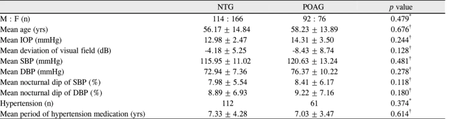
Table 1. Demographics of patients in NTG and POAG
NTG POAG p value
M : F (n) 114 : 166 92 : 76 0.479*
Mean age (yrs) 56.17 ± 14.84 58.23 ± 13.89 0.676†
Mean IOP (mmHg) 12.98 ± 2.47 14.31 ± 3.50 0.244†
Mean deviation of visual field (dB) -4.18 ± 5.25 -8.43 ± 8.74 0.128†
Mean SBP (mmHg) 115.95 ± 11.02 120.63 ± 13.24 0.481†
Mean DBP (mmHg) 72.94 ± 7.36 76.37 ± 10.22 0.278†
Mean nocturnal dip of SBP (%) 7.98 ± 5.54 8.41 ± 6.17 0.118†
Mean nocturnal dip of DBP (%) 8.89 ± 6.93 9.22 ± 7.16 0.180†
Hypertension (n) 112 61 0.374*
Mean period of hypertension medication (yrs) 7.33 ± 4.28 7.03 ± 3.47 0.614†
NTG = normal tension glaucoma; POAG = priamry open angle glaucoma; SBP = systolic blood pressure; DBP = diastolic blood pressure.
*Chi-square test; †Student t-test.
아래의 공식과 같이 평균 동맥압(mean arterial blood pressure, MAP)과 안압에 의해 결정되고,19,21MAP는 수축 기 혈압(systolic blood pressure, SBP)과 이완기 혈압 (diastolic blood pressure, DBP)에 의해 결정된다.17,18
Ocular perfusion pressure = 2/3 × MAP IOP MAP = DBP + 1/3(SBP DBP)
혈압은 24시간 항상 변화하며 주간보다는 야간의 혈압이 더 낮다.24,25 야간에는 카테콜아민(catecholamine)이 적게 분비되고 이것은 교감신경의 활동을 떨어뜨려 야간에 혈압 의 저하를 일으키며, 이로 인해 심박동수(heart rate, HR), 심박출량(cardiac output) 및 말초 혈관의 저항을 떨어뜨리 는 결과를 초래한다.26따라서 24시간 중 한 시점에서 측정 한 혈압은 임상적으로 크게 의미가 없고, 24시간 동안 측정 한 혈압에서 주간보다 야간의 혈압이 낮을 때 안혈류양은 감소되고 시신경에 허혈성 손상을 초래하여 녹내장성 시야 결손의 진행을 야기할 수 있다.27-30
이에 본 연구에서는 앞에서 언급한 여러 방법들에 비해 비교적 덜 침습적이고 쉽게 시행할 수 있는 24시간 활동 혈 압을 측정하여 야간혈압하강(nocturnal dip)이 녹내장성 시 야 결손의 진행에 미치는 영향과 고혈압 치료와의 연관성 을 알아보고자 하였다.
대상과 방법
본원에서 2년 이상 관찰 중인 NTG 환자 140명(280안) 과 POAG 환자 84명(168안)을 대상으로 하였다. NTG 환 자군과 POAG 환자군의 성별, 연령 및 고혈압 유무를 조사 하였으며, 고혈압으로 진단받은 환자들은 모두 고혈압 약물 을 복용하였고 그 치료기간을 조사하였다(Table 1).
24시간 활동 혈압 측정은 전자혈압계인 24시간 활동 혈
압 측정기(TONOPORT V. GM Medical System, Germ- any)를 이용하여 비우세완(non-dominant arm)에서 측정 하였다. 224명을 대상으로 평균 1.17회 시행하였으며, 주 간 혈압(오전 7시~오후 10시)은 30분 간격으로, 야간 혈 압(오후 10시~오전 7시)은 1시간 간격으로 측정하였다.
SBP, DBP, HR, MAP를 측정하여 최고치, 최저치, 평균치 를 조사하였으며, NTG 환자군과 POAG 환자군의 유의한 차이는 없었다(Table 1).
야간혈압하강(nocturnal dip)은 주간 혈압의 평균치에 비 하여 야간 혈압의 평균치가 얼마나 감소하는가를 나타낸 것으로, 다음의 식으로 구한다.31-33
Nocturnal dip = (Average of day time blood pressure Average of night time blood pressure) / Average of day time blood pressure × 100
본 연구 초기에 목표 안압을 유지하는 녹내장 환자들에 서 시야 결손의 진행을 보이는 여러 환자들에서 10% 이상 의 야간혈압저하(nocturnal dip)를 보이는 경우가 많았으 며, 그리고 여러 연구들26,29,34에서의 기준을 참고하여 야간 혈압저하(nocturnal dip)가 10% 미만인 경우를 non-dipper, 10% 이상인 경우를 dipper로 정의하였다.
시야검사는 자동시야검사(Humphrey field analyzer, 24-2 Swedish Interactive Thresholding Algorithm (SITA) standard program, Carl-Zeiss Meditec, Dublin, CA, USA)를 이용하여 적어도 2년 이상 경과 관찰한 환자 중 최 소 6개월 간격으로 4회 이상 시행하였으며, the Advanced Glaucoma Intervention Study (AGIS) method를 이용하여 AGIS score상 적어도 2 unit 이상의 감소가 있거나 mean deviation (MD)이 2dB 이상의 감소가 있을 때를 시야 결손 의 진행이라고 정의하였다.35,36 또한 false-positive error 와 false-negative error가 33% 미만이고 fixation loss도
Table 2. Association of dipper and V/F progression in NTG and POAG
V/F progression Chi-square test
Yes No p value* Odd ratio Relative risk
SBP in NTG <0.000
4.951 (CI†: 2.448~10.015)
3.816 (CI†: 2.103~6.925)
Dipper 27 67
Non-dipper 14 172
DBP in NTG 0.002
3.093 (CI†: 1.481~6.456)
2.650 (CI†: 1.384~5.077)
Dipper 30 112
Non-dipper 11 127
SBP in POAG 0.116
2.071 (CI†: 0.824~5.202)
1.881 (CI†: 0.848~4.173)
Dipper 11 51
Non-dipper 10 96
DBP in POAG 0.128
2.050 (CI†: 0.802~5.242)
1.875 (CI†: 0.82~4.287)
Dipper 13 65
Non-dipper 8 82
V/F = visual field; SBP = systolic blood pressure; NTG = normal tension glaucoma; DBP = diastolic blood pressure; POAG = priamry open angle glaucoma.
*Statistical significance: p<0.05; †CI: confidence interval, CI was calculated at 95 percentile.
Table 3. Association of hypertension and V/F progression in NTG and POAG
V/F progression Chi-square test
Yes No p value* Odd ratio Relative risk
NTG 0.112
1.708 (CI†: 0.878~3.323)
1.575 (CI†: 0.896~2.768)
HTN Treatment group 21 91
Normal BP group 20 148
POAG 0.249
1.711 (CI†: 0.681~4.299)
1.595 (CI†: 0.719~3.537)
HTN Treatment group 10 51
Normal BP group 11 96
V/F = visual field; NTG = normal tension glaucoma; HTN = hypertension; POAG = priamry open angle glaucoma; BP = blood pressure.
*statistical significance: p<0.05; †CI: confidence interval, CI was calculated at 95 percentile.
20% 미만일 때의 시야 검사를 적절한 것으로 보았다.
안압 검사는 낮 시간에 한 명의 동일 검사자가 골드만 압 평 안압계를 이용하여 3회 측정한 평균치를 사용하였고, NTG 환자군와 POAG 환자군 간의 유의한 차이는 보이지 않았다.
Chi-square test를 이용하여 대상군의 분포의 통계적 유 의성을 검증을 통해 야간혈압하강, 시야 결손의 진행, 그리 고 고혈압 치료의 연관성을 분석하여 그 상대위험도 또한 조사하였으며, Student t-test를 이용하여 야간혈압하강 (nocturnal dip)이 녹내장성 시야 결손의 진행에 미치는 영 향에 대해 살펴보았다(SPSS 12.0).
결 과
24시간 활동 혈압이 10% 이상의 야간혈압하강(noct- urnal dip)을 보인 dipper 환자에서 수축기 혈압이 하강된 환자는 NTG군의 94안, POAG군에서는 62안, 이완기 혈압 이 하강된 환자는 NTG군에서 142안, POAG군에서는 78안 으로 나타났다. 이 중 시야 결손이 진행한 경우는 수축기 혈압이 하강된 환자는 NTG군은 27안, POAG군은 11안이
었고, 이완기 혈압이 하강된 경우는 NTG군은 30안, POAG 군은 13안으로 나타났다(Table 2).
NTG는 수축기와 이완기의 dipper가 녹내장성 시야 결손 의 진행에 영향을 미쳤으며(SBP: p<0.000, DBP: p= 0.002), POAG는 수축기와 이완기 모두에서 그 유의성이 없었다(SBP: p=0.116, DBP=0.128) (Table 2).
이를 근거로 하여 신뢰구간(confidence interval, CI) 95%에서 relative risk와 odd ratio를 살펴보았다. NTG 환 자군에서는 dipper와 녹내장성 시야 결손의 진행이 동반된 경우는 그렇지 않은 군에 비해 수축기 혈압에서 4.951 (CI:
2.448~10.015)의 odd ratio와 3.816배(CI: 2.103~6.925) 의 relative risk를 보였고, 이완기 혈압에서는 3.093 (CI:
1.481~6.456)의 odd ratio와 2.65배(CI: 1.384~5.077)의 relative risk를 보였다(Table 2). POAG 환자군에서는 수 축기 혈압에서 2.071 (CI: 0.824~5.202)의 odd ratio와 1.881배(CI: 0.848~4.173)의 relative risk를 보였고, 이완 기 혈압에서는 2.05 (CI: 0.802~5.242)의 odd ratio와 1.875배(CI: 0.82~4.287)의 relative risk를 보였다(Table 2).
고혈압 유무와 시야 결손의 진행과의 관계에서는 NTG, POAG 모두에서 통계학적 유의성을 보이지 않았으나
Table 4. Association of hypertension and dipper in NTG and POAG
Dipper Chi-square test
Yes No p value* Odd ratio Relative risk
SBP in NTG 0.001
2.430 (CI†: 1.462~4.040)
1.779 (CI†: 1.282~2.469) HTN treatment group 51 61
Normal BP group 43 125
DBP in NTG <0.000
10.625 (CI†: 6.054~18.647)
3.750 (CI†: 2.688~5.232) HTN treatment group 80 32
Normal BP group 62 106
SBP in POAG 0.068
1.822 (CI†: 0.953~3.481)
1.445 (CI†: 0.979~2.131) HTN treatment group 28 33
Normal BP group 34 73
DBP in POAG <0.000
3.170 (CI†: 1.639~6.132)
1.996 (CI†: 1.355~2.940) HTN treatment group 33 28
Normal BP group 45 62
SBP = systolic blood pressure; NTG = normal tension glaucoma; DBP = diastolic blood pressure; HTN = hypertension; POAG = priamry open angle glaucoma.
*Statistical significance: p<0.05; †CI: confidence interval, CI was calculated at 95 percentile.
Table 5. The mean of nocturnal dip & 24 hours blood pressure in NTG & POAG
The mean of nocturnal dip (%) The mean of blood pressure (mmHg) V/F progression
p value* V/F progression
p value*
Yes No Yes No
SBP in NTG 0.001 0.392
Dipper 15.66 ± 4.02 13.21 ± 2.70 118.27 ± 11.05 121.28 ± 10.77
Non-dipper 3.63 ± 5.42 5.10 ± 3.55 115.28 ± 10.93 113.56 ± 10.38
DBP in NTG <0.000 0.245
Dipper 16.57 ± 4.23 14.07 ± 3.30 73.84 ± 8.11 74.40 ± 7.15
Non-dipper 1.81 ± 3.54 3.11 ± 3.94 75.11 ± 8.69 71.25 ± 6.97
SBP in POAG 0.144 0.175
Dipper 18.03 ± 5.23 13.93 ± 3.45 122.30 ± 8.51 123.15 ± 19.10
Non-dipper 3.25 ± 3.06 4.91 ± 3.84 126.51 ± 11.88 118.49 ± 9.11
DBP in POAG 0.104 0.139
Dipper 18.03 ± 5.48 14.90 ± 3.71 81.25 ± 3.16 79.55 ± 11.74
Non-dipper 2.75 ± 3.24 3.95 ± 4.35 76.55 ± 10.44 73.07 ± 8.52
SBP = systolic blood pressure; NTG = normal tension glaucoma; DBP = diastolic blood pressure; POAG = priamry open angle glaucoma.
*Student t-test, statistical significance: p<0.05.
(Table 3), 고혈압 유무와 dipper와의 상관성을 조사한 결 과 NTG군에서 수축기와 이완기 모두 통계학적 유의성을 보였으며, 수축기의 경우 1.779배(CI: 1.282~2.469), 이완 기의 경우 3.750배(CI: 2.688~5.232)의 relative risk를 나 타내었다(Table 4).
NTG 환자군에서는 수축기와 이완기 혈압의 야간혈압하 강(nocturnal dip)은 녹내장성 시야 결손의 진행이 있는 경 우에 녹내장성 시야결손의 진행이 없는 경우보다 통계학적 으로 유의하게 크게 나타났으나, POAG 환자군에서는 야간 혈압하강이 녹내장성 시야 결손의 진행에 영향을 미치지 않았다(Table 5).
또한 NTG와 POAG에서 녹내장성 시야 결손의 진행이 있는 군과 없는 군에서 수축기와 이완기의 24시간 평균혈 압(mmHg)에 대해 비교분석하였으나, 통계학적으로 유의
하지 않았다(Table 5).
고 찰
혈압이 녹내장성 시신경 손상과 시야에 미치는 영향에 대하여 많은 이견이 있으나,37-39본 연구에서 조사한 바로 는 녹내장 환자에서 시야 결손의 진행한 군은 진행하지 않 은 군에 비해 24시간 수축기와 이완기 평균 혈압(mmHg) 이 상대적으로 낮았으나 녹내장 환자의 시야 결손의 진행 에 직접적인 영향을 미치지 않는 것으로 조사되었다.
Leske et al39,40은 POAG와 NTG 환자에서 24시간 혈압 의 동태, 안압, 안관류압 및 시신경유두의 녹내장성 변화를 관찰하여 이들이 녹내장성 시야 결손 진행에 미치는 상대 위험도(relative risk)를 조사하였는데, 혈압이 이상적으로
조절이 되고 있는 고혈압 환자에서는 relative risk가 0.49 배임에 반해서, 수축기 관류압(perfusion pressure)이 101 mmHg 미만인 경우는 relative risk가 2.6배, 이완기 관류압 이 55 mmHg 미만인 경우는 relative risk가 3.2배, MAP가 42 mmHg 미만인 경우는 relative risk가 3.1배였다고 보고 하였고, 정상보다 낮은 관류압을 보인 개방각 녹내장 환자 에서 3배의 상대위험도가 있었다고 보고하였다.39,40 그리 고, Tielsch et al41은 POAG 환자에서 이완기 관류압과 녹 내장 위험도와의 관계를 조사하였는데, 30 mmHg 미만의 이완기 관류압은 50 mmHg 이상일 때보다 6.22배의 녹내 장 발생 위험이 있었다고 보고하였으며,41 The Egna- Neumarkt Study10에서도 낮은 이완기 관류압이 녹내장의 시신경 변화와 녹내장성 시야 결손의 진행에 영향을 미쳤 다고 보고하였다. 본 연구에서는 관류압의 중요한 요소인 야간혈압하강(nocturnal dip)과 시야 결손의 진행 유무를 조사하여, dipper와 녹내장성 시야 결손의 진행을 동반한 군은 그렇지 못한 군에 비해 NTG군에서는 relative risk가 약 3배였고, POAG군에서 relative risk가 약 1.8배였다. 앞 서 언급한 관류압에 관한 다른 연구들에서는 NTG와 POAG 에서 그리고 특히 이완기에 대한 연구 결과가 의미가 있었 던 것에 비해, 본 연구에서는 NTG의 수축기 dipper에서 녹 내장의 진행에 영향을 주었으며, 추후 관류압에 대한 연구 조사도 필요할 것으로 보인다.
일반적으로 항고혈압제가 안관류압을 감소시킨다고 알 려져 있다. 고혈압 치료를 받고 있는 녹내장 환자군이 정상 혈압을 가진 녹내장 환자군에 비해 녹내장성 시신경손상이 더 진행하였다던지,42고혈압 치료를 받는 정상인에서 이완 기 혈압이 90 mmHg 이하인 경우에 시신경 유두함몰비가 증가되었으며,43 고혈압 치료를 받고 있는 개방각 녹내장 환자에서 이완기 관류압이 50 mmHg 이하인 군에서는 50 mmHg 이상인 군에 비해 녹내장 발생률이 4배 이상이었다 는 연구도 있다.44본 연구에서는 혈압약 복용 유무와 시야 결손의 진행과의 관계는 없으나, 고혈압 치료를 받고 있는 경우 NTG에서 수축기에서는 약 2배, 이완기에서는 약 4배 정도 dipper를 유발하였으며, 이러한 dipper는 시야 결손의 진행에 약 3배의 위험을 가지는 것으로 나타났다. 이는 이 전 연구들과는 달리 POAG에서보다는 NTG에서 dipper, 즉 안관류압의 감소가 시야 결손의 진행에 영향을 주며, 고혈 압 약물 복용이 영향을 미칠 수 있음을 시사하고 있다.
NTG와 POAG의 야간혈압하강(nocturnal dip)의 차이와 녹내장의 진행에 대한 측면에서, NTG에서 POAG보다 혈압 의 일중변동과 야간혈압하강(nocturnal dip)의 폭이 더 컸 었고 녹내장성 시신경 변화와 녹내장성 시야 결손의 진행 을 더 악화시켰다는 연구가 있는가 하면,45 NTG와 POAG
의 야간혈압하강(nocturnal dip)의 유의한 차이는 없으나 SBP가 90 mmHg 이하인 경우가 상대적으로 NTG에서 많 았다는 보고도 있다.46본 연구에서는 NTG와 POAG 간의 야간혈압하강(nocturnal dip)의 차이는 보이지 않았고, NTG에서만 dipper가 녹내장성 시야 결손의 진행을 야기하 였다.
어느 정도의 야간혈압하강(nocturnal dip)이 녹내장의 진 행에 영향을 주는지에 대한 연구는 다양하다. 20% 이상의 야간혈압하강(nocturnal dip)이 NTG와 POAG의 시신경 유 두함몰비를 증가시켰다는 보고가 있으며,4710% 이상의 야 간혈압의 하강(nocturnal dip)은 녹내장성 시야 결손의 진 행을 야기하였다는 연구도 있다.34,48,49저자들은 10% 이상 의 야간혈압하강(nocturnal dip)을 dipper로 보았고, dipper 는 NTG군에서 녹내장성 시야 결손의 진행을 유발하였다.
정상인과 NTG 환자에서 야간혈압하강(nocturnal dip)의 유의한 차이는 없었으나, 10% 이상의 야간혈압하강(nocturnal dip)을 보인 NTG군에서는 녹내장성 시야 결손이 진행을 보였다는 연구도 있으며,49정상인에서 50세 미만의 연령군 보다 50세 이상의 고연령군에서 주간과 야간의 안혈류 속 도의 차가 더 컸다는 보고도 있다.50 이는 추후 본 연구의 결과를 바탕으로 정상인과의 비교 분석이나 연령에 따른 비교 분석 또한 필요함을 시사하고 있다.
안혈류와 녹내장과의 관계에 대해서도 많은 연구가 있다.
POAG에서 20% 이상의 야간혈압하강(nocturnal dip)이 있 는 경우 구후혈류 속도의 감소를 보였다는 보고가 있고,51 POAG에서 시야결손의 진행이 있는 경우는 없는 경우에 비 해 구후혈류 속도가 감소되었다는 연구도 있다.21또한, 정 상인에서의 시신경유두의 혈류 속도가 주간에 비해 야간에 감소되었다던지,27정상인에 비해 POAG와 안고혈압증(ocular hypertension, OHT)에서 혈류속도가 현저히 저하되었다는 보고도 있다.52 그리고, POAG 환자에서 안혈류 속도가 높 은 군보다 낮은 군에서 시야 결손이 진행한 경우가 더 많이 나타났다는 연구가 있다.53 이러한 연구 결과는 본 연구에 서 안관류압이나 안혈류를 직접적으로 조사하지는 못하였 으나, dipper는 야간에 감소하는 안혈류를 반영하는 인자임 을 고려할 때, 시야 결손의 진행을 유발하는 결과에 근거가 될 수 있겠다. 또한, 안관류압을 조사하기 위해서는 야간에 24시간의 안압 변동을 측정해야 하지만, 환자를 안압만을 측정하기 위하여 입원시켜야 하고, 2시간마다 환자를 깨워 안압 측정을 하였을 때 측정된 안압과 24시간 활동 혈압의 신뢰도가 떨어질 위험성이 있다. 저자들은 이러한 위험성을 피하고, 안관류압을 감소시킬 뿐만 아니라 안혈류를 떨어뜨 리는 야간혈압하강(nocturnal dip)을 조사하기 위해 24시 간 활동 혈압 검사를 시행하였다.
NTG와 POAG 환자에서 고혈압, 심혈관 질환, 당뇨병과 뇌혈류 질환 등의 전신질환과 상당히 밀접한 연관이 있
다.54,55그리고 NTG 환자에서 혈액의 endothelin-1 (ET-1)
이증가되었다는 보고가 있고,5610% 이상의 야간혈압하강 (nocturnal dip)이 있을 때 혈장(plasma) 내 D-dimer, plasminogen, activator inhibitor-1, von Willebrand factor, soluble intercellular adhesion molecule-1, interleukin-6 의 수치가 낮아, 세포 결합, 염증, 및 혈액 응고의 장애를 보 였다는 연구도 있다.57 이는 향후 시야 결손의 진행에 영향 을 줄 수 있는 여러 안압 외적인 인자에 대한 연구가 필요 함을 보여주고 있다.
녹내장의 진행에서 안압의 역할은 무엇보다도 중요하다.
그러나 목표안압 이하로 안압이 일정하게 유지됨에도 불구 하고 시신경 녹내장성 변화와 녹내장성 시야 결손의 진행 이 있다면 분명 안압 외적인 인자, 특히 야간혈압하강 (nocturnal dip)을 유념해야 할 것이며, 이러한 야간혈압하 강(nocturnal dip)은 고혈압 치료와 관련이 크다. 녹내장 환 자의 24시간 활동 혈압 검사는 목표안압 이하로 안압이 비 교적 잘 유지된 녹내장 환자에서 시야 결손의 진행의 원인 규명과 녹내장의 치료에도 도움을 줄 것으로 생각하며, 과 도한 고혈압 치료가 과연 녹내장 치료에 도움을 주는 것인 지에 대해 충분히 고려해야 한다. 그리고, 반복적인 24시간 활동 혈압 검사를 통해 낮은 혈압과 야간혈압하강(noc- turnal dip)의 호전과 악화에 대한 분석도 중요할 것으로 생 각된다.
참고문헌
1) Van Buskirk EM, Cioffi GA. Glaucomatous optic neuropathy. Am J Ophthalmol 1992;113:447-52.
2) Levene RZ. Low tension glaucoma: a critical review and new material. Surv Ophthalmol 1980;24:621-64.
3) Flammer J, Orgul S. Optic nerve blood-flow abnormalities in glaucoma. Prog Retin Eye Res 1998;17:267-89.
4) Leske MC, Heijl A, Hussein M, et al; Early Manifest Glaucoma Trial Group. Factors for glaucoma progression and the effect of treatment: the early manifest glaucoma trial. Arch Ophthalmol 2003;121:48-56.
5) Collaborative Normal-Tension Glaucoma Study Group. Compar- ison of glaucomatous progression between untreated patients with normal-tension glaucoma and patients with therapeutically re- duced intraocular pressures. Am J Ophthalmol 1998;126:487-97.
6) Lichter PR, Musch DC, Gillespie BW, et al; CIGTS Study Group.
Interim clinical outcomes in the Collaborative Initial Glaucoma Treatment Study comparing initial treatment randomized to medi- cations or surgery. Ophthalmology 2001;108:1943-53.
7) The Advanced Glaucoma Intervention Study Investigators. The Advanced Glaucoma Intervention Study (AGIS): 7. The relation- ship between control of intraocular pressure and visual field
deterioration. Am J Ophthalmol 2000;130:429-40.
8) Becker B. Diabetes mellitus and primary open-angle glaucoma.
Am J Ophthalmol 1971;71:1-16.
9) Mcleod SD, West SK, Quigley HA, Fozard JL. A longitudinal study of the relationship between intraocular and blood pressure.
Invest Ophthalmol Vis Sci 1990;31:2361-6.
10) Bonomi L, Marchini G, Marraffa M, et al. Vascular risk factor for primary open angle glaucoma: The Egna-Neumarkt Study.
Ophthalmology 2000;107:1287-93.
11) Drance SM, Douglas GR, Wijsman K, et al. Response of blood flow to warm and cold in normal and low-tension glaucoma patients. Am J Ophthalmol 1988;105:35-9.
12) Gasser P, Flammer J. Blood-cell velocity in the nailfold capillaries of patients with normal-tension and high-tension glaucoma. Am J Ophthalmol 1991;111:585-8.
13) Pradalier A, HamardP, Sellem E, Bringer L. Migraine and glauco- ma: an epidemiologic survey of French ophthalmologists. Cephalalgia 1998;18:74-6.
14) Broadway DC, Drance SM. Glaucoma and vasospasm. Br J Ophthalmol 1998;82:862-70.
15) Leske MC. Ocular perfusion pressure and glaucoma: clinical trial and epidemiologic findings. Curr Opin Ophthalmol 2009;20:73 -8.
16) Hayreh SS. Progress in the understanding of the vascular etiology of glaucoma. Curr Opin Ophthalmol 1994;5:26-35.
17) Flammer J, Orgül S, Costa VP, et al. The impact of ocular blood flow in glaucoma. Prog Retin Eye Res 2002;21:359-393.
18) Kaiser HJ, Schoetzau A, Stümpfig D, Flammer J. Blood-flow ve- locities of the extraocular vessels in patients with high-tension and normal-tension primary open-angle glaucoma. Am J Ophthalmol 1997;123:320-7.
19) Hayreh SS. The blood supply of the optic nerve head and the eval- uation of it: myth and reality. Prog Retin Eye Res 2001;20:563-93.
20) Harris A, Rechtman E, Siesky B, et al. The role of optic nerve blood flow in the pathogenesis of glaucoma. Ophthalmol Clin North Am 2005;18:345-53.
21) Gherghel D, Orgül S, Gugleta K, et al. Relationship between ocu- lar perfusion pressure and retrobulbar blood flow in patients with glaucoma with progressive damage. Am J Ophthalmol 2000;
130:597-605.
22) Sehi M, Flanagan JG, Zeng L, et al. Relative change in diurnal mean ocular perfusion pressure: a risk factor for the diagnosis of primary open-angle glaucoma. Invest Ophthalmol Vis Sci 2005;
46:561-6.
23) Hayreh SS. Duke-elder Lecture. Systemic arterial blood pressure and the eye. Eye 1996;10:5-28.
24) Millar-Craig MW, Bishop CN, Raftery EB. Circadian variation of blood pressure. Lancet 1978;1:795-7.
25) Pickering T. Recommendation for use of home (self) and ambula- tory blood-pressure monitoring. Am J Hypertens 1996;9:1-11.
26) Joe SG, Park SB, Kook MS, et al. Twenty-four hour blood pressure pattern in patients with normal tension glaucoma in the habitual position. Korean J Ophthalmol 2009;23:32-9.
27) Osusky R, Rohr P, Schotzau A, Flammer J. Nocturnal dip in the op- tic nerve head perfusion. Jpn J Ophthalmol 2000;44:128-31.
28) Mozaffarieh M, Grieshaber MC, Flammer J. Oxygen and blood flow: players in the pathogenesis of glaucoma. Molecular Vision 2008;14:224-33.
29) Choi J, Jeong J, Cho H, Kook MS. Effect of nocturnal blood pres-
sure reduction on circadian fluctuation of mean ocular perfusion pressure: a risk factor for normal tension glaucoma. Invest Ophthalmol Vis Sci 2006;47:831-6.
30) Choi J, Kim KH, Jeong J, et al. Circadian fluctuation of mean ocu- lar perfusion pressure is a consistent risk factor for normal tension glaucoma. Invest Ophthalmol Vis Sci 2007;48:104-11.
31) Plange N, Kaup M, Daneljan L, et al. 24-h blood pressure monitor- ing in normal tension glaucoma: night-time blood pressure variability. J Hum Hypertens 2006;20:137-42.
32) Graham SL, Drance SM, Wijsman K, et al. Ambulatory blood pressure monitoring in glaucoma. The nocturnal dip. Ophthalmol- ogy 1995;102:61-9.
33) Hirotsu C, Ohta E, Hirose N, Shimizu K. Profile analysis of 24-hours measurements of blood pressure. Biometrics 2003;59:
907-15.
34) Collignon N, Dewe W, Guillaume S, Collignon-Brach J. Ambula- tory blood pressure monitoring in glaucoma patients. The noctur- nal systolic dip and its relationship with disease progression. Int Ophthalmol 1998;22:19-25.
35) The Advanced Glaucoma Intervention Study Investigators.
Advanced Glaucoma Intervention Study. 2. Visual field test scor- ing and reliability. Ophthalmology 1994;101:1445-55.
36) Kim J, Dally LG, Ederer F, et al. The Advanced Glaucoma Intervention Study Investigators. The Advanced Glaucoma Intervention Study (AGIS): 14. Distinguishing progression of glaucoma from visual field fluctuations. Ophthalmology 2004;111:2109-16.
37) Deokule S, Weinreb RN. Relationships among systemic blood pressure, intraocular pressure, and open-angle glaucoma. Can J Ophthalmol 2008;43:302-7.
38) Leske MC, Heijl A, Hyman L, et al. EMGT Group. Predictors of long-term progression in the early manifest glaucoma trial.
Ophthalmology 2007;114:1965-72.
39) Leske MC, Wu SY, Hennis A, et al. BESs Study Group. Risk fac- tors for incident open-angle glaucoma: the Barbados Eye Studies.
Ophthalmology 2008;115:85-93.
40) Leske MC, Wu SY, Nemesure B, Hennis A. Incident open-angle glaucoma and blood pressure. Arch Ophthalmol 2002;120:954-9.
41) Tielsch JM, Katz J, Sommer A, et al. Hypertension, perfusion pres- sure, and primary open-angle glaucoma. A population-based assessment. Arch Ophthalmol 1995;113:216-21.
42) Punjabi OS, Stamper RL, Bostrom AG, Lin SC. Does treated sys- temic hypertension affect progression of optic nerve damage in glaucoma suspects? Curr Eye Res 2007;32:153-160.
43) Topouzis F, Coleman AL, Harris A, et al. Association of blood pressure status with the optic disc structure in nonglaucoma sub-
jects: the Thessaloniki Eye Study. Am J Ophthalmol 2006;142:
60-7.
44) Hulsman CA, Vingerling JR, Hofman A, et al. Blood pressure, ar- terial stiffness, and open-angle glaucoma: the Rotterdam study.
Arch Ophthalmol 2007;125:805-12.
45) Riccadonna M, Covi G, Pancera P, et al. Autonomic system activ- ity and 24-hour blood pressure variations in subjects with normal- and high-tension glaucoma. J Glaucoma 2003;12:156-63.
46) Yazici B, Usta E, Erturk H, Dilek K. Comparison of ambulatory blood pressure values in patients with glaucoma and ocular hypertension. Eye 2003;17:593-8.
47) Tokunaga T, Kashiwagi K, Tsumura T, et al. Association between nocturnal blood pressure reduction and progression of visual field defect in patients with primary open-angle glaucoma or normal tension glaucoma. Jpn J Ophthalmol 2004;48:380-5.
48) Graham SL, Drance SM. Nocturnal hypotension role in glaucoma progression. Surv Ophthalmol 1999;43:S10-6.
49) Kashiwagi K, Hosaka O, Kashiwagi F, et al. Systemic circulatory parameters: comparison between patients with normal tension glaucoma and normal subjects using ambulatory monitoring. Jpn J Ophthalmol 2001;45:388-96.
50) Kida T, Liu JHK, Weinreb RN. Effect of aging on nocturnal blood flow in optic nerve head and macula in healthy human eyes. J Glaucoma 2008;17:366-71.
51) Gherghel D, Orgül S, Gugleta K, Flammer J. Retrobulbar blood flow in glaucoma patients with nocturnal over-dipping in systemic blood pressure. Am J Ophthalmol 2001;132:641-7.
52) Fuchsjäger-Mayrl G, Wally B, Georgopoulos M, et al. Ocular blood flow and systemic blood pressure in patients with primary open-angle glaucoma and ocular hypertension. Invest Ophthalmol Vis Sci 2004;45:834-9.
53) Emre M, Orgül S, Gugleta K, Flammer J. Ocular blood flow alter- ation in glaucoma is related to systemic vascular dysregulation. Br J Ophthalmol 2004;88:662-6.
54) Hennis A, Wu SY, Nemesure B, Leske MC; Barbados Eye Studies Group. Hypertension, diabetes, and longitudinal changes in intra- ocular pressure. Ophthalmology 2003;110:908-14.
55) Girkin CA, Kannel WB, Friedman DS, Weinreb RN. Glaucoma risk factor assessment and prevention: lessons from coronary heart disease. Am J Ophthalmol 2004;138:S11-8.
56) Grieshaber MC, Mozaffarieh M, Flammer J. What is the link be- tween vascular dysregulation and glaucoma? Surv Ophthalmol 2007;52:S144-54.
57) von Känel R, Jain S, Mills PJ, et al. Relation of nocturnal blood pressure dipping to cellular adhesion, inflammation and hemo- stasis. J Hypertens 2004;22:2087-93.
=ABSTRACT=
Correlation Between Nocturnal Dip and Progression of Glaucoma
Hong Ryung Seo, MD, Won Yeol Ryu, MD, Sae Heun Rho, MD, PhD
Department of Ophthalmology, Dong-A University College of Medicine, Pusan, Korea
Purpose: To investigate the effect of nocturnal dip influence on the progression of glaucomatous visual field defect.
Methods: We performed 24hr ABPM and V/F tests on patients diagnosed with NTG (140 patients, 280 eyes) and POAG (84 patients, 168 eyes). Nocturnal dips below 10% were classified as non-dippers, and those above 10% were noted as dippers. The correlations among nocturnal dip, progression of glaucomatous visual field defect, and hypertension treat- ment were examined.
Results: In NTG, dippers in both systolic and diastolic blood pressure furthered glaucomatous visual field defects, with a relative risk of approximately three times that of non-dippers. Hypertension treatment was not influenced by the pro- gression of glaucomatous visual field defect but was influenced by dips in the systolic and diastolic pressures in NTG and in diastolic pressure in POAG. Nocturnal dips were more frequent in the group with progression of the visual field com- pared to those in the group with non-progression of the visual field in NTG.
Conclusions: Dipper caused a progression of glaucomatous visual field defects in NTG and was influenced by hyper- tension treatment in NTG and POAG. Performing 24hr ABPM should be helpful for glaucoma patients with progression of a glaucomatous visual field defect even when the IOP is less than the target pressure. In addition, hypertension treatment should be considered an important factor in the treatment of glaucoma.
J Korean Ophthalmol Soc 2010;51(11):1471-1478
Key Words: Dipper, Hypertension treatment, Nocturnal dip, Progression of visual field defect, 24-hour blood pressure monitoring
Address reprint requests to Sae Heun Rho, MD, PhD
Department of Ophthalmology, Dong-A University Medical Center
#3 Dongdaesin-dong, Seo-gu, Busan 602-715, Korea
Tel: +82-51-240-5227, Fax: +82-51-254-1987, E-mail:[email protected]