INTRODUCTION
The external ear is prone to trauma or exposure to ultraviolet (UV) light, these factors can lead to external ear injury or skin cancer. The injury of external ear, including laceration, avulsion, human or animal bite and skin cancer result from UV light can lead to external ear defects.
These defects could be reconstructed by skin graft, local flap or regional flap, but these techniques have disadvantages, such as asymmetry of the ears, color mismatching, skin graft of donor site, and need for a secondary operation.1-4
Retroauricular skin is similar to the facial skin in skin texture and color match. Because of these qualities, the retroauricular
skin is widely used as donor site of the full thickness skin graft, local flap based on superior auricular artery connected with posterior auricular artery, and posterior auricular artery to reconstruct defects of the periauricular region, forehead region, and the nose.5
The aim of this study is to review the surgical method, its application area, advantage and pitfall of the reconstruction of the external ear with inferior based retroauricular flaps, used for one stage external ear reconstruction.
MATERIALS AND METHODS
In a single-institution, retrospective review, we identified 8
The External Auricular Reconstruction with Inferior Based Retroauricular Flap Including the Posterior Auricular Artery
Jong Hwan Choi, Sae Hwi Ki1,*
Department of Plastic Surgery, Inha University Hospital, 1Department of Plastic Surgery, Inha University School of Medicine, Incheon, Korea
CC This is an open-access article distributed under the terms of the Creative Commons Attribution Non-Commercial License (http://creativecommons.org/licenses/by-nc/4.0) which permits unrestricted noncommercial use, distribution, and reproduction in any medium, provided the original work is properly cited.
Copyright © 2016 by the Korean Society for Microsurgery. All Rights Reserved.
Received December 3, 2015 Revised December 30, 2015 Accepted January 9, 2016
*Correspondence to: Sae Hwi Ki Department of Plastic Surgery, Inha University School of Medicine, 100 Inha- ro, Nam-gu, Incheon 22212, Korea Tel: +82-32-890-3619
Fax: +82-32-890-2918 E-mail: [email protected]
Financial support: None.
Conflict of interest: None.
Purpose: The external ear is a common area of trauma on the body prone to exposure of ultraviolet light, which can lead to skin cancer. Thus, variable techniques have been developed and used for reconstruction of the external ear. The aim of this study is to review the surgical method, its area of application, as well as advantages and pitfalls of reconstruction of the external ear with inferior based retroauricular flaps.
Materials and Methods: Eight patients underwent external ear reconstruction with inferior based retroauricular flap for external ear defects in our institute from September 2012 to June 2015. According to the area of the defect, patients were classified as middle 1/3 (n=4), inferior 1/3 (n=2), superior auroculo-cephalic sulcus (n=1), and external auditory canal (n=1).
Results: All of the flaps survived the operation and there was no marginal necrosis. Mean size of the defect was 2.8×1.8 cm and mean size of the retroauricular flap was 5×2 cm. For insetting of the flap, a subcutaneous tunneling technique was used in 6 cases and rotation without subcutaneous tunneling was used in 2 cases. Transient paresthesia occurred in 3 cases. Two cases recovered within 3 months but one case did not recover until 6 months.
Conclusion: The inferior based retroauricular flap is an available technique in external ear reconstruction with one stage operation.
Key Words: Posterior auricular flap, Posterior auricular artery, Ear reconstruction
ARMS
Archieves of Reconstructive Microsurgery http://dx.doi.org/10.15596/ARMS.2016.25.1.1patients with external ear defect who underwent reconstruction by inferior based retroauricular flap from September 2012 to June 2015. The causes of the external ear defects were trauma, amputation, bite injury, infection, complication of the middle ear surgery, and skin cancer excision.
The cases of the small defect reconstruction with skin graft, wedge excision or V-Y advancement flap and large defect including nearly total external ear reconstruction, and the microtia reconstruction were excluded from the study.
Patient information, operative method and findings were corrected from the medical record.
Flap design
Handheld Doppler was used for tracing the posterior auricular artery, and the artery was marked on the retroauricular region (Fig. 1A). After the size of the external ear defect was
measured, the flap was designed on the retroauricular skin and marked posterior auricular artery was included in the flap.
The base of the flap was placed on the ear defect. The anterior incision line of the flap was extended to inferior margin of the ear defect, and the posterior incision line of the flap was shortened comparing with anterior incision. The difference of the length of the both incision lines was for primary closure of the donor site after the rotation of this flap via subcutaneous tunneling with 90 degrees to reconstruct the external ear defect.
Operative technique
The flap was elevated from the distal portion at the level of subcutaneous tissue using meticulous dissection technique.
Dissection was deepened to the fascia of auricularis posterior muscle. The flap was elevated from the distal to the proximal side over the muscle. During elevation of the flap, posterior
A B C
D E F
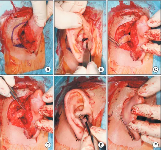
Fig. 1. Flap design, elevation and insetting. (A) Posterior auricular artery was marked by Handheld Doppler tracing on the retroauricular region. Posterior auricular artery was included in the designed flap. The flap was designed on the retroauricular skin, considered to size of external auditory meatus defect. (B) The defect on posterior and inferior wall of the external auditory meatus. (C) The flap was elevated over the fascial of auricularis posterior muscle. (D) Subcutaneous tunnel was made from the flap to skin of external auditory meatus. Skin of the flap that located under the subcutaneous tunnel was deepithelized. (E, F) The flap was inset on the defect and donor site was repaired primarily.
auricular artery was included in the proximal site of this flap.
The elevated flap was rotated 90 degrees to defect and checked the overlapped area between the flap and auricular skin. Under the auricular skin of overlapped area, subcutaneous dissection was done and subcutaneous tunnel was made. The skin of the overlapped area of the flap was deepithelized (Fig. 1D). The flap was rotated 90 degrees as clockwise or count clockwise and flap was passed through the subcutaneous tunnel. The flap was inset on the defect with 5-0, 6-0 nylon suture, donor site was repaired primarily with PDS 5-0, nylon 5-0.
Stitch out was performed after 7 to 14 days.
RESULTS
All of the eight patients were male and the mean age was 38.6 years (range, 6 to 59 years). The most common site of the external ear defect was middle 1/3 which were 4 cases. In addition, patient group was consisted of 2 cases of inferior 1/3 and 1 case of superior auriculo-cephalic sulcus reconstruction and 1 case of external auditory canal. The defect were caused by trauma which were 2 cases, 2 cases of human bite and 1 case of skin cancer excision and 1 case of congenital anomaly. The mean size of the ear defect was 2.8×1.8 cm and mean size of the retroauricular flap was 5×2 cm.
For insetting of the flap, the subcutaneous tunneling technique was used for 6 cases and rotation without subcutaneous tunneling was used for 2 cases. Transient paresthesia occurred in 3 cases due to partial injury of the retroauricular nerve. Two cases were recovered within 3 months but one case did not
recover until 6 months.
All of the flap was totally survived and there was no marginal necrosis. Initial arterial insufficient problem occurred during insetting in one case, therefore secondary closure was done after 3 weeks (Table 1).
Case 1
A 28-year-old male patient, who had previous chronic otitis media operation, came to for external meatoplasty in our department and mastoidectomy with tympanoplasty in the department of the otolaryngology due to narrow external auditory canal by aggravation of chronic otitis media.
There were 4.0×1.5 cm2 sized defect on posterior and inferior wall of the external auditory meatus. The defect was reconstructed with inferior based retroauricular flap, that size was 6.0×1.5 cm2. The proximal side of the flap was deepithelized and the flap was rotated to external meatus of the ear. The flap placed in posterior and inferior wall of the external auditory meatus with skin graft on the remaining area. Donor site was primary closured (Fig. 1).
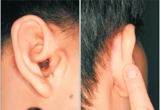
After 8 weeks, passage of the external auditory meatus and area of the tympanoplasty have been maintained (Fig. 2).
Case 2
A 53-year-old male patient slipped down from the stairs and bumped his right ear on the floor. Superior 1/3 of the right ear was amputated. In emergence room, the ear stump was sutured to defect of the ear as composite graft, but most of the stump was necrotized and debridement was done. The size of
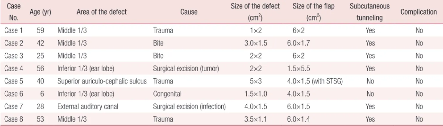
Table 1. Details of 8 cases of external ear reconstructions Case
No. Age (yr) Area of the defect Cause Size of the defect
(cm2)
Size of the flap (cm2)
Subcutaneous
tunneling Complication
Case 1 59 Middle 1/3 Trauma 1×2 6×2 Yes No
Case 2 42 Middle 1/3 Bite 3.0×1.5 6.0×1.7 Yes No
Case 3 25 Middle 1/3 Bite 2×2 6×2 Yes No
Case 4 56 Inferior 1/3 (ear lobe) Surgical excision (tumor) 2×2 1.5×5.5 Yes No
Case 5 40 Superior auriculo-cephalic sulcus Trauma 5×3 4.0×1.5 (with STSG) No No
Case 6 6 Inferior 1/3 (ear lobe) Congenital 1.5×1.0 4.0×1.5 No No
Case 7 28 External auditory canal Surgical excision (infection) 4.0×1.5 6.0×1.5 Yes No
Case 8 53 Middle 1/3 Trauma 3.5×1.1 6.0×1.4 Yes No
All cases of patient are male.
STSG: split-thickness skin graft.
the external ear defect was 3.5×1.1 cm2 and reconstruction was done with inferior based retroauricular flap, that size was 6.0×1.4 cm2. For insetting of the flap, subcutaneous tunneling was done under the posterior auricular skin.
When the flap was positioned and sutured to the defect of the ear, the flap was pale. So, donor site was closed primary, but the external ear defect and subcutaneous tunneling area were closed with key suture at the margin without excess skin tension (Fig. 3).
After 3 weeks, secondary closure was performed (Fig. 4).
DISCUSSION
The causes of external ear defect from skin defect to full thickness defect are from trauma, tumor excision and congenital, etc. The external ear defect could be classified to superior 1/3, middle 1/3, inferior 1/3, and combine form, by location.
A B C D
Fig. 3. Flap design, elevation and insetting. (A) The flap was designed on the retroauricular skin, considered to size of external ear defect. (B) The flap was elevated over the fascial of auricularis posterior muscle. (C) The elevated flap was rotated 90 degrees to defect and subcutaneous tunnel was made.
(D) The flap was inset on the defect and donor site was repaired primarily.
Fig. 2. Outpatient department follow-up after 4 weeks.
The defect of helical rim, is introduced in 1967 by Antia and Buch,2 which can be reconstructed most easily by advancing the helix in both directions. This technique is effective to reconstruction of the superior 1/3 and middle 1/3 defect, but it could lead to asymmetry of the ears, due to the size of the reconstructed ear to be smaller than normal ear. To correction of asymmetry, wedge excision technique of normal ear is introduced. The Antia-Buch technique is an excellent reconstructive technique in small to intermediate helical rim defect of less than 3 cm in length, but limited in larger defect.1,6
The ear defect of the superior 1/3 could be successfully reconstructed with a contralateral concha cartilage graft7 or with the entire concha by chondrocutaneous composite flap based on an anterior pedicle of the crus helix.8 But, chodrocutaneous flap is restricted to large concha defect.
The middle 1/3 auricular defect can be repaired by cartilage graft, Dieffenbach flap or Converse’s tunnel procedure.9 Converse’s tunnel procedure is also effective in moderated- sized defect, all two methods have advantage to preserve retroauricular sulcus, but have disadvantages to need the secondary division stage, requiring skin graft to mastoid region in need. If the auriculo-cephalic sulcus skin is intact, retroauricular area can be used as fine-caliber tube flap providing a superb site for tube construction.1,5
Vascular patterns have been studied as anatomical base of these operation techniques, auricles are supplied by branches of superficial temporal artery and posterior auricular artery.10 According to the earlier studies, superficial temporal artery
mostly supply anterior surface of auricle and posterior auricular artery supply posterior surface, the interconnection between two arteries make the ear potentially well vascularized.
Superficial temporal artery mainly makes 3 branches to anterior auricular surface, posterior auricular artery makes 3 to 5 branches to the posterior auricular surface, and some articles reported that posterior auricular artery is a dominant artery of the auricle and the middle region has a most sufficient arterial network.10,11
Recently, one stage methods by perforator flap, base on a superficial temporal artery or a posterior auricular artery, have been reported, and used for reconstruction of other part of the face as well as periauricular region, and widely used because of sufficient interconnection among the arteries constructing flap with antegrade and retrograde.12,13 But these techniques have disadvantages that complicated, time consuming, and distorted auriculo-cephalic sulcus. Using a free flap or a regional flap, there are disadvantages such as, technically difficult, a risk of flap failure, mostly need the two stage operation, and discolormatch with auricular skin, especially free flap.5,14
Comparing with previous described reconstruction methods of the ear defects, the inferior retroauricular flap based posterior auricular artery is technically simple and hemodynamic stable compare to regional flap and free flap. The reconstructed ear with this method has symmetry and nearly similar shape to the normal ear, because this method supplies the sufficient tissue from periauricular area for the auricular defects. The inferior based retroauricular flap has additional advantages including,
A B C D
Fig. 4. Secondary closure and outpatient department (OPD) follow-up of the flap. (A, B) Secondary closure of raw surface of the flap after 3 weeks. (C, D) OPD follow-up after 7 weeks.
one stage operation and normal shape of the auriculo-cephalic sulcus (Fig. 4B).
Comparing with previous retroauricular flap method, this flap is elevated suprafascial layer and conchal perichondrium undisturbed, which differs from previous reports from other investigators, who made an incision down to the periosteum and raised the island pedicle flap.15 And, different lengths between the anterior and the posterior incision of this flap could lead to tension free closure of the donor site, rectangular rotation without dog ear, and normal auricular-cephalic sulcus shape.
Our technique has better color matching and mentioned above, technically simple and stable. But, this technique also has disadvantages, such as pedicle compression by subcutaneous tunneling as mentioned above in Case 1 and limited use in larger defect.
This technique is useful in reconstruction of middle 1/3, and have an ideal rotation arc to external ear defect of the middle 1/3. It also could reconstruct inferior 1/3 and external auditory canal by the size and the location of construction.
But, in upper 1/3, this flap is limited due to the arc of the rotation limitation. As transient ischemia, might be caused by kingking of subcutaneous pedicle or excessive subcutaneous skin tension, during the operation has occurred in one case of reconstruction of middle 1/3 area, was released within 30 minutes after suture of the donor site. But, this finding would suggest a subcutaneous tunneling technique could be harmful in flap survival due to excessive skin tension, sometimes.
The limitation of this study is that the number of cases is insufficient, requiring studies for more cases, and furthermore, comparing other flaps used currently is necessary.
CONCLUSION
The inferior based retroauricular flap is an available technique in external ear reconstruction, it could use the one stage operation method of reconstruction of external ear defect from auriculo-cephalic sulcus of superior side to the lower-third, especially middle-third and ear lobule.
REFERENCES
1. Brent B. The acquired auricular deformity. A systematic approach to its analysis and reconstruction. Plast Reconstr Surg 1977;59:475-85.
2. Antia NH, Buch VI. Chondrocutaneous advancement flap for the marginal defect of the ear. Plast Reconstr Surg 1967;39:472- 7.
3. Dujon DG, Bowditch M. The thin tube pedicle: a valuable technique in auricular reconstruction after trauma. Br J Plast Surg 1995;48:35-8.
4. Steffanoff DN. Auriculo-mastoid tube pedicle for otoplasty. Plast Reconstr Surg (1946) 1948;3:352-60.
5. Youn S, Kim YH, Kim JT, Ng SW. Successful reconstruction of a large helical rim defect using retroauricular artery perforator- based island flap. J Craniofac Surg 2011;22:635-7.
6. Ellabban MG, Maamoun MI, Elsharkawi M. The bi-pedicle post-auricular tube flap for reconstruction of partial ear defects.
Br J Plast Surg 2003;56:593-8.
7. Adams WM. Construction of upper half of auricle utilizing composite concha cartilage graft with perichondrium attached on both sides. Plast Reconstr Surg (1946) 1955;16:88-96.
8. Davis J. Reconstruction of the upper third of the ear with a chondrocutaneous composite flap based on the crus helix. In:
Tanzer RC, Edgerton MT, editors. Symposium on reconstruction of the auricle. St. Louis: C.V. Mosby Co.; 1974. p. 247.
9. Converse JM. Reconstruction of the auricle. I. Plast Reconstr Surg Transplant Bull 1958;22:150-63.
10. Pinar YA, Ikiz ZA, Bilge O. Arterial anatomy of the auricle:
its importance for reconstructive surgery. Surg Radiol Ana 2003;25:175-9.
11. Imanishi N, Nakajima H, Aiso S. Arterial anatomy of the ear.
Okajimas Folia Anat Jpn 1997;73:313-23.
12. Song R, Song Y, Qi K, Jiang H, Pan F. The superior auricular artery and retroauricular arterial island flaps. Plast Reconstr Surg 1996;98:657-67.
13. Pinho C, Choupina M, Silva P, Ferreira P, Guimarães I, Amarante J. A new retroauricular flap for facial reconstruction.
Br J Plast Surg 2003;56:599-602.
14. Pascal S, Deveze A, Casanova D, Philandrianos C. Treatment of a mastoid defect by free anterolateral thigh flap. Eur Ann Otorhinolaryngol Head Neck Dis 2015. doi: 10.1016/
j.anorl.2015.08.034 [Epub].
15. Nagaoka M, Noguchi Y, Kawashima Y, Ito T, Koda H, Kitamura K. Long-term result of meatoplasty using inferiorly based retroauricular island pedicle flap for external auditory canal stenosis. Auris Nasus Larynx 2016;43:382-6.