INTRODUCTION
Early and complete recanalization of an occluded artery is probably the most effective way to reduce mortality and neurologic deficits in acute stroke patients.
Plasminogen activators such as tissue-type plasminogen activator (t-PA) and urokinase have been widely used to restore the blood flow to the ischemic brain, and have shown that they are effective in acute stroke patients.1,2
However, many patients still remain disabled because of hemorrhagic transformation as well as thrombolysis failure or deterioration after recanalization. Actually, it is known that recanalization is achieved in only 30-70% of stroke patients with thrombolytic treatment.3
Few studies have examined the biomarkers that may be related to thrombolysis failure in stroke.4,5 However, it is important to rapidly detect subjects who might be unsuitable for conventional fibrinolytic therapy prior to thrombolytic therapy because they may be managed with
Plasma Fibrinolysis Inhibitor Levels in Acute Stroke Patients with Thrombolysis Failure
Seo Hyun Kim, M.D.†, Sang Won Han, M.D.‡, Eun Hee Kim, M.S., Dong Joon Kim, M.D.*, Kyung Yul Lee, M.D.§, Dong Ik Kim, M.D.*, Ji Hoe Heo, M.D., Ph.D.
Departments of Neurology and Diagnostic Radiology*, National Core Research Center for Nanomedical Technology, Brain Research Institute, Yonsei University College of Medicine, Yonsei University Wonju College of Medicine†; Sanggye Paik Hospital‡; Yongdong Severance Hospital§
Background and Purpose: Thrombolytics-induced recanalization fails in a significant portion of patients with ischemic stroke, which is partly due to the resistance of clots to lysis by thrombolytic agents. The pretreatment level of endogenous fibrinolysis inhibitors may affect such thrombolysis failure.
Methods: We studied 43 stroke patients whose arterial recanalization had been evaluated by angiography, and whose blood had been obtained prior to the administration of thrombolytic agents. Plasma samples from 34 healthy volunteers were used as normal controls. Plasminogen activator inhibitor type 1 (PAI-1) and thrombin-activatable fibrinolysis inhibitor (TAFI) levels were quantified using an enzyme-linked immunosorbent assay.
Results: Arteries were recanalized [Thrombolysis in Myocardial Infarction (TIMI) grade 2 or 3] in 30 patients, but not (TIMI grade 0 or 1) in the other 13. The plasma PAI-1 level was significantly higher in patients without recanalization (nonrecanalization) than in those with recanalization and in normal controls. The TAFI levels did not differ among the groups.
Conclusions: The pretreatment PAI-1 levels are increased in acute stroke patients with thrombolysis failure.
J Clin Neurol 1(2):142-147, 2005
Key Words : Fibrinolytic agents, Acute stroke, Plasminogen activator inhibitor type 1, Thrombin-activatable fibrinolysis inhibitor
Received : July 7, 2005 / Accepted : September 15, 2005 / Address for correspondence : Ji Hoe Heo, M.D., Ph.D.
Department of Neurology, Yonsei University College of Medicine, 134 Shinchon-dong, Seodaemoon-gu, Seoul, 120-752, Korea Tel: +82-2-2228-1605, Fax: +82-2-393-0705, E-mail: jhheo@yumc.yonsei.ac.kr
*This work was supported by KOSEF through the National Core Research Center for Nanomedical Technology (R15-2004-024-00000-0).
an alternative or additive strategy such as platelet glyco- protein IIb/IIIa receptor antagonists or mechanical clot removal.6,7
The action of endogenous fibrinolysis inhibitors may influence the success or failure of clot lysis, and inter- individual variation in the plasma levels of the fibrino- lysis inhibitors may influence the individual suscepti- bility to the fibrinolytic treatment. Although increased endogenous fibrinolytic inhibitor levels such as plasmi- nogen activator inhibitor type 1 (PAI-1) are associated with thrombolysis failure and poor outcome in patients with acute myocardial infarction,8 little is known about PAI-1 as a biomarker of thrombolysis failure in stroke patients. In this study, we examined the pretreatment plasma levels of two well-known endogenous fibrinolysis inhibitors, PAI-1 and thrombin-activatable fibrinolysis inhibitor (TAFI), and investigated their potential association with thrombolysis failure in acute stroke patients who receive thrombolytic treatment.
MATERIALS AND METHODS
1. Patients
Among a total 106 stroke patients who received thrombolytics over a 4-year period, 43 consecutive patients whose arterial recanalization could be evaluated by post-thrombolysis angiography (39 by catheter angio- graphy, 3 by MR angiography, and 1 by CT angio- graphy) and whose blood could be obtained before administering the thrombolytic agents were enrolled in this study. The exclusions were due to not performing angiography in 8 patients and the inability to obtain blood samples in 55 patients. The demographic charac- teristics of sex and age, risk factors for stroke, laboratory data, and the initial National Institutes of Health Stroke Scale (NIHSS) score did not differ between the 43 included and 63 excluded patients (P<0.05). Seventeen patients were treated with intravenous (IV) t-PA, 11 with intra-arterial (IA) urokinase, and 15 with combined IV t-PA and IA urokinase. The indication and regimen for IV, IA, or combined IV and IA treatment, and the outcome measurements have been reported previously.9,10
Briefly, IV t-PA was indicated when the planned infusion could be initiated within 3 hours after symptom onset, and IA urokinase was administered to patients showing no early clinical responses to IV t-PA at the end of t-PA infusion or to those who could be treated within 3-6 hours after symptom onset. The institutional review board approved this study, and informed consent was obtained from the patient or the patient's represen- tative.
The patency of the occluded arteries was assessed using the Thrombolysis in Myocardial Infarction (TIMI) grading system,11 and the patients were grouped into nonrecanalization (TIMI grade 0 or 1) and recanalization (TIMI grade 2 or 3).
2. Blood sampling
On their arrival at hospital, blood was drawn from the patients into a heparinized tube at the time of the initial blood sampling for the emergent laboratory workup.
Control blood samples were obtained from volunteers aged >40 years at the time of their annual institutional health examinations. The examinations included routine history taking, a physical examination, blood pressure measurements, chest x-ray, electrocardiography, and blood tests including hemoglobin, fasting sugar, and total cholesterol. Those with a previous history of hyper- tension, diabetes, stroke, coronary artery diseases, inflam- matory diseases, or malignancies were excluded. Those with a systolic blood pressure >140 mmHg, a diastolic blood pressure >90 mmHg, a fasting blood sugar >140 mg/dl, or a total cholesterol >240 mg/dl were also excluded. The control blood samples were obtained from 34 volunteers (17 men and 17 women) with a mean age of 48 years.
The blood samples obtained from the patients and volunteers were immediately refrigerated and centrifuged at 900Xg for 15 minutes at 4℃, and the plasma were then stored at -80℃ until analysis.
3. Measurement of plasma PAI-1 and TAFI levels
The PAI-1 and TAFI antigen levels were determined using a commercially available enzyme-linked immuno-
sorbent assay (ELISA) kit (Zymutest PAI-1 Ag and Zymutest TAFI Ag, Hyphen BioMed, Andresy, France) according to the manufacturer's instructions.
4. Statistical analysis
The levels of plasma PAI-1 and TAFI were compared among the groups for significant differences using the Kruskal-Wallis test. The Mann-Whitney U test and Fisher's exact test were used to compare the variables between the nonrecanalization and recanalization groups.
Logistic regression was used to determine independent factors associated with the nonrecanalization group. The 95% confident intervals (CIs) were calculated, and differences were considered statistically significant when P<0.05. Statistical analyses were performed with SPSS (version 10.0) software.
RESULTS
Recanalization was evaluated by conventional catheter
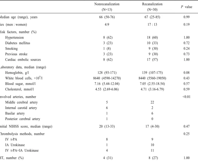
Table 1. Demographic characteristics of the patient groups
Nonrecanalization (N=13)
Recanalization
(N=30) P value
Median age (range), years 66 (50-76) 67 (25-85) 0.99
Sex (men : women) 4:9 17 : 13 0.19
Risk factors, number (%) Hypertension Diabetes mellitus Smoking Previous stroke Cardiac embolic sources
8 (62) 3 (23) 1 (8) 3 (23) 8 (62)
18 (60) 10 (33) 9 (30) 9 (30) 17 (57)
1.00 0.72 0.24 0.73 1.00 Laboratory data, median (range)
Hemoglobin, g/l White blood cells, ×109/l Blood sugar, mmol/l Cholesterol, mmol/l
128 (93-171) 8640 (4590-14270)
7.16 (5.44-12.04) 4.53 (2.69-6.06)
139 (107-175) 8440 (5560-19050)
7.05 (2.55-18.54) 4.71 (3.16-6.79)
0.08 0.43 0.57 0.59 Involved arteries, number
Middle cerebral artery Internal carotid artery Basilar artery
Posterior cerebral artery
5 6 1 1
22 2 6 0
<0.01
Initial NIHSS score, median (range) 20 (13-33) 17 (4-30) 0.47
Thrombolysis methods, number IV t-PA
IA Urokinase IV t-PA+IA Urokinase
8 1 4
9 10 11
0.25
HT, number (%) 4 (31) 8 (27) 1.00
HT: hemorrhagic transformation, NIHSS: national institutes of health stroke scale, IV t-PA: intravenous tissue-type plasminogen activator, IA: intra-arterial.
For comparison between groups, the Mann-Whitney U test was used for age, laboratory data, and NIHSS score, and Fisher's exact test was used for sex, risk factors, involved arteries, and HT.
angiography in 39 of the 43 enrolled patients. Among 17 patients treated with IV t-PA, catheter angiography was performed for IA thrombolysis immediately after infusion of t-PA in 11 patients, but IA urokinase was not administered because the artery was occluded at the proximal carotid level (3 patients) or the artery was opened or occluded at the distal branch (8 patients). The remaining six patients received diagnostic investigations within 48 hours after symptom onset (two by catheter angiography, three by MR angiography, and one by CT angiography). The median time from symptom onset to angiography was 4 hours and 10 minutes. Recanalization was achieved in 30 of the 43 patients. An occlusion at the middle cerebral artery or the basilar artery was recanalized more often (P<0.01). Otherwise, there were no significant differences between the two groups in the risk factors for stroke, the initial laboratory data, the severity of the neurological deficits based on the initial NIHSS score, the thrombolysis method, and the frequency of hemorrhagic transformation (Table 1).
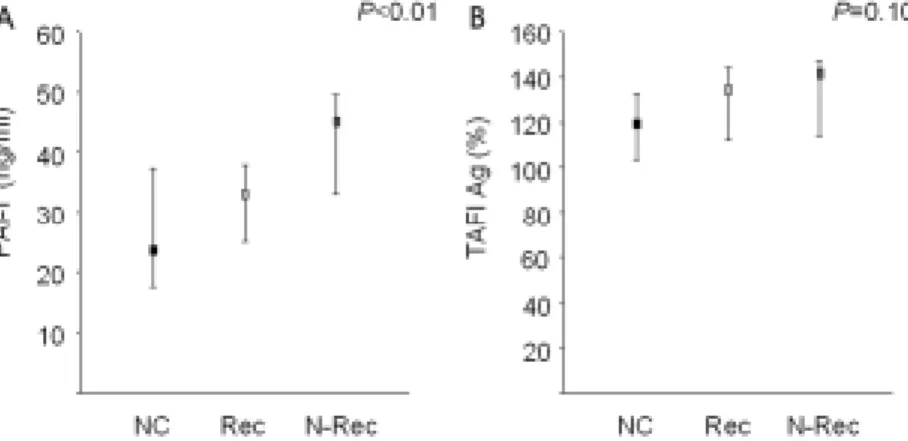
The plasma PAI-1 and TAFI levels, which were measured by ELISA, were compared between the two thrombolytics-treated groups and the controls. The time interval from symptom onset to blood sampling did not differ between the two thrombolytics-treated groups (medians of 98 minutes and 80 minutes in the non- recanalization and recanalization groups, respectively;
P=0.84). The PAI-1 levels differed significantly among the three groups, and was highest in the nonrecanali-
zation group: medians of 45.2 ng/ml (interquartile range [IQR], 32.5-50.0 ng/ml), 33.1 ng/ml (IQR, 24.9-38.1 ng/ml), and 23.6 ng/ml (IQR, 17.5-37.5 ng/ml) in the nonrecanalization, recanalization, and control groups, respectively (P=0.007) (Fig. 1-A). However, the TAFI levels did not differ significantly among the groups (P=0.097) (Fig. 1-B).
Logistic regression showed that only the plasma PAI-1 levels were independently associated with recanalization (odds ratio, 0.916; 95% CI, 0.844-0.994).
DISCUSSION
In this study, the plasma PAI-1 levels were higher in patients with acute stroke, and higher pretreatment plasma PAI-1 levels were associated with thrombolysis failure. But the plasma TAFI levels did not differ between the control and patient groups. One study showed that increased plasma PAI-1 levels predicted clot lysis resistance in stroke patients treated with t-PA.12 In that study, evidence of successful thrombolysis was based on transcranial Doppler investigations, which is consistent with our determination of the success or failure of thrombolysis in a more definitive and direct manner by angiography.
PAI-1 is reportedly increased in blood obtained within 1-7 days after symptom onset in acute stroke patients.13-15 The findings in the present study further demonstrate
Figure 1. Plasma plasminogen activator inhibitor type 1 (PAI-1) (A) and thrombin-activatable fibrinolysis inhibitor (TAFI) (B) levels in each group as measured by an enzyme-linked immunosorbent assay. The PAI-1 level was significantly higher in the nonrecanalization group (N-Rec) than in the normal control (NC) and recanalization (Rec) groups. Medians and interquartile ranges are shown.
that plasma PAI-1 levels are increased during the very early stage of stroke, because all samples were obtained from patients who were eligible for thrombolytic therapy.
T-PA is rapidly neutralized by PAI-1, which binds to the active site of t-PA and forms inactive complexes.16,17 Therefore, the presence of a pretreatment plasma PAI-1 in excess of endogenous and/or exogenous t-PA or urokinase may depress their fibrinolytic activity.17
The thrombus composition has been suggested as one of the important causes of thrombolytic resistance, and platelet-rich thrombi are disposed to resistance to throm- bolysis.18,19 Possible sources of increased PAI-1 in acute stroke are the endothelium, atherosclerotic plaques, and platelets, which are major sources of circulating PAI-1.16 The latent PAI-1 in alpha-granules of platelets is converted into the active form and is released upon platelet activation and aggregation. Therefore, large amounts of active PAI-1 are released from fresh platelet- rich thrombi, which contribute to the thrombolytic resistance.17,20 In a murine carotid injury model that induced platelet-rich thrombi in wild-type and PAI-1- deficient mice, PAI-1 was found to be a major determi- nant of the resistance of platelet-rich arterial thrombi to lysis by t-PA.21 In contrast, the inactivation of PAI-1 using a monoclonal antibody against PAI-1 accelerated the lysis of the platelet-rich thrombi in the mesenteric arteriole of rats.12 All these findings indicate an important role of PAI-1 in inducing resistance to thrombolytics via platelet-mediated mechanisms, and suggest that an increased plasma PAI-1 level can be used as a biomarker for the prediction of thrombolysis failure in stroke patients.
Although TAFI acts as a fibrinolytic inhibitor, the plasma TAFI levels were neither increased in stroke patients nor associated with thrombolysis failure. Previous studies on coronary artery disease are also contro- versial.22 It has been suggested that, in contrast to PAI-1, genetic factors are much more important determinants of the plasma TAFI levels than environmental factors,23 which may explain the absence of an elevated plasma TAFI in the present study.
REFERENCES
1. The National Institute of Neurological Disorders and Stroke rt-PA Stroke Study Group. Tissue plasminogen activator for acute ischemic stroke. N Engl J Med 1995;333:1581-1587.
2. Furlan A, Higashida R, Wechsler L, Gent M, Rowley H, Kase C, et al. Intra-arterial prourokinase for acute ischemic stroke. The PROACT II study: a randomized controlled trial. Prolyse in Acute Cerebral Thrombo- embolism. JAMA 1999;282:2003-2011.
3. Mohr J, Choi D, Grotta J, Weir B, Wolf PA. Stroke:
Pathophysiology, diagnosis, and management. Philadel- phia: Churchill Livingstone; 2004.
4. Heo JH, Kim SH, Lee KY, Kim EH, Chu CK, Nam JM.
Increase in plasma matrix metalloproteinase-9 in acute stroke patients with thrombolysis failure. Stroke 2003;
34:e48-50.
5. Ribo M, Montaner J, Molina CA, Arenillas JF, Santamarina E, Alvarez-Sabin J. Admission fibrinolytic profile predicts clot lysis resistance in stroke patients treated with tissue plasminogen activator. Thromb Haemost 2004;91:1146-1151.
6. Abciximab Emergent Stroke Treatment Trial (AbESTT) Investigators. Emergency administration of abciximab for treatment of patients with acute ischemic stroke: results of a randomized phase 2 trial. Stroke 2005;36:880-890.
7. Smith WS, Sung G, Starkman S, Saver JL, Kidwell CS, Gobin YP, et al. Safety and efficacy of mechanical embolectomy in acute ischemic stroke: results of the MERCI trial. Stroke 2005;36:1432-1438.
8. Sinkovic A. Pretreatment plasminogen activator inhibitor-1 (PAI-1) levels and the outcome of thrombolysis with streptokinase in patients with acute myocardial infarction.
Am Heart J 1998;136:406-411.
9. Heo JH, Lee KY, Kim SH, Kim DI. Immediate reocclusion following a successful thrombolysis in acute stroke: a pilot study. Neurology 2003;60:1684-1687.
10. Lee KY, Kim DI, Kim SH, Lee SI, Chung HW, Shim YW, et al. Sequential combination of intravenous recombinant tissue plasminogen activator and intra-arterial urokinase in acute ischemic stroke. Am J Neuroradiol 2004;25:1470-1475.
11. Chesebro JH, Knatterud G, Roberts R, Borer J, Cohen LS, Dalen J, et al. Thrombolysis in Myocardial Infarction (TIMI) trial, Phase I: A comparison between intravenous tissue plasminogen activator and intravenous streptokinase.
Clinical findings through hospital discharge. Circulation 1987;76:142-154.
12. Rupin A, Martin F, Vallez MO, Bonhomme E, Verbeuren TJ. Inactivation of plasminogen activator inhibitor-1 accelerates thrombolysis of a platelet-rich thrombus in rat mesenteric arterioles. Thromb Haemost 2001;86:1528- 1531.
13. Lindgren A, Lindoff C, Norrving B, Astedt B, Johansson BB. Tissue plasminogen activator and plasminogen activator inhibitor-1 in stroke patients. Stroke 1996;27:
1066-1071.
14. Fisher M, Francis R. Altered coagulation in cerebral ischemia. Platelet, thrombin, and plasmin activity. Arch Neurol 1990;47:1075-1079.
15. Wang J, Li J, Liu Q. Association between platelet activation and fibrinolysis in acute stroke patients.
Neurosci Lett 2005;384:305-309.
16. Dobrovolsky AB, Titaeva EV. The fibrinolysis system:
regulation of activity and physiologic functions of its main components. Biochemistry (Mosc) 2002;67:99-108.
17. Huber K. Plasminogen activator inhibitor type-1 (part one): basic mechanisms, regulation, and role for thrombo- embolic disease. J Thromb Thrombolysis 2001;11:183-193.
18. Jang IK, Gold HK, Ziskind AA, Fallon JT, Holt RE, Leinbach RC, et al. Differential sensitivity of erythrocyte- rich and platelet-rich arterial thrombi to lysis with recombinant tissue-type plasminogen activator. A possible explanation for resistance to coronary thrombolysis.
Circulation 1989;79:920-928.
19. Cannon CP. Overcoming thrombolytic resistance: rationale and initial clinical experience combining thrombolytic
therapy and glycoprotein IIb/IIIa receptor inhibition for acute myocardial infarction. J Am Coll Cardiol 1999;34:
1395-1402.
20. Levi M, Biemond BJ, van Zonneveld AJ, ten Cate JW, Pannekoek H. Inhibition of plasminogen activator inhibitor-1 activity results in promotion of endogenous thrombolysis and inhibition of thrombus extension in models of experimental thrombosis. Circulation 1992;
85:305-312.
21. Zhu Y, Carmeliet P, Fay WP. Plasminogen activator inhibitor-1 is a major determinant of arterial thrombolysis resistance. Circulation 1999;99:3050-3055.
22. Juhan-Vague I, Morange PE, Aubert H, Henry M, Aillaud MF, Alessi MC, et al. Plasma thrombin-activatable fibrinolysis inhibitor antigen concentration and genotype in relation to myocardial infarction in the north and south of Europe. Arterioscler Thromb Vasc Biol 2002;22:867- 873.
23. Morange PE, Juhan-Vague I, Scarabin PY, Alessi MC, Luc G, Arveiler D, et al. Association between TAFI antigen and Ala147Thr polymorphism of the TAFI gene and the angina pectoris incidence. The PRIME study (Prospective Epidemiological Study of MI). Thromb Haemost 2003;89:554-560.