The risk of lymphedema after
postoperative radiation therapy in endometrial cancer
Devarati Mitra,1 Paul J. Catalano,2,3 Nicole Cimbak,1 Antonio L. Damato,1 Michael G. Muto,4 Akila N. Viswanathan1
1Department of Radiation Oncology, Dana-Farber Cancer Institute and Brigham & Women’s Hospital, Boston, MA, USA
2Department of Biostatistics and Computational Biology, Dana-Farber Cancer Institute, Boston, MA, USA
3Department of Biostatistics, Harvard School of Public Health, Boston, MA, USA
4Department of Gynecologic Oncology, Dana-Farber Cancer Institute and Brigham & Women’s Hospital, Boston, MA, USA
ABSTRACT
Objective: Lower extremity lymphedema adversely affects quality of life by causing discomfort, impaired mobility and increased risk of infection. The goal of this study is to investigate factors that influence the likelihood of lymphedema in patients with endometrial cancer who undergo adjuvant radiation with or without chemotherapy.
Methods: A retrospective chart review identified all stage I–III endometrial cancer patients who had a hysterectomy with or without complete staging lymphadenectomy and adjuvant radiation therapy between January 2006 and February 2013. Patients with new-onset lymphedema after treatment were identified. Logistic regression was used to find factors that influenced lymphedema risk.
Results: Of 212 patients who met inclusion criteria, 15 patients (7.1%) developed new-onset lymphedema. Lymphedema was associated with lymph-node dissection (odds ratio [OR], 5.6; 95% CI, 1.01 to 105.5; p=0.048) and with the presence of pathologically positive lymph nodes (OR, 4.1; 95% CI, 1.4 to 12.3; p=0.01). Multivariate logistic regression confirmed the association with lymph-node positivity (OR, 3.2; 95% CI, 1.0007 to 10.7; p=0.0499) when controlled for lymph-node dissection. Median time to lymphedema onset was 8 months (range, 1 to 58 months) with resolution or improvement in eight patients (53.3%) after a median of 10 months.
Conclusion: Lymph-node positivity was associated with an increased risk of lymphedema in endometrial cancer patients who received adjuvant radiation. Future studies are needed to explore whether node-positive patients may benefit from early lymphedema-controlling interventions.
Keywords: Endometrial Neoplasms; Lymphedema; Positive Lymph Nodes
Original Article
Received: Apr 17, 2015 Revised: Aug 16, 2015 Accepted: Sep 13, 2015 Correspondence to Devarati Mitra
Department of Radiation Oncology, Brigham & Women’s Hospital, 75 Francis Street, Boston, MA 02115, USA.
E-mail: [email protected] Copyright © 2016. Asian Society of Gynecologic Oncology, Korean Society of Gynecologic Oncology
This is an Open Access article distributed under the terms of the Creative Commons Attribution Non-Commercial License (http://
creativecommons.org/licenses/by-nc/4.0/) which permits unrestricted non-commercial use, distribution, and reproduction in any medium, provided the original work is properly cited.
ORCID Devarati Mitra
http://orcid.org/0000-0002-2225-164X Antonio L. Damato
http://orcid.org/0000-0002-6558-812X
Funding
Dr. Viswanathan receives funding from National Institutes of Health R21 167800.
Conflict of Interest
No potential conflict of interest relevant to this article was reported.
This study was presented in part at the American Society for Therapeutic Radiation Oncology Annual Meeting in San Francisco on September 15, 2014.
INTRODUCTION
Lower-extremity lymphedema is a known possible complication of endometrial cancer treatment that adversely affects quality of life [1,2]. Prior single-institution retrospective studies have reported lymphedema rates that vary greatly from 2% to 47% [1-8]. Notably, the number of lymph nodes removed varied greatly among these studies, with the lowest rates of lymphedema (2.4% to 11.4%) reported in studies where most patients had fewer than 10 lymph nodes removed, and the highest rates (27.2% to 47%) reported in studies where most patients had more than 20 lymph nodes removed [3-6,8,9]. A recent survey of the Australian National Endometrial Cancer Study Group found that 13% of 1,243 patients treated for endometrial cancer developed self-reported or physician-reported lymphedema. In that study, the absolute risk of lymphedema in patients with no lymph nodes removed was 8% or less whereas the risk in patients with more than 15 lymph nodes removed was over 30% [10].
In this study and in others, a variety of other risk factors for lymphedema after endometrial cancer treatment have also been suggested, including radiation, chemotherapy, and
nonsteroidal anti-inflammatory drug use; however, the strength of correlation and magnitude of these effects are typically less pronounced than the effect of lymph node dissection [6,8,10].
Whether factors other than lymphadenectomy may increase the risk of lymphedema has not been well established. In addition, once lymphedema develops, the expected clinical course is not well understood. The goal of the current study is to investigate patient, disease, and treatment factors that may be associated with lymphedema development in endometrial cancer patients who received adjuvant external beam radiotherapy and vaginal brachytherapy after hysterectomy, and to characterize the clinical course of this complication.
MATERIALS AND METHODS
Retrospective chart review identified 222 patients with stage I–III endometrial cancer who received adjuvant radiation after hysterectomy between January 2006 and February 2013, were not on a trial of bevacizumab, and had more than 1 year of follow-up data available. Of these, 10 patients had disease recurrence less than 3 months after radiation therapy and were therefore excluded, resulting in the data from 212 patients being used for analysis.
The medical record of each patient was examined to collect patient characteristics (including age, body mass index [BMI] and Charlson comorbidity index), disease characteristics (including stage, grade, myometrial invasion, lymphovascular invasion, pelvic washings, and positive lymph nodes found upon surgery), detailed treatment parameters (including number of lymph nodes removed, total external beam radiation dose including a boost if present, vaginal brachytherapy dose, length of radiation treatment, and type of chemotherapy regimen), and disease outcomes (including disease recurrence and mortality).
All patients underwent computed tomography-based planning with custom immobilization.
External beam radiation was administered in daily treatments 5 days per week in 1.8 Gy daily fractions to the pelvis with 15 MV photons; some patients also received concurrent 1.2 to 1.5 Gy to the whole abdomen (anterior-posterior/posterior-anterior technique or intensity modulated radiation therapy [IMRT]) for 20 to 25 fractions with 6 MV. Patients with para-
aortic nodal disease received IMRT consisting of 7 to 9 fields, using 6 MV photons with dose limits as previously described [11].
The medical record was also used to identify patients with new-onset lower extremity lymphedema, as described by the physician or reported by the patient. The medical records of each patient with lymphedema were carefully examined to confirm that the onset of this symptom was subsequent to cancer treatment and not attributable to an independent pre- existing etiology, such as heart failure. Due to the retrospective nature of this study, precise measurements of lymphedema were not available. However, it is standard practice in our clinic to ask all patients who have had endometrial-cancer treatment whether they have noted new lower-extremity swelling as well as to formally evaluate this by both visual inspection and palpation.
For patients who developed new-onset lymphedema, the medical record was used to track the clinical course of this complication. Specifically recorded were: time from treatment completion to onset; subsequent progression, improvement or resolution (as documented by a physician at follow-up visits); time to resolution (if present); and the documented interventions used to treat lymphedema.
Statistical analyses were performed using JMP (SAS Institute, Cary, NC, USA). For each demographic, disease and treatment parameter, univariate logistic regression was used to estimate the odds ratio (OR) of association with lymphedema. For parameters that had a statistically significant (p<0.05) association with lymphedema, a multivariate logistic model was created using stepwise selection. In the final analysis, parameters were considered significant with a two-sided p<0.05. This study was approved by the Institutional Review Board of the Dana Farber/Brigham & Women’s Cancer Center.
RESULTS
1. Patient, disease, and treatment characteristics
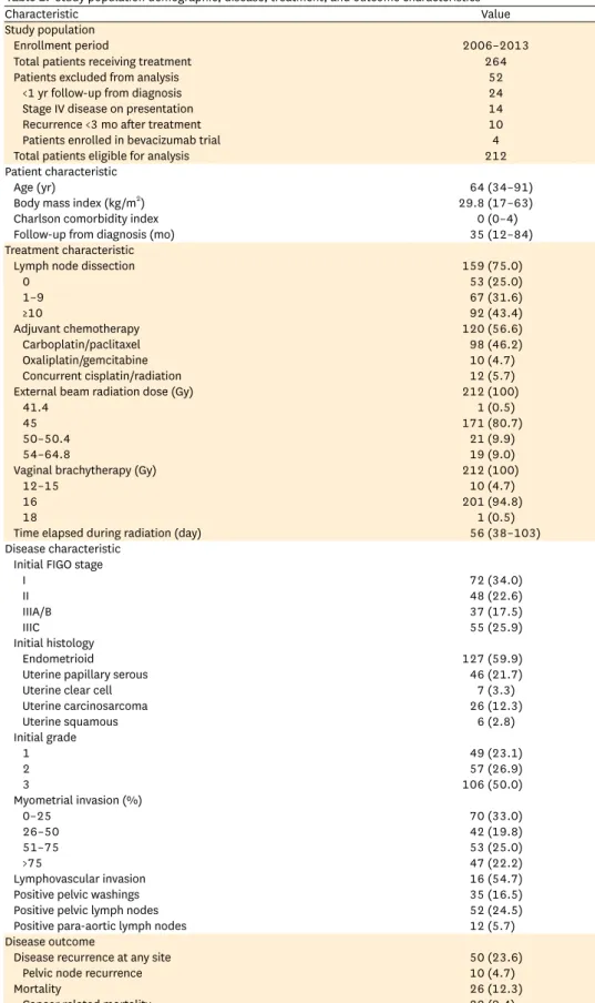
The records of the 212 endometrial cancer patients who received adjuvant radiotherapy after hysterectomy and met the inclusion criteria were reviewed. Patient, disease, and treatment characteristics as well as disease outcomes are described in Table 1. The population represented a wide spectrum of intermediate- to high-risk disease. Most patients had either endometrioid or papillary serous histology (81.6%), which manifested as mostly grade 2–3 disease (76.9%). Approximately half of patients had lymphovascular invasion and half had deep (more than 50%) myometrial invasion. Pelvic lymph-node dissection was performed in 75% (n=159) with a median of 6.5 (range, 0 to 58) pelvic lymph nodes removed. Notably, only 11.3% (n=24) had more than 20 lymph nodes removed. All patients received adjuvant radiation, consisting most commonly of 45 Gy external beam radiotherapy (80.7%) followed by 16 Gy (4 fractions of 4 Gy) vaginal brachytherapy (94.8%). Adjuvant chemotherapy was received by slightly more than half of patients, with carboplatin and taxol being used most commonly. Disease recurred in 23.6% of patients, with many having simultaneously detected recurrence at multiple sites. Overall, no patients had an isolated vaginal recurrence, 4.7% had recurrence in a pelvic lymph node, 9.0% had regional recurrence (para-aortic or pelvic side- wall), and 17.0% had recurrence at a distant site.
Table 1. Study population demographic, disease, treatment, and outcome characteristics
Characteristic Value
Study population
Enrollment period 2006–2013
Total patients receiving treatment 264
Patients excluded from analysis 52
<1 yr follow-up from diagnosis 24
Stage IV disease on presentation 14
Recurrence <3 mo after treatment 10
Patients enrolled in bevacizumab trial 4
Total patients eligible for analysis 212
Patient characteristic
Age (yr) 64 (34–91)
Body mass index (kg/m2) 29.8 (17–63)
Charlson comorbidity index 0 (0–4)
Follow-up from diagnosis (mo) 35 (12–84)
Treatment characteristic
Lymph node dissection 159 (75.0)
0 53 (25.0)
1–9 67 (31.6)
≥10 92 (43.4)
Adjuvant chemotherapy 120 (56.6)
Carboplatin/paclitaxel 98 (46.2)
Oxaliplatin/gemcitabine 10 (4.7)
Concurrent cisplatin/radiation 12 (5.7)
External beam radiation dose (Gy) 212 (100)
41.4 1 (0.5)
45 171 (80.7)
50–50.4 21 (9.9)
54–64.8 19 (9.0)
Vaginal brachytherapy (Gy) 212 (100)
12–15 10 (4.7)
16 201 (94.8)
18 1 (0.5)
Time elapsed during radiation (day) 56 (38–103)
Disease characteristic Initial FIGO stage
I 72 (34.0)
II 48 (22.6)
IIIA/B 37 (17.5)
IIIC 55 (25.9)
Initial histology
Endometrioid 127 (59.9)
Uterine papillary serous 46 (21.7)
Uterine clear cell 7 (3.3)
Uterine carcinosarcoma 26 (12.3)
Uterine squamous 6 (2.8)
Initial grade
1 49 (23.1)
2 57 (26.9)
3 106 (50.0)
Myometrial invasion (%)
0–25 70 (33.0)
26–50 42 (19.8)
51–75 53 (25.0)
>75 47 (22.2)
Lymphovascular invasion 16 (54.7)
Positive pelvic washings 35 (16.5)
Positive pelvic lymph nodes 52 (24.5)
Positive para-aortic lymph nodes 12 (5.7)
Disease outcome
Disease recurrence at any site 50 (23.6)
Pelvic node recurrence 10 (4.7)
Mortality 26 (12.3)
Cancer related mortality 20 (9.4)
Values are presented as number, median (range), or number (%).
FIGO, International Federation of Gynecology and Obstetrics.
2. Onset and management of lymphedema
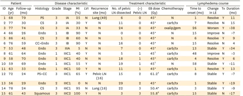
With a median follow-up time of 35 months (range, 12 to 84 months), 7.1% of patients (n=15) developed new-onset lower-extremity lymphedema. Table 2 describes the detailed demographic, disease, and treatment characteristics of each patient who developed lymphedema as well as the clinical course of this symptom. The median time to onset of documented lymphedema, measured from the last day of radiation therapy, was 8 months (range, 1 to 58 months) with six patients (40.0%) having complete resolution of their lymphedema a median of 10 months (range, 4 to 15 months) later, two patients (13.3%) having some improvement, six patients (40.0%) having stable symptoms, and one patient (6.7%) experiencing worsening lymphedema over time. Two-thirds (n=10) of the patients with lymphedema were documented to have received compression stockings and 3 were also documented to have received therapeutic lower-extremity massage. Information regarding the details of the massage technique were not available. However, no correlation was found between documented intervention and improvement or resolution of symptoms. There was no difference in the characteristics of patients who had resolution of symptoms and those who did not; characteristics examined included BMI, age, Charlson comorbidity index and time interval from treatment completion to lymphedema onset.
3. Incidence and prevalence of lymphedema
The incidence and prevalence of lymphedema over time is illustrated in Fig. 1. Of the 15 patients who developed lymphedema after treatment, 80.0% (n=12) developed the symptom within 18 months of completing therapy. Three additional patients developed new lymphedema later (26, 48, and 58 months after treatment completion). An early peak prevalence of lymphedema occurred by 15 months after treatment followed by a decline that plateaued at 25 months and subsequently slowly rose at much later time points to a final peak prevalence of 4.3% at 5 years after treatment.
Table 2. Detailed characteristics of patients with new-onset lymphedema
Patient Disease characteristic Treatment characteristic Lymphedema course
ID Age
(yr) Follow-up
(mo) Histology Grade Stage MI
(%) LVI Recurrence
site (mo) No. of pelvic LN dissected (+)
Pelvic LNEB dose
(Gy) Chemotherapy Time to
onset (mo) Change
in LE Tx Duration (mo)
1 68 79 PS 3 IA 25 N Lung (49) 6 0 45* N 1 Resolve Y 11
2 77 30 CS 3 IA 30 Y N 11 0 45* carb/tx 7 Resolve N 15
3 77 74 CS 3 IA 33 N N 24 0 45* oxalip/gem 26 Worsen Y >38
4 66 26 Endo 1 IB 90 Y N 0 0 45* N 15 Improve N >7
5 86 41 CS 3 IB 60 N N 1 0 45* N 6 Resolve Y 9
6 76 59 CC+Endo 3 IB 90 Y N 16 0 45* N 15 Resolve N 4
7 53 48 Endo 3 IIIA 5 N N 7 0 45* carb/tx 13 Stable Y >34
8 61 64 Endo 1 IIIC1 40 Y N 5 1 45† carb/tx 48 Improve N >3
9 58 70 Endo 2 IIIC1 40 N N 18 1 45* carb/tx 4 Resolve Y 6
10 59 69 Endo 1 IIIC1 55 Y N 19 1 45† N 58 Stable Y >11
11 63 33 Endo 1 IIIC1 50 Y N 11 1 45* carb/tx 1 Resolve Y 13
12 72 24 PS+CC 3 IIIC1 65 Y Pelvic LN
(16) 15 2 61.2‡ carb/tx 8 Stable Y >7
13 56 29 Endo 2 IIIC1 0 N N 29 2 45* carb/tx 1 Stable Y >19
14 78 24 CS 3 IIIC1 95 N Lung (16) 22 3 50.4* carb/tx 3 Stable Y >9
15 61 43 Squamous 3 IIIC2 100 Y N 3 3 55.8‡ carb/tx 15 Stable N >17
carb/tx, carboplatin/paclitaxel; CC, clear cell; CS, carcinosarcoma; EB, external beam radiotherapy; Endo, endometrioid; LE, lymphedema; LN, lymph nodes;
LVI, lymphovascular invasion; MI, myometrial invasion; N, no or absent; oxalip/gem, oxaliplatin/gemcitabine; PS, papillary serous; Tx, lymphedema treatment; Y, yes or present; (+), tumor positive.
*4-field pelvis. †Whole abdomen anterior-posterior/posterior-anterior. ‡Intensity modulated radiation therapy to pelvis and para-aortic nodes.
4. Risk factors for lymphedema
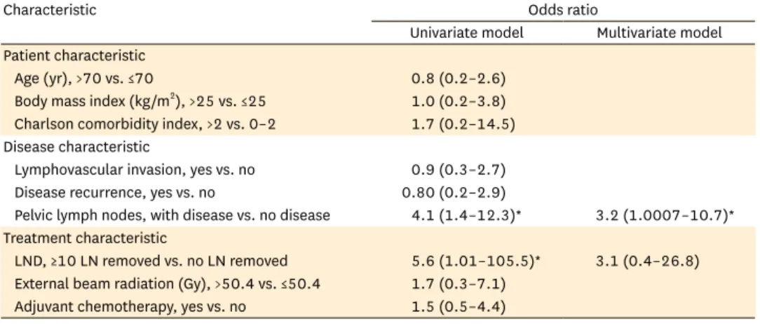
Univariate logistic regression was performed to investigate possible factors that influence the risk of lymphedema after endometrial-cancer treatment. Table 3 illustrates the ORs of lymphedema by disease and treatment characteristic tested. Having a more complete pelvic lymph node dissection (defined as 10 or more lymph nodes removed) was associated with an increased risk of lymphedema when compared to no dissection (OR, 5.6; 95% CI, 1.01 to 105.5; p=0.048). However, a less complete lymph-node dissection (defined as 1 to 9 lymph nodes removed) was not associated with increased risk (p=0.14). The cut-off of 10 lymph nodes as the break-point for analysis was chosen because the mean number of lymph nodes removed in our study was 9.7; 56.6% of patients who had any nodes dissected had 1 to 9 lymph nodes removed and 43.4% had 10 or more removed (Table 1). Of the 15 patients who developed lymphedema, 1 (6.7%) had no lymph nodes removed, 5 (33.3%) had fewer than 10 lymph nodes removed, and 9 (60.0%) had 10 or more lymph nodes removed.
Table 3. Predictors of lymphedema by univariate and multivariate models
Characteristic Odds ratio
Univariate model Multivariate model Patient characteristic
Age (yr), >70 vs. ≤70 0.8 (0.2–2.6)
Body mass index (kg/m2), >25 vs. ≤25 1.0 (0.2–3.8) Charlson comorbidity index, >2 vs. 0–2 1.7 (0.2–14.5) Disease characteristic
Lymphovascular invasion, yes vs. no 0.9 (0.3–2.7) Disease recurrence, yes vs. no 0.80 (0.2–2.9)
Pelvic lymph nodes, with disease vs. no disease 4.1 (1.4–12.3)* 3.2 (1.0007–10.7)*
Treatment characteristic
LND, ≥10 LN removed vs. no LN removed 5.6 (1.01–105.5)* 3.1 (0.4–26.8) External beam radiation (Gy), >50.4 vs. ≤50.4 1.7 (0.3–7.1)
Adjuvant chemotherapy, yes vs. no 1.5 (0.5–4.4) LN, lymph node; LND, lymph node dissection.
*Represents odds ratio for which p<0.05.
Fig. 1. Incidence and prevalence of lymphedema. Bar graph shows the percent of patients with new onset lymphedema during each 6-month interval after adjuvant therapy completion. Line graph shows the prevalence of lymphedema during the same time period.
4.5 (%)
0.0
Months after treatment
Patients with lymphedema
4.0 3.5 3.0 2.5 2.0 1.5 1.0 0.5
1–6 7–12
13–18 19–24
25–30 31–36
37–42 43–48
49–54 55–60
Incidence Prevalance
The presence of at least one tumor-positive pelvic lymph node was also associated with increased lymphedema risk (OR, 4.1; 95% CI, 1.4 to 12.3; p=0.01). Specifically, 4.3% of patients with negative pelvic lymph nodes developed lymphedema whereas 15.7% of patients with positive pelvic lymph nodes did. However, the presence of more positive lymph nodes did not increase the likelihood of lymphedema (OR, 1.08 per additional positive lymph node;
95% CI, 0.79 to 1.34). Of note, the presence of positive pelvic lymph nodes was not associated with an increased risk of disease recurrence after treatment (p=0.40).
A multivariate regression model was constructed to include lymph node positivity and lymph node dissection as possible predictors of lymphedema risk. In this model, lymph node positivity continued to exhibit a statistically significant association with lymphedema (OR, 3.2; 95% CI, 1.0007 to 10.7; p=0.0499) while the association of lymph-node dissection with lymphedema risk was no longer statistically significant (p=0.27).
Notably, in this population of patients with relatively high-risk endometrial cancer, including 43.4% (n=92) with stage III disease and 40.1% (n=85) with non-endometrioid histology, disease recurred in only 23.6% (n=50), with isolated pelvic recurrence occurring in only 1.4% (n=3).
DISCUSSION
This series found that patients who have at least one pathologically positive lymph node at diagnosis have a higher risk of lymphedema, even when controlling for pelvic lymph node dissection. While this observation has not been previously reported in studies of endometrial cancer, there is precedent in the breast cancer literature, where the number of positive axillary lymph nodes has been found to be associated with the risk of upper-extremity lymphedema, independent of the number of lymph nodes dissected [12]. In a study from Mayo Clinic where 591 endometrial cancer patients responded to a mail-in survey and 17% reported a diagnosis of lymphedema, the authors found an association between stage III–IV disease (vs. stage I–II disease) and lymphedema risk, though this did not hold up with multivariate analysis [8].
While the mechanism by which lymph-node positivity increases the risk of lymphedema is not known, recent molecular imaging techniques have shown that in the axilla, the presence of tumor cells in sentinel lymph nodes can impede lymphatic flow [13]. This observation suggests that the presence of tumor cells in the pelvic lymph nodes might also disrupt the normal lymphatic architecture, resulting in altered lymphatic drainage from the lower extremities. However, given that there is no association between disease recurrence and lymphedema risk, it seems unlikely that the mechanism by which lymph-node positivity predisposes patients to lymphedema is by serving as a nidus for disease recurrence, acting by mass effect to obstruct lymphatic flow. Rather, we hypothesize that the presence of tumor cells in the lymphatic space may cause irreversible changes to the extracellular environment, thereby altering the local architecture. The presence of positive pelvic lymph nodes likely increases the probability of microscopic tumor implants in the pelvic lymphovascular bed that were not observed during surgical staging. Adjuvant radiation may effectively kill these microscopic tumor cells leading to regions of lymphovascular fibrosis, further increasing the risk of lymphedema. The surprising observation that there is no association between pelvic lymph-node positivity and risk of disease recurrence likely speaks to the efficacy of adjuvant radiotherapy in treating even high-risk patients.
Several previous studies with conflicting results have investigated a possible association between radiation treatment and lymphedema. Some have suggested that postoperative radiotherapy increases the risk of lymphedema. Notably, however, the majority of patients in those studies had more than 20 nodes removed, which may be higher than typical of many institutions [4,6,8,14]. In contrast, neither van de Poll-Franse et al. [15], with a median of 13 nodes (range, 1 to 42 nodes) removed, nor Achouri et al. [5], with a mean of 12.0 nodes (standard deviation, 5.9) removed, found a relationship between adjuvant radiotherapy and lymphedema risk using either physician-reported or patient-reported outcome data. While all patients in our series received adjuvant radiation, the dose or length of treatment time did not have a significant effect on lymphedema rates.
While we found only a weak association of lymphedema with lymph-node dissection on univariate analysis, the average number of nodes dissected in our patient population is lower (mean, 9.7) than that of series reporting an increased association. For example, in two Japanese series with higher rates (27.2% to 37.8%) of lymphedema, 0.7% to 9.8% of patients received adjuvant radiation and more than 90% had 20 or more lymph nodes removed [4,6].
Interestingly, the few patients from the Japanese series with fewer than 10 lymph nodes removed had a lymphedema rate of only 10% [6]. In one series from Memorial Sloan-Kettering, the lymphedema rate was found to be 3.4% and was limited exclusively to women who had more than 10 lymph nodes removed [3]. Our current series reports a mean of 9.7 lymph nodes removed and a 7.1% lymphedema rate. Similarly, a report by Achouri et al. [5] found that in the context of a mean 12.0 lymph nodes removed, the lymphedema rate was 11.4%.
Given that in this series only 11.3% of patients had more than 20 lymph nodes removed, the less extensive dissection may account for the weaker association between dissection and lymphedema. By not having lymph node dissection be an overwhelming explanatory variable, this study is uniquely positioned to find the novel factor of nodal positivity influencing lymphedema risk. Despite the relatively limited lymph node dissection performed in this population of high-risk patients, the outcomes in terms of lymphedema (7%) and disease recurrence (23.6%) remained quite strong.
In this study, more than 50% of lymphedema cases (n=8) improved or resolved by 15 months.
No association was found between documented interventions to treat the new-onset lymphedema and improvement or resolution of the symptoms. Notably, one-third of patients with lymphedema did not receive any intervention. Of those who did, most tried
compression stockings.
The literature on lower extremity lymphedema treatment and its efficacy is not yet robust. A systematic review of the literature for secondary lower extremity lymphedema management in cancer patients was unable to reach any conclusions about best practice recommendations [16]. In general, the most commonly recommended intervention is compression stockings, which have shown some efficacy for decades [17-20]. However, more recent work has questioned whether class-II compression stockings have much effect [21]. Some studies suggest intermittent pneumatic compression may be more effective in mobilizing stagnant lymphatic fluid [18,22]. The best data seem to support complex decongestion therapy, but this intensive regimen of physical therapy, massage, and other adjuncts has significant resource demands and is not available to all patients [23-25]. The role of physical therapy for lower extremity lymphedema is not well defined. Various surgical interventions are also
possible, including lymphatico-venous anastomoses; however, given their invasive nature, these interventions are typically undertaken in only the most problematic and refractory cases [26,27]. Future studies into early and systematic implementation of interventions including exercise and compression devices in endometrial-cancer patients found to have lymph-node positivity may be effective in decreasing the burden of posttreatment lymphedema.
This analysis was performed on a retrospectively collected single-institution dataset; as such, the results and conclusions described are hypothesis-generating rather than definitive.
In addition, it was not the practice at our institution during the period under review to formally measure the degree of lymphedema during patient visits despite the existence of multiple validated methods for doing so [28]. As a result, we relied on the documentation of physician- or patient-reported lymphedema after treatment. Similarly, the resolution, improvement or progression of lymphedema was recorded only as qualitatively reported by physician or patient and documented in the medical record. For this reason the precise numbers documented may be only an approximation of the “true” lymphedema rate and course. However, our study is not unique is this respect and it is reassuring that multiple prior studies of endometrial-cancer patients treated similarly to our own have reported very similar overall rates of lymphedema [3,5].
In conclusion, this study found that 7.1% of 212 endometrial-cancer patients receiving adjuvant pelvic radiotherapy with or without chemotherapy developed new-onset lower-extremity lymphedema after completion of treatment. The presence of at least one pathologically positive node at diagnosis was associated with an increased risk of lymphedema, even when controlling for pelvic lymph-node dissection. These findings are significant in suggesting that patients with stage III disease may benefit from more aggressive monitoring and potentially prophylactic measures to limit the risk of long-term lymphedema.
However, future studies are needed to determine which such interventions are likely to be most effective and whether targeting their implementation to the highest risk groups will lower overall post-treatment lymphedema rates.
ACKNOWLEDGMENTS
Thank you to Barbara Silver for reviewing the manuscript.
REFERENCES
1. Rowlands IJ, Beesley VL, Janda M, Hayes SC, Obermair A, Quinn MA, et al. Quality of life of women with lower limb swelling or lymphedema 3-5 years following endometrial cancer. Gynecol Oncol 2014;133:314-8.
PUBMED | CROSSREF
2. Nunns D, Williamson K, Swaney L, Davy M. The morbidity of surgery and adjuvant radiotherapy in the management of endometrial carcinoma. Int J Gynecol Cancer 2000;10:233-8.
PUBMED | CROSSREF
3. Abu-Rustum NR, Alektiar K, Iasonos A, Lev G, Sonoda Y, Aghajanian C, et al. The incidence of symptom- atic lower-extremity lymphedema following treatment of uterine corpus malignancies: a 12-year experi- ence at Memorial Sloan-Kettering Cancer Center. Gynecol Oncol 2006;103:714-8.
PUBMED | CROSSREF
4. Tada H, Teramukai S, Fukushima M, Sasaki H. Risk factors for lower limb lymphedema after lymph node dissection in patients with ovarian and uterine carcinoma. BMC Cancer 2009;9:47.
PUBMED | CROSSREF
5. Achouri A, Huchon C, Bats AS, Bensaid C, Nos C, Lécuru F. Complications of lymphadenectomy for gyne- cologic cancer. Eur J Surg Oncol 2013;39:81-6.
PUBMED | CROSSREF
6. Todo Y, Yamamoto R, Minobe S, Suzuki Y, Takeshi U, Nakatani M, et al. Risk factors for postoperative lower-extremity lymphedema in endometrial cancer survivors who had treatment including lymph- adenectomy. Gynecol Oncol 2010;119:60-4.
PUBMED | CROSSREF
7. Menderes G, Azodi M, Schwartz P, Silasi DA. Comparison of lymphedema incidence between 2 lymph- adenectomy techniques in patients with uterine cancer undergoing robotic staging. Int J Gynecol Cancer 2015;25:160-5.
PUBMED | CROSSREF
8. Yost KJ, Cheville AL, Al-Hilli MM, Mariani A, Barrette BA, McGree ME, et al. Lymphedema after surgery for endometrial cancer: prevalence, risk factors, and quality of life. Obstet Gynecol 2014;124:307-15.
PUBMED | CROSSREF
9. Todo Y, Kato H, Kaneuchi M, Watari H, Takeda M, Sakuragi N. Survival effect of para-aortic lymphadenec- tomy in endometrial cancer (SEPAL study): a retrospective cohort analysis. Lancet 2010;375:1165-72.
PUBMED | CROSSREF
10. Beesley VL, Rowlands IJ, Hayes SC, Janda M, O’Rourke P, Marquart L, et al. Incidence, risk factors and estimates of a woman’s risk of developing secondary lower limb lymphedema and lymphedema-specific supportive care needs in women treated for endometrial cancer. Gynecol Oncol 2015;136:87-93.
PUBMED | CROSSREF
11. Townamchai K, Poorvu PD, Damato AL, DeMaria R, Lee LJ, Berlin S, et al. Radiation dose escalation using intensity modulated radiation therapy for gross unresected node-positive endometrial cancer. Pract Radiat Oncol 2014;4:90-8.
PUBMED | CROSSREF
12. Morcos B, Ahmad FA, Anabtawi I, Sba’ AM, Shabani H, Yaseen R. Development of breast cancer-related lymphedema: is it dependent on the patient, the tumor or the treating physicians? Surg Today 2014;44:100-6.
PUBMED | CROSSREF
13. Alitalo K. The lymphatic vasculature in disease. Nat Med 2011;17:1371-80.
PUBMED | CROSSREF
14. Biglia N, Librino A, Ottino MC, Panuccio E, Daniele A, Chahin A. Lower limb lymphedema and neurolog- ical complications after lymphadenectomy for gynecological cancer. Int J Gynecol Cancer 2015;25:521-5.
PUBMED | CROSSREF
15. van de Poll-Franse LV, Pijnenborg JM, Boll D, Vos MC, van den Berg H, Lybeert ML, et al. Health related quality of life and symptoms after pelvic lymphadenectomy or radiotherapy vs. no adjuvant regional treatment in early-stage endometrial carcinoma: a large population-based study. Gynecol Oncol 2012;127:153-60.
PUBMED | CROSSREF
16. Leung EY, Tirlapur SA, Meads C. The management of secondary lower limb lymphoedema in cancer patients: a systematic review. Palliat Med 2015;29:112-9.
PUBMED | CROSSREF
17. Yasuhara H, Shigematsu H, Muto T. A study of the advantages of elastic stockings for leg lymphedema. Int Angiol 1996;15:272-7.
PUBMED
18. Zaleska M, Olszewski WL, Durlik M. The effectiveness of intermittent pneumatic compression in long- term therapy of lymphedema of lower limbs. Lymphat Res Biol 2014;12:103-9.
PUBMED | CROSSREF
19. Brambilla L, Tourlaki A, Ferrucci S, Brambati M, Boneschi V. Treatment of classic Kaposi’s sarcoma-asso- ciated lymphedema with elastic stockings. J Dermatol 2006;33:451-6.
PUBMED | CROSSREF
20. Sawan S, Mugnai R, Lopes Ade B, Hughes A, Edmondson RJ. Lower-limb lymphedema and vulval cancer:
feasibility of prophylactic compression garments and validation of leg volume measurement. Int J Gynecol Cancer 2009;19:1649-54.
PUBMED | CROSSREF
21. Stuiver MM, de Rooij JD, Lucas C, Nieweg OE, Horenblas S, van Geel AN, et al. No evidence of benefit from class-II compression stockings in the prevention of lower-limb lymphedema after inguinal lymph node dissection: results of a randomized controlled trial. Lymphology 2013;46:120-31.
PUBMED
22. Muluk SC, Hirsch AT, Taffe EC. Pneumatic compression device treatment of lower extremity lymphedema elicits improved limb volume and patient-reported outcomes. Eur J Vasc Endovasc Surg 2013;46:480-7.
PUBMED | CROSSREF
23. Kim SJ, Park YD. Effects of complex decongestive physiotherapy on the oedema and the quality of life of lower unilateral lymphoedema following treatment for gynecological cancer. Eur J Cancer Care (Engl) 2008;17:463-8.
PUBMED | CROSSREF
24. Liao SF, Li SH, Huang HY. The efficacy of complex decongestive physiotherapy (CDP) and predictive factors of response to CDP in lower limb lymphedema (LLL) after pelvic cancer treatment. Gynecol Oncol 2012;125:712-5.
PUBMED | CROSSREF
25. Yamamoto R, Yamamoto T. Effectiveness of the treatment-phase of two-phase complex decongestive physiotherapy for the treatment of extremity lymphedema. Int J Clin Oncol 2007;12:463-8.
PUBMED | CROSSREF
26. Maegawa J, Hosono M, Tomoeda H, Tosaki A, Kobayashi S, Iwai T. Net effect of lymphaticovenous anastomosis on volume reduction of peripheral lymphoedema after complex decongestive physiotherapy.
Eur J Vasc Endovasc Surg 2012;43:602-8.
PUBMED | CROSSREF
27. Mihara M, Hara H, Iida T, Todokoro T, Yamamoto T, Narushima M, et al. Antegrade and retrograde lymphatico-venous anastomosis for cancer-related lymphedema with lymphatic valve dysfuction and lymphatic varix. Microsurgery 2012;32:580-4.
PUBMED | CROSSREF
28. Stanton AW, Badger C, Sitzia J. Non-invasive assessment of the lymphedematous limb. Lymphology 2000;33:122-35.
PUBMED