Original Article
Prognostic factors of secondary cytoreductive surgery for patients with recurrent epithelial ovarian cancer
Jaeman Bae1, Myong Cheol Lim2, Jae-Ho Choi2, Yong-Joong Song2, Kyoung-Soo Lee2, Sokbom Kang2, Sang-Soo Seo2, Sang-Yoon Park2
1Department of Obstetrics and Gynecology, Konkuk University Medical Center, Seoul,
2Center for Uterine Cancer, Research Institute and Hospital, National Cancer Center, Goyang, Korea
Objective: The objective of this study was to identify the prognostic factors of secondary cytoreductive surgery on survival in patients with recurrent epithelial ovarian cancer.
Methods: The medical records of all patients who underwent secondary cytoreductive surgery between May 2001 and October 2007 at the National Cancer Center, Korea were reviewed. Univariate and multivariate analyses were executed to evaluate the potential variables for overall survival.
Results: In total, 54 patients met the inclusion criteria. Optimal cytoreduction to <0.5 cm residual disease was achieved in 87% of patients who had received secondary cytoreductive surgery. Univariate analysis revealed that site of recurrence (median survival, 53 months for the largest tumors in the pelvis vs. 24 months for the largest tumors except for the pelvis; p=0.007), progression free survival (PFS) (median survival, 43 months for PFS≥12 months vs.
24 months for PFS<12 months; p=0.036), and number of recurrence sites (median survival, 49 months for single recurred tumor vs 29 months for multiple recurred tumors; p=0.036) were significantly associated with overall survival. On multivariate analysis, prognostic factors that correlated with improved survival were site of recurrence (p=0.013), and PFS (p=0.043).
Conclusion: In the author’s analysis, a significant survival benefit was identified for the recurred largest tumors within the pelvis and PFS≥12 months. Secondary cytoreductive surgery should be offered in selected patients and large prospective studies are needed to define the selection criteria for secondary cytoreductive surgery.
Key Words: Secondary cytoreductive surgery, Recurrent ovarian cancer, Prognostic factor
Received April 3, 2009, Revised June 6, 2009, Accepted June 9, 2009
Address reprint requests to Sang-Yoon Park
Center for Uterine Cancer, Research Institute and Hospital, National Cancer Center, 809, Madu 1-dong, Ilsan-gu, Goyang 411-351, Korea Tel: 82-31-920-2381, Fax: 82-31-920-1238
E-mail: [email protected]
This study was awarded a best poster presentation prize at 23rd annual meeting of Korean Society of Gynecologic Oncology and Colposcopy.
INTRODUCTION
Ovarian cancer represents 25% of all malignancies of the fe- male genital tract but is the most common cause of death among women who develop gynecologic malignancies. The majority of patients with epithelial ovarian cancer have ad- vanced-stage disease at the time of diagnosis. At least 60% of advanced ovarian caner (stage III, IV) patients who meet clin- ical complete remission after complete primary therapy will ultimately develop a recurrent tumor and will require further treatment.1
The role and theoretical bases of cytoreductive surgery are
well established in the treatment of primary epithelial ovarian cancer. The prognostic effect of primary surgical cytor- eduction was first reported by Griffiths, who found improved survival in patients with no residual tumor after primary sur- gery, compared to patients with persistent tumor load.2 Many investigators have since reproduced and confirmed this ob- servation, and a meta-analysis summarizing data from 1989 to 1998 revealed that maximal cytoreduction was one of the most powerful determinants of survival in patients with ad- vanced epithelial ovarian cancer.3 Although randomized in- vestigations evaluating the role of primary cytoreductive sur- gery are lacking due to the difficulties involved in conducting such trials, the value of debulking a large tumor mass during primary surgery for ovarian cancer has been generally ac- cepted, and primary cytoreductive surgery followed by che- motherapy is considered to be a standard treatment procedure for patients with advanced ovarian cancer.
Patients with recurrent ovarian cancers can be recruited to secondary cytoreduction or salvage chemotherapy. Theoreti- cally, the favorable effects of cytoreductive surgery may also be expected in patients with recurrent epithelial ovarian
cancer. Recently, several investigators reported on the advan- tages of secondary cytoreductive surgery in recurrent epi- thelial ovarian cancer patients. However, the lack of random- ized trial data in this field makes it difficult to determine whether such surgery is superior to salvage chemotherapy alone. In addition, prognostic factors affecting the outcomes of such surgery are yet to be firmly identified. Therefore, the objectives of this study were to identify the prognostic factors of secondary cytoreductive surgery and to define the selection criteria associated with improved survival in patients with re- current epithelial ovarian cancer.
MATERIALS AND METHODS 1. Inclusion criteria
Data from 72 patients with recurrent ovarian cancer patients who underwent secondary cytoreductive surgery at the National Cancer Center between May 2001 and October 2007 were retrospectively evaluated. Cytoreductive surgeries were performed in patients who had progression free survival (PFS) ≥6 months, Gynecologic Oncology Group performance status ≤2, and no radiographic findings of extra-abdominal metastasis or unresectable intra-abdominal tumors (peritoneal carcinomatosis, multiple liver metastasis, involvement of porta hepatis, involvement of pancreatic head, involvement of abdominal wall, and involvement of para-aortic lymph node above renal vein). Patients who did not meet these criteria un- derwent salvage chemotherapy. Exclusion criteria were those patients who received more than 3 chemotherapeutic agents, 3rd or 4th cytoreductive surgery, and pathology other than ep- ithelial ovarian cancer. In total, 54 patients met the above criteria. All patients underwent primary cytoreductive sur- gery followed by intravenous chemotherapy with plati- num-based regimens. Diagnosis of recurrence was estab- lished clinically by pelvic examination, imaging studies (ultrasound, computed tomography, magnetic resonance imaging of the pelvis and abdomen, and/or positron emission tomography) and serological tests for tumor markers. Data were obtained from the cancer registry and patient medical records.
2. Surgical procedures
A thorough evaluation using imaging techniques was per- formed in order to assess the disease extent prior to surgery.
Surgical cytoreduction was not undertaken if the lesion was deemed unresectable following consultation with the radiologist. Patients underwent mechanical and antibiotic bowel preparation for 2 days prior to surgery. Informed con- sent was obtained from all patients.
Surgery with a therapeutic rather than diagnostic purpose was performed to remove all visible tumor tissue. To achieve this goal, aggressive surgical measures were applied, includ- ing extensive intestinal resection, splenectomy, peritonec- tomy, diaphragmatic stripping or resection, abdominal wall
resection and low anterior resection or urinary tract excision, and hepatectomy (with the aid of a hepatic surgeon). We pre- ferred surgical resection rather than use of an argon beam co- agulator and cavitron ultrasound surgical aspirator. All proce- dures were performed by a single gynecological oncologist with an assistant at the fellowship level in gynecologic oncology. The largest dimension of the largest residual tumor after surgery was agreed upon among the attending surgeons as being either none, ≤0.5 cm, 0.5-1.0 cm, 1.0-2.0 cm, or
>2 cm. The location of the residual tumor was recorded on a diagram. After recovering from surgery, all patients received individualized salvage chemotherapy based on the initial treatment, response to prior chemotherapy, PFS, and antici- pated ability to tolerate the toxicity of salvage chemotherapy.
Our chemotherapy principle was paclitaxel-carboplatin as 1st line, topotecan (with or without cisplatin) as 2nd line, doce- taxel (with or without cisplatin) as 3rd line, and vinorelbine as 4th line chemotherapy.
3. Definitions
PFS was defined as the time from the date of primary cytor- eductive surgery to the date of recurrence. Cytoreductive sur- gery was defined as optimal if the largest dimension of the largest residual tumor measured ≤0.5 cm, and suboptimal if it measured >0.5 cm. Overall survival time was calculated from the date of secondary cytoreductive surgery to death or the date censored.
4. Statistical methods
The Kaplan-Meier method was used to calculate survival curves, and differences in survival were tested using the log-rank test. Cox’s regression model was used to perform multivariate analysis to evaluate the survival benefit of secon- dary cytoreductive surgery when adjusted for other favorable prognostic variables. P-values less than 0.05 were considered to indicate a significant difference. Data were analyzed using SPSS ver. 12.0 (SPSS Inc., Chicago, IL, USA).
RESULTS 1. Patient characteristics
Patient characteristics are summarized in Table 1. The me- dian age at recurrence was 54 years, with a range of 31 to 72 years. There were five platinum-resistant patients who re- curred within 6 months of the end of initial adjuvant chemo- therapy. More than half of patients had (59.3%) were stage III disease at the time of primary cytoreduction. Thirty nine pa- tients (72.2%) were serous carcinoma, 2 (3.7%) were mucinous carcinoma, 4 (7.4%) were endometrioid carcinoma, and 3 (5.6%) were clear cell carcinoma. Three patients (5.5%) were grade 1 disease, 15 (27.8%) were grade 2 disease, and 20 (37.0%) were grade 3 disease. Of the 54 patients, only 10 (18.5%) pa- tients received primary cytoreduction at the National Cancer Center and 44 patients (81.5%) underwent primary cytor-
Table 1. Characteristics of patients
Characteristic no. (%)
Median age (range) Initial FIGO stage I
II III IV Unknown Tumor histology Serous Mucinous Endometrioid Clear cell Others Tumor grade 1
2 3 Unknown
Residual tumor after primary cytoreduction No residual
<1 cm >1 cm Unknown
Median PFS (mon, range) Median CA-125 (U/ml)*
54.0 (31-72)
8 (14.8) 8 (14.8) 32 (59.3) 3 (5.6) 3 (5.6)
39 (72.2) 2 (3.7) 4 (7.4) 3 (5.6) 6 (11.2)
3 (5.5) 15 (27.8) 20 (37.0) 16 (29.6)
14 (25.9) 4 (7.4) 1 (1.9) 35 (64.8) 24.0 (6-122)
102 (6-2350) PFS: progression free survival
*before 2nd cytoreduction
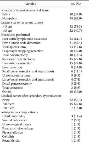
Table 2. Outcomes and findings of secondary cytoreduction
Variable no. (%)
Location of largest recurrent disease Pelvis
Non-pelvis
Largest size of recurrent tumors <3 cm
≥3 cm
Procedures performed
Para-aortic lymph node dissection Pelvic lymph node dissection Total splenectomy
Diaphragm stripping/resection Total omentectomy
Supracolic omentectomy Low anterior resection Liver resection
Small bowel resection and anastomosis Ureteroureterostomy
Large bowel resection and anastomosis Distal pancreatectomy
Total colectomy Others
Residual tumor after secondary cytoreduction None
<0.5 cm ≥0.5 cm
Postoperative complications Febrile morbidity Wound dehiscence Ureterovaginal fistula Pancreatic juice leakage Pleural effusion Cellulitis Rectal fistula
20 (37.0) 34 (63.0)
32 (59.3) 22 (40.7)
33 (61.1) 31 (57.4) 23 (42.6) 20 (37.0) 18 (33.3) 15 (27.8) 15 (27.8) 8 (14.8) 6 (11.1) 5 (9.3) 4 (7.4) 3 (5.6) 3 (5.6) 20 (37.0)
32 (59.3) 15 (27.8) 7 (13.0)
3 (11.4) 2 (5.7) 1 (1.9) 1 (1.9) 1 (1.9) 1 (1.9) 1 (1.9) eduction at other hospitals. Therefore, there was insufficient
data regarding residual tumor after primary cytoreduction in the latter group. The median PFS after primary cytoreductive surgery was 24 months, with a range of 6 to 122 months. The median CA-125 before secondary cytoreductive surgery was 102 U/ml, with a range of 6 to 2,350 U/ml (Table 1).
2. Surgery outcomes
Of the 54 patients, complete resection of all visible tumor tissue was achieved in 32 (59.3%) patients, and residual tu- mor with a largest dimension of less than 0.5 cm in 15 (27.8%) patients. Thus, optimal resection was achieved in 47 (87.0%) patients.
The outcome and findings of secondary cytoreduction are summarized in Table 2. At secondary cytoreductive surgery, 33 (66.1%) patients underwent para-aortic lymph node dis- section, 31 (57.4%) underwent pelvic lymph node dissection, 23 (42.6%) patients underwent splenectomy, 20 (37.0%) un- derwent diaphragm stripping or resection, 33 (66.1%) patients underwent excision of retained omental tissue, 15 (27.8%) pa- tients underwent low anterior resection (prophylactic colos- tomy and ileostomy performed in 1 and 2 patients, re- spectively), 8 (14.8%) patients underwent liver resection, 6 (11.1%) patients underwent small bowel resection and anasto- mosis, 4 (7.4%) patients underwent large bowel resection and
anastomosis, 3 (5.6%) patients underwent total colectomy, 5 (9.3%) patients underwent ureteroureterostomy, 3 (5.6%) pa- tients underwent distal pancreatectomy with splenectomy, 2 (3.7%) patient underwent partial bladder excision, 1 (1.9%) patient underwent abdominal wall resection, and 1 (1.9%) pa- tient underwent a partial ostectomy of the pubic bone.
All patients received the first cycle of chemotherapy before discharge. Postoperative complications occurred in 11 (20.4%) patients, comprising of febrile morbidity in 3 (11.4%) pa- tients, wound dehiscence in 2 (5.7%) patients, ureterovaginal fistula in 1 (1.9%) patient, pancreatic juice leakage in 1 (1.9%) patient, pleural effusion in 1 (1.9%) patient, cellulitis in 1 (1.9%) patient, deep vein thrombosis in 1 (1.9%) patient, and rectal fistula in 1 (1.9%) patient. There were no post- operative deaths (Table 2).
3. Survival
The median follow up was 31 months (range, 1 to 77 months), and the median survival was 42 months (95% CI, 18.3 to 75.3 months). The median overall survival time and
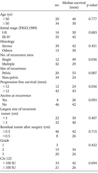
Table 3. Univariate analysis for variables influencing overall survival no. Median survival
(mon) p-value*
Age (yr) <50 ≥50
Initial stage (FIGO,1989) I-II
III-IV Histology Serous Others
No. of recurrence sites Single
Multiple Site of recurrence Pelvis
Non-pelvis
Progression free survival (mon) <12
≥12
Ascites at recurrence Yes
No
Largest size of recurrent tumor (cm)
<3 ≥3
Residual tumor after surgery (cm) ≤0.5
>0.5 Grade 1 2 3 CA-125 <100 IU ≥100 IU
20 34
16 35
39 15
12 42
20 34
12 42
8 46
22 32
46 8
3 15 20
33 21
46 30
30 42
42 30
49 29
53 24
24 43
26 42
30 40
42 26
34 26
42 26
0.777
0.683
0.451
0.036
0.007
0.036
0.093
0.407
0.715
0.432
0.694
*p-value by log-rank test for univariate analysis
Fig. 1. Overall survival according to the site of recurrence.
Fig. 2. Overall survival according to progression free survival.
PFS: progression free survival
Table 4. Multivariate analysis of overall survival
Risk ratio p-value*
Site of recurrence Pelvis
Non-pelvis
Progression free survival (mon) <12
≥12
Ascites at recurrence Yes
No
No. of recurrence sites Single
Multiple
1 4.972
1 0.346
1 0.786
1 2.421
0.013
0.043
0.630
0.419
*p-value by Cox proportional hazards regression analysis progression-free survival time was 42.0 months (95% CI,
32.46 to 47.47 months) and 13.0 months (95% CI, 9.14 to 16.86 months), respectively. Factors influencing overall sur- vival after recurrence were analyzed using univariate analyses (Table 3). Comparison of the median survivals of the patients with only 1 recurrence and the patients with 2 or more recur- rences revealed a significant difference (49 vs. 29 months, p=0.036). The median survival of the patients with only pel- vic recurrence was longer than that of the patients with non-pelvic recurrence (53 vs. 24 months, p=0.007) (Fig. 1).
The PFS also significantly affected the overall survival dura- tion with a median survival of 24 months for a PFS of less than 12 months, and 43 months for a PFS of 12 months or greater (p=0.036) (Fig. 2). The outcome of secondary cytoreduction was not a significant factor for survival (42 months for pa- tients with optimal debulking and 26 months for patients with suboptimal debulking) (p=0.715). Other variables, such
as age, CA-125, histology, grade, initial FIGO stage, and larg- est size of recurrent tumor, did not affect the overall survival significantly on univariate analysis.
On multivariate analysis, we used 4 variables. Those were
three variables (PFS, number of recurrence sites, and site of recurrence) found to be significant on the univariate analysis, and 1 more variable (ascites) that showed borderline sig- nificance (p=0.093). Site of recurrence (p=0.013) and PFS (p=0.043) were significantly associated with the overall sur- vival after secondary cytoreduction (Table 4).
DISCUSSION
The majority of patients will ultimately develop recurrent disease and will require further treatment despite high initial response rates to primary treatment in advanced ovarian cancer. Lack of curative options for recurrent disease means the goal of treatment is to maximize overall and disease-free survival and quality of life. Therefore, approaches such as sec- ondary cytoreductive surgery, new second-line chemo- therapeutic agents, hormonal therapy and immunotherapy have become the subject of research interest. Among these modalities, secondary cytoreductive surgery and second-line chemotherapy are the mainstays of treatment. Although most recurrent ovarian cancer patients receive second-line chemo- therapy, the response rate and the benefits do not reach those of first-line chemotherapy. In addition, the role of secondary cytoreductive surgery remains controversial.
Previous studies have involved a variety of surgical proce- dures. Although a standard technique for secondary cytor- eductive surgery has been suggested by Chen et al.,4 there is at present no general agreement. In particular, Eisenkop et al.5 performed aggressive procedures such as pelvic exenteration to achieve complete surgical resection of carcinomas. We also aimed for complete cytoreduction, and applied aggressive sur- gical procedures such as multiple bowel resection, partial hepatectomy, splenectomy, and partial ostectomy. We ach- ieved optimal cytoreduction in a considerable proportion of patients with a few major perioperative complications which were easily controlled. The morbidity rates associated with the secondary cytoreductive surgery in this study were consid- ered acceptable, and there were no postoperative deaths.
Although there is a limitation that this study was conducted by a single gynecologic oncologist and therefore it cannot be reproducible in other institutions, these outcomes support the hypothesis that the benefits of secondary cytoreductive surgery outweigh the potential deficits. We conclude that ag- gressive surgical procedures to achieve optimal resection of recurrent tumors should be considered in planning secondary cytoreductive surgery for selected patients with recurrent epi- thelial ovarian cancer.
The prognostic role and feasibility of secondary cytor- eductive surgery has been examined by several investigators.5-18 However, most such studies are retrospective or non-random- ized prospective studies and include only small numbers of patients. In addition, these studies differ in terms of treat- ment methods, study populations and the definition of opti- mal cytoreduction, and include limited details regarding pre-
and postoperative chemotherapy.19
Despite the apparent shortcomings associated with previous studies, some consistent findings appear to exist regarding the prognostic impact of secondary cytoreductive surgery for recurrent epithelial ovarian cancer. The two most important factors affecting survival appear to be the largest dimension of residual tumor after secondary cytoreductive surgery, and the PFS from primary treatment to recurrence. Most previous studies reported better survival in optimal surgery groups.5,7-18,20 Whether the results reflect the skills of the surgical team or merely the inherent biology of the tumor remains unclear. In the present study, overall survival difference between the group of optimal cytoreductive surgery and the group of sub- optimal surgery after secondary cytoreductive surgery was not significant statistically. We postulate that there were only 8 patients who underwent suboptimal cytoreduction and this limitation is responsible for these results.
While most previous studies have shown improved survival in groups with a longer PFS, some have reported that the PFS had no impact on prognosis.5,7-9,11-15,17,18,20 In the present study, a longer PFS improved overall survival after secondary cytoreductive surgery. To our knowledge, there has been no studies that have showed that the site of recurrence affected survival in patients with recurrent epithelial ovarian cancer.
The observed relation between the site of recurrent tumor and survival may reflect the difficulty of complete cytoreduction and the inherent aggressive biology of the tumor.
In terms of previous studies, while the largest dimension of residual tumor and PFS were often found to influence surviv- al, other factors were also identified, albeit less consistently.
These factors include the use of postoperative chemotherapy, absence of ascites at recurrence, use of preoperative induction chemotherapy, a smaller number of recurrent lesions, fewer prior chemotherapy combinations, younger age, good per- formance status, and absence of liver metastasis.5-7,11,13,14,16-18,20
In the present study, univariate analysis showed that the num- ber of recurrence sites, site of recurrence, and PFS were sig- nificant factors which affected the overall survival duration.
Multivariate analysis identified the site of recurrence and PFS as factors which affected overall survival. Because this study was retrospective, it has limitations in terms of se- lection bias from the individual selection of patients and inter- pretation of the data. There were only 10 patients (18.5%) who had undergone primary cytoreductive surgery at the National Caner Center, while the remaining patients were re- ferred from other hospitals after primary cytoreductive surgery. Because the limitation of primary cytoreductive sur- gery data, we were unable to incorporate the potential impact of this factor on overall survival.
In summary, the current study found that patients with re- current disease after a PFS of more than 12 months and pa- tients with tumors located within the pelvis may benefit from optimal secondary cytoreductive surgery. Maximizing the benefits of secondary cytoreductive surgery appears to require
prudent patient selection and surgical approaches designed to ensure optimal cytoreduction. In the present study, such ag- gressive surgical techniques were associated with acceptable levels of morbidity and mortality. Ideally, to evaluate both the role and the selection criteria for secondary cytoreductive sur- gery a large, multi-institutional, and prospective study should be mandated.
REFERENCES
1. Burke TW, Morris M. Secondary cytoreductive surgery for ovar- ian cancer. Obstet Gynecol Clin North Am 1994; 21: 167-78.
2. Griffiths CT. Surgical resection of tumor bulk in the primary treatment of ovarian carcinoma. Natl Cancer Inst Monogr 1975;
42: 101-4.
3. Bristow RE, Tomacruz RS, Armstrong DK, Trimble EL, Montz FJ. Survival effect of maximal cytoreductive surgery for ad- vanced ovarian carcinoma during the platinum era: a meta- analysis. J Clin Oncol 2002; 20: 1248-59.
4. Chen LM, Karlan BY. Recurrent ovarian carcinoma: is there a place for surgery? Semin Surg Oncol 2000; 19: 62-8.
5. Eisenkop SM, Friedman RL, Spirtos NM. The role of secondary cytoreductive surgery in the treatment of patients with re- current epithelial ovarian carcinoma. Cancer 2000; 88: 144-53.
6. Ayhan A, Gultekin M, Taskiran C, Aksan G, Celik NY, Dursun P, et al. The role of secondary cytoreduction in the treatment of ovarian cancer: Hacettepe University experience. Am J Obstet Gynecol 2006; 194: 49-56.
7. Berek JS, Hacker NF, Lagasse LD, Nieberg RK, Elashoff RM.
Survival of patients following secondary cytoreductive surgery in ovarian cancer. Obstet Gynecol 1983; 61: 189-93.
8. Segna RA, Dottino PR, Mandeli JP, Konsker K, Cohen CJ.
Secondary cytoreduction for ovarian cancer following cisplatin therapy. J Clin Oncol 1993; 11: 434-9.
9. Zang RY, Zhang ZY, Li ZT, Chen J, Tang MQ, Liu Q, et al. Effect of cytoreductive surgery on survival of patients with recurrent epithelial ovarian cancer. J Surg Oncol 2000; 75: 24-30.
10. Munkarah A, Levenback C, Wolf JK, Bodurka-Bevers D,
Tortolero-Luna G, Morris RT, et al. Secondary cytoreductive surgery for localized intra-abdominal recurrences in epithelial ovarian cancer. Gynecol Oncol 2001; 81: 237-41.
11. Scarabelli C, Gallo A, Carbone A. Secondary cytoreductive sur- gery for patients with recurrent epithelial ovarian carcinoma.
Gynecol Oncol 2001; 83: 504-12.
12. Tay EH, Grant PT, Gebski V, Hacker NF. Secondary cytor- eductive surgery for recurrent epithelial ovarian cancer. Obstet Gynecol 2002; 99: 1008-13.
13. Zang RY, Li ZT, Tang J, Cheng X, Cai SM, Zhang ZY, et al.
Secondary cytoreductive surgery for patients with relapsed epi- thelial ovarian carcinoma: who benefits? Cancer 2004; 100:
1152-61.
14. Onda T, Yoshikawa H, Yasugi T, Yamada M, Matsumoto K, Taketani Y. Secondary cytoreductive surgery for recurrent epi- thelial ovarian carcinoma: proposal for patients selection. Br J Cancer 2005; 92: 1026-32.
15. Gungor M, Ortac F, Arvas M, Kosebay D, Sonmezer M, Kose K.
The role of secondary cytoreductive surgery for recurrent ovar- ian cancer. Gynecol Oncol 2005; 97: 74-9.
16. Gronlund B, Lundvall L, Christensen IJ, Knudsen JB, Hogdall C. Surgical cytoreduction in recurrent ovarian carcinoma in pa- tients with complete response to paclitaxel-platinum. Eur J Surg Oncol 2005; 31: 67-73.
17. Chi DS, McCaughty K, Diaz JP, Huh J, Schwabenbauer S, Hummer AJ, et al. Guidelines and selection criteria for secon- dary cytoreductive surgery in patients with recurrent, plati- num-sensitive epithelial ovarian carcinoma. Cancer 2006; 106:
1933-9.
18. Tebes SJ, Sayer RA, Palmer JM, Tebes CC, Martino MA, Hoffman MS. Cytoreductive surgery for patients with recurrent epithelial ovarian carcinoma. Gynecol Oncol 2007; 106: 482-7.
19. Munkarah AR, Coleman RL. Critical evaluation of secondary cytoreduction in recurrent ovarian cancer. Gynecol Oncol 2004;
95: 273-80.
20. Salani R, Santillan A, Zahurak ML, Giuntoli RL 2nd, Gardner GJ, Armstrong DK, et al. Secondary cytoreductive surgery for localized, recurrent epithelial ovarian cancer: analysis of prog- nostic factors and survival outcome. Cancer 2007; 109: 685-91.