Although the vast majority of spinal deformity surgeries are still done in adults and in the adolescents who are past the growth spurt, an increasing number of surgeries are being performed on younger patients with spinal deformities, and this is due to the better understanding of the natural histo- ry of spinal diseases and the improved surgical techniques.
While the main goal of surgical intervention in these younger patients remains the same, i.e., the cessation of the deform- ing progress, modern surgical techniques tend to put an em- phasis on actively correcting the existing deformity to restore the spinal balance and the biomechanics, in addition to stabi-
lizing the deformity via a solid arthrodesis.
To fulfill these objectives, the use of internal fixation has become an integral part of the deformity surgery, and this similar to surgical correction that is performed in the older patient population. However, due to the biological charac- teristics that are unique to the pediatric population, there has been much debate concerning the necessity of employing internal fixation as the most suitable fixation devices for the pediatric population.
Spinal pedicle screws were first introduced by Boucher1)in the 1950s and they were popularized by Roy-Camille10)in the 1960s. They offer a secure vertebral grip that achieves improved control of the instrumented segments and rigid internal immobilization. Due to these advantages, spinal pe- dicle screws are gaining increasing popularity for the manage- ment of spinal deformities. However, for the younger pedi-
583 583
Pedicle Screw Fixation in Pediatric Spinal Deformities - Results for patients under 10 years old -
Jin-Hyok Kim, M.D., Se-Il Suk, M.D., Ewy-Ryong Chung, M.D., Sung-Soo Kim, M.D., You-Min Oh, M.D., Jae-Min Jeon, M.D., and Yun-Seok Choi, M.D.
Seoul Spine Institute, Inje University Sanggye-Paik Hospital, Seoul, Korea
583 583 Address reprint requests to
Sung-Soo Kim, M.D.
Seoul Spine Institute, Inje University Sanggye Paik Hospital, 761-1 Sanggye-7-dong, Nowon-gu, Seoul 139-707, Korea Tel: +82.2-950-1288, Fax: +82.2-934-6342
E-mail: toetotoe [email protected]
Purpose: We wanted to determine the efficacy of performing pedicle screw fixation to treat pediatric spinal deformities and we also wanted to evaluate its long-term effects on the growing spine.
Materials and Methods: Thirty-eight consecutive spinal deformity patients (25 congenital, 9 idiopathic and 4 other etiologies) under 10 years old at the time of the surgery who underwent pedicle screw instrumen- tation were reviewed after a minimum follow up of 2 years (range: 2 to 7 years). To evaluate the effect of the pedicle screws on the growing spine, a thin slice CT scan was performed in 27 patients (72%) at the last fol- low up. The patients were treated by posterior fusion with segmental pedicle screw fixation being per- formed in 21 patients, vertebral column resection combined with segmental pedicle screw fixation was done in 16 patients and combined anterior and posterior correction was done in 1 patient.
Results: The frontal correction was 67.2% in the posterior fusion group, 71.5% in the posterior resection group and 64.7% in the patients who underwent combined anterior and posterior correction. A mean correc- tion of 20°was obtained in the sagittal plane. A total of 341 pedicle screws were inserted (an average of 8.9 screws per patient). The complications were comprised of 7 screw malpositions (2.1%), 1 loss of fixation (screw pull-out), 1 recurrence of deformity and one superficial infection. There were no significant neurologi- cal or vascular complications. Any Symptoms or radiological evidence suggestive of spinal stenosis were not detected in any of the patients.
Conclusion: Pedicle screw fixation may be used with the same efficacy for pediatric spinal deformities, and even for the patients under 10 years old, without causing any hazard of iatrogenic spinal stenosis.
Key Words: Spine deformity, Pediatric, Pedicle screw fixation
atric population, there has been few studies that have docu- mented the long term results regarding the effectiveness and safety of the pedicle screw fixation and the potential complica- tions of employing pedicle screws in the immature spines that have continued vertebral growth4)and small, plastic pedicles, i.e., iatrogenic spinal stenosis and the crankshaft phenomenon3). This retrospective study was carried out to determine the efficacy of performing pedicle screw fixation for correcting spinal deformity correction in the younger pediatric popula- tion, and to evaluate the long-term effects of pedicle screw fixation on the growing spine.
MATERIAL AND METHOD
Three-hundred-fifty pediatric spinal deformity patients (the patients were less than 18 years old) underwent posterior pedicle screw instrumentation, and 38 consecutive patients who were less than 10 years of age at the time of surgery were analyzed after a minimum follow up of 2 years (range: 2 to 7 years) to determine the effect of pedicle screw fixation on the actively growing spinal column.
There were 16 males and 22 females with a mean age of 6.9 years (range: 2.7 to 10 years) at the time of their surgical treatment. The triradiate cartilage was open in all the patients.
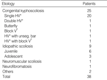
The etiologic diagnosis was congenital scoliosis/kyphosis in 25 patients, idiopathic scoliosis in 9 patients, neuromuscular scoliosis from the postnatal form of cerebral palsy in 1 patient, neurofibromatosis in 1 patient, postinfectious deformity in 1 patient and postlaminectomy kyphosis that has occurred following resection of neuroblastoma in 1 patient. The pa- tients with congenital deformities had an average of 5.7 years
(range: 2.7 to 10.0 years) at the time of the surgery. The pa- thology of congenital spinal deformities were single hemiver- tebra in 20 patients, double hemivertebrae in 1 patient, but- terfly vertebra in 1 patient, hemivertebra with an unsegment- ed bar in 1 patient, block vertebra in 1 patient and hemiver- tebra with block vertebra in 1 patient (Table 1). The patients with idiopathic deformities had an average of 9.7 years (range:
9.1 to 10.0 years) at the time of the surgery. Three of these patients with idiopathic deformities were diagnosed with juvenile form and 6 of them were diagnosed with adolescent form.
The patients were reviewed using the medical records, the preoperative and postoperative standing AP radiographs, and the lateral radiographs; these were all taken at 2 weeks, 6 months, 12 months, 24 months and at the final follow-up for the determination of the spinal maturity, the deformity correction and the spinal balance in the coronal and the sagit- tal planes. The deformity was measured on all the radiographs by the Cobb’s method with using the end vertebrae that was determined on the preoperative standing radiographs. Trunk balance was determined by the distance of the T1 plumb line from the center sacral line on the standing AP radiographs.
A deviation greater than 20 mm was considered decompen- sation.
The crankshaft phenomenon was evaluated at the final fol- low-up for the patients who were treated with only posterior fusion and had Risser 4 or 5. This phenomenon was defined as the progression of the Cobb angle of more than 10°during postoperative follow-up if no other specific cause of progres- sion (pseudoarthrosis or adding-on) was found.
The position of the screws was checked on postoperative radiographs at first postoperative exam and this was confir- med with the CT scans if possible. The occurrence of screw- induced iatrogenic stenosis was evaluated using the thin slice CT scans that were taken during the first postoperative week and at the last follow up. The spinal canal was considered stenotic when there was a visible constriction of the dura in the instrumented area with obliteration of the epidural fat or when the anteroposterior or interpedicular distance of the instrumented vertebrae was significantly less than those of the uninstrumented vertebrae that lay above or below the instrumented area.
All radiographic evaluations were carried out in a double blind fashion by the third and the fourth authors, and the
HV*, hemivertebra; V�, vertebra.
Etiology Patients
Congenital kyphoscoliosis 25
Single HV* 20
Double HV* 1
Butterfly 1
Block V� 1
HV* with unseg. bar 1
HV* with block V� 1
Idiopathic scoliosis 9
Juvenile 6
Adolescent 3
Neuromuscular scoliosis 1
Neurofibromatosis 1
Others 2
Total 38
Table 1.Etiologic diagnosis
mean values of the measurement were used for the analysis.
Surgical techniques
All the surgeries were done by the second author (SI Suk).
The pedicle screw instrumentation was carried out by the following technique.
The spine was approached via a posterior midline incision that exposed the tip of the transverse processes. Following bilateral facetectomy and removal of the cartilages, the pre- sumed pedicle entry points were decorticated with a rongeur.
Guide pins were then inserted into the exposed cancellous bone, and the intraoperative PA and lateral radiographs were taken to determine the position of the guide pins relative to the ideal pedicle entry points. When the entry points were determined, a low speed drill was used to enter the pedicle.
A safe entry was determined when the hole was surrounded by bone and resistance was met in all direction on probing.
When a sound entry was confirmed, the hole was sequential- ly enlarged with larger drill bits until the hole assumed the size equal to the minor diameter of the inserted screw. Fol- lowing a second probing to confirm the bony containment of the pedicle path, the pedicle screw was inserted.
With the pedicle screws in position, they were connected to the longitudinal members and instrumental correction of the deformity was carried out as planned.
Following the completion of the correction, all the screws were tightly locked to the rods and the constructs were stabi- lized by cross-linking of the longitudinal members by means of transfixators.
RESULT
For congenital deformities, the mean preoperative primary curve was 43°(range: 35°to 75°) in the coronal plane with a mean preoperative coronal imbalance of 13 mm (range: 0 to 25 mm) and 28°(range: 7°to 60°) in the sagittal plane with a mean sagittal imbalance of 12 mm (range: 0 to 25 mm). For the idiopathic scoliosis. the curve pattern was King type II in 3 patients, type IV in 2 patients and type V dou- ble thoracic in 4 patients. The mean preoperative major tho- racic curve was 61°(range: 43°to 85°) in the coronal plane with a mean preoperative coronal imbalance of 18 mm (range:
5 to 25 mm) and sagittal imbalance of 17 mm (range: 3 to 45 mm). Four patients had a significant hypokyphosis. The mean magnitude of the proximal thoracic curve in patients diagnosed as King type V was 38°(range: 30°to 52°) with flexibility of 42.2%.
The treatment was comprised of posterior fusion with seg- mental pedicle screw fixation in 21 patients, vertebral col- umn resection combined with segmental pedicle screw fix- ation in 16 patients (15 for the congenital deformities and 1 for a postlaminectomy kyphoscoliosis), and combined anteri- or and posterior correction was done in 1 patient (one case of juvenile idiopathic scoliosis). An average of 5.2 levels (range:
1 to 15) were fused with a mean of 8.9 screws per patient (range: 4 to 25 screws).
1. Deformity correction
In the posterior fusion group, the deformity was corrected to 16°(range: 3°to 41°) in the coronal plane, which showed
Fig. 1.(A, B) A 9.8 year-old male with idiopathic scoliosis of King type IV. The magnitude of the thoracic and lumbar fractional curve was 75°and 35°, respectively. This patient was treated by posterior fusion with segmental pedicle screw fixation. (C, D) The postopeative 2- year follow-up standing radiographs. The thoracic and lumbar curves were corrected to 26°and 7°, respectively.
A B C D
T3
75
T11
35
T3
26
T11
7
a correction of 67.2%. In the sagittal plane the mean kypho- sis was 13°(range: 2°to 42°), which showed a correction of 19°following the surgery. At the final follow up, the defor- mity in the coronal plane was 18°(range: 3°to 45°), which showed a loss of correction of 7.2%. In the sagittal plane, the deformity was 15°(range: 4°to 58°), which showed a loss of correction of 2°. The spine was balanced in all the patients.
There was no significant progression of the deformity (Fig. 1, Table 2). For this study group, 12 patients (8 idiopathic defor- mities and 4 congenial deformities) was evaluated as Risser 4 or 5 at the final follow up. None of the patients showed aggravation of the deformity that was attributable to the crankshaft phenomenon.
In the posterior vertebral resection group, the mean preop- erative coronal deformity of 45°(range: 36°to 75°) was cor- rected to 13°(range: 3°to 34°), which showed a correction
of 71.5%. The mean preoperative kyphosis of 35°(range: 25° to 70°) was corrected to 11°(range: 2°to 42°), which showed a correction of 24°following the surgery. At the final follow up, the deformity in the coronal plane was 15°(range: 3°to 39°), which showed a loss of correction of 9.2%. In the sagit- tal plane, the deformity was 14°(range 4-58°), which showed a loss of correction of 3°. The spine was balanced in all the patients. There was no significant progression of the deformi- ty (Fig. 2, Table 2).
For the patient treated by combined anterior release and posterior instrumentation, the preoperative scoliosis of 85° was corrected to 30°, which showed a correction of 64.7%.
At the final follow up, the curve was 38°with 9.4% loss of correction. There was no significant progression of the defor- mity (Table 2).
2. Complications
A total of 341 screws (156 in the congenital patients, 139 in the idiopathic patients and 46 in the other type patients)
Patients Screws (%)
Screw malposition 5 7 (2.1)
Lateral 3 (0.9)
Superior 2 (0.6)
Inferior 2 (0.6)
Medial 0
Pedicle fracture 4 6 (1.8)
Screw fixation failure 1
Defomity recurrence 1
Infection 1
Table 3.Complications Table 2.Deformity correction
PF*, posterior fusion; PVCR�, posterior vertebral column resection;
Ant+P�, anterior release and posterior instrumentation; Preop�, pre- operative; IMPO‖, immediate postoperative (2 weeks postoperation);
IM corr¶, immediate postoperative correction; Final FU**, final follow- up; LOC��, loss of correction.
Preop�(°) 49 32 45 35 85 50 32
IMPO‖(°) 16 13 13 11 30 16 12
IM corr¶ 67.2% 19° 71.5% 24° 64.7% 67.2% 20°
Final FU** (°) 18 15 15 14 38 19 15
LOC�� 7.2% 2° 9.2% 3° 9.4% 7.9% 3°
PF* group (n=21)
kypho- sis scol- iosis
PVCR� group (n=16)
Ant+PI� (n=1)
Total (n=38) scol-
iosis kypho-
sis scol- iosis
kypho- sis scol- iosis
Fig. 2.(A, B) A 5.4 year-old female with a congenital kyphoscoliosis due to hemivertebra at L1. The magnitude of the coronal and sagittal angle was 40°and 34°, respectively. This patient was treated by posterior vertebral column resection with monosegmental fusion. (C, D) The postoperative 2-year follow-up standing radiographs. The coronal and sagittal angles were corrected to 17°and 5°, respectively.
A B C D
T12 L2 40
-34
T12
L2 L2
-5T12 RMJ
were inserted with a mean of 8.9 screws per patient (range:
4 to 25). The diameter of the screws ranged from 4.0 to 5.0 mm (86 were 4.0 mm, 188 were 4.5 mm, 64 were 5.0 mm and 4 were 6.0 mm). There were 7 screw malpositions (2.1%) in 5 patients. The malpositions were lateral for 3 screws (0.9
%), superior for 2 screws (0.6%) and inferior for 2 screws (0.6%) (Table 3). There were no medial malpositions. The screw malpositions were more common in the congenital deformities than in the other types of deformities.
Intraoperative fractures of the pedicle-lamina occurred in 6 pedicles (1.8%). All but 2 were detected intraoperatively.
Those detected fractures occurred during the final stage of inserting the pedicle screws, but none of them caused gross intraoperative loosening that necessitated removal of the sc- rews. Those intraoperatively undetected fractures occurred during the course of applying compression over the screws and they were recognized on a postoperative CT scan.
There were no major neurological or visceral complications that were attributable to the pedicle screws. There was 1 fix- ation failure in a congenital kyphoscoliosis patient who was treated by posterior vertebral column resection. In this pa- tient, the intraoperative pedicle fracture occurred in the upper most instrumented vertebra during the course of applying compression over the pedicle screws. As the patient was very obese, the screw cutout was detected only on the postoper- ative CT scan that was performed several days later. Because there were no neurological symptoms, she was immobilized in a plaster of Paris body jacket for 3 months. However, on removal of the body jacket, the deformity recurred with pull- out of the proximal screws. Revision was carried out by per- forming proximal extension of the fusion and replacing the pulled out pedicle screws with larger diameter screws. There was 1 recurrence of deformity in a congenital kyphoscoliosis patient who was treated by posterior fusion due to an inap- propriately short fusion. It was revised by performing prox- imal and distal extension of the fusion. A superficial infection occurred in a congenital scoliosis patient who was treated by posterior vertebral column resection, and the infection was treated by incision and drainage. The wound healed unevent- fully in 2 weeks.
3. Iatrogenic spinal stenosis
A thin slice CT scan was performed at the last follow up for 27 patients (72%). Compared to the CT scans taken dur-
ing the preoperative period and the CT scans taken during the immediate postoperative period, the shape of the spinal canal at the last follow up was neither deformed nor narrow.
The AP and transverse diameters were 18 mm (range: 16 to 20 mm) and 20 mm (range: 18 to 21 mm), respectively, in the immediate postoperative period, and AP and transverse diameters were 19 mm (range: 17 to 20 mm) and 22 mm (range: 20 to 23 mm), respectively, at the last follow up and there was no statistically significant difference. The AP and transverse diameters of the uninstrumented areas were simi- lar to the levels in the adjacent instumented areas. There was no patient displaying indentation of the dura due to the sc- rews or any patient who showed symptoms of neurogenic claudication that would be suggestive of spinal stenosis.
DISCUSSION
The spinal pedicle screws are a penetrating type anchor with resistance that spans to the middle and anterior column, and this enables a cantilever beam fixation in various force application modes and it offers a rigid bony grip that enables control of all three vertebral columns. Due to these biome- chanical advantages, it has become more popular to use for correcting spinal deformities, both in adults and in the pedi- atric population.
Nevertheless, the pedicle screws are not as popular to use in the pediatric population before the growth spurt as in adults or in adolescents due to the fear of technical difficul- ties that might be encountered in placing the screws into small, immature pedicles, and there is also the potential of causing an iatrogenic stenosis of the spinal canal by transfix- ation of the neurocentral junction located at the posterior third of the vertebral bodies1,5). The ability of the posterior pedicle screw fixation to resist the crankshaft phenomenon is also undetermined, and this is thought to be the main cause of deformity progression in the immature spine that is treat- ed without an anterior growth arrest procedure4). This retro- spective review was performed to shed some light on these controversies.
As a whole, the reported risk of pedicle screw misplacement ranges from 3 to 40% with 0 to 41% of the neurological complications being attributable to the misplaced screws in surgeries that involve pedicle screw instrumentation2,8,13,14). The reported incidence of screw misplacement in the pedi- atric population ranges from 0.3 to 25% with screw related
neurological complication being noted in 0 to 0.9%2,7). Though there has never been a study specifically reporting on the complications of pedicle screw placement in patients before the onset of their growth spurt, it is not too difficult to imagine a higher rate of misplacement-related compli- cations, when considering the small size of the pedicles and the higher proportion of congenital deformities that occurs for the surgical candidates in this age group. However, our study revealed that screw misplacement in this age group is not more common than in the older age group. This may be attributed to our screw insertion technique that employs intraoperative radiographic control to determine the exact entry point, and also the gradual enlargement of the pilot hole with sequential drilling. The other factor that reduced malposition of the screws was that the pedicle screws were inserted into the normal vertebra after the resection of the deformed vertebra (resection of the hemivertebra during posterior column resection).
Intraoperative fracture of the pedicle in this age group is mainly caused by the gross discrepancy between the diame- ter of the pedicle and the diameter of the inserted screws.
Although screws with diameters up to 115% of the pedicle diameter may be inserted without causing a fracture of the pedicle in the immature spine, fractures may occur even with the smallest diameter screws that are commercially available (4.0 mm) due to the small size of the immature pedicles2,12). However, due to the plasticity of the immature cortical bone in young patients that allows significant deformation, com- minuted fracture of the pedicle that renders the screw grossly unstable is a very rare event in this age group. In addition, the pedicle screws offer significant pull-out strength even with the split fracture of the pedicle by their grip into the vertebral body. These fractures mostly occur during the inser- tion process of the pedicle screws, especially during the final drive into the pedicles, and the fracture may be prevented by performing generous decortication of the entry points and enlarging the proximal portion of the pedicle path with using a drill slightly larger than the minor diameter of the employ- ed screw. As the screws are quite stable, it is not necessary to remove them unless there is gross instability. Neverthe- less, fractures of the pedicles will render the screws unstable to longitudinally directed forces (e.g. compression and dis- traction) and it may necessitate extending the fusion level if the fractures occur bilaterally at the end of the pedicle screw
construct. Fracture of the pedicles may also occur during the course of applying a compressive or distractive force to the spinal column via the inserted screws, and this occurred for one patient in the study. This complication is more prone to occur in the smaller pedicles when using oversized pedicle screws that are greater than 80% of the pedicle diameter and this may be attributed to the microfractures of the pedicle cortex that occurs during the plastic deformation, which weakens the pedicle wall. As these cutouts significantly re- duce the holding power of the pedicle screws, extending the instrumentation/fusion to secure additional points of fixation often necessary, as was done in our patient.
The neurocentral junctions are two obliquely oriented car- tilage plates that lay between the anterior and posterior ossi- fication centers of the vertebral body, and these plates are res- ponsible for the growth of the spinal canal. Hence, spinal stenosis may result from premature closure of the synchon- drosis or from premature osseous fusion of the anterior and the posterior elements. Although the neurocentral junctions usually close at the age of 3 to 6 years, the time for the spinal canal to reach its adult size seems to have regional variation.
The proximal part of the vertebral column tends to mature before the caudal parts do.
The spinal canal at L3, 4 is 70% of the adult size at birth and it reaches the fully mature adult size by one year of age, while the canal of L5 is about 50% of the adult size at this age and reaches the adult size by 5 years5,9).
Being a penetrating type anchor that spans all three colu- mns of the spine, the pedicle screws essentially cross the neu- rocentral junction that is located at the posterior third of the vertebral body and may cause closure of the synchondrosis.
However, iatrogenic fusion of the neurocentral junction does not invariably result in retardation of the canal growth or iatrogenic stenosis as the development of any stenosis is main- ly dependant upon the size of the spinal canal at the time of the violation11). The absence of iatrogenic spinal stenosis in our series may be attributable to the patient’ age at the time of operation being greater than 2 years which is when the spinal canal has nearly reached its adult size, and to the fact that most of the screws were inserted in the upper lumbar spine and the thoracic spine, which both mature relatively earlier than the lower lumbar spine.
Another potential complication of lengthy fusion in the immature patients, the crankshaft phenomenon, is attributed
to the continued growth of the anterior column in the pres- ence of a posterior tether, which causes rotation of the fused segments3). It mainly occurs in the immature patients below the age of 10 years who have open triradiate cartilage.
Though the standard method of preventing such a compli- cation is adding an anterior arthrodesis at the time of poste- rior instrumentation, there have been suggestions, via animal studies, that using a stiff pedicle anchored construct may prevent the crankshaft phenomenon by overpowering the deforming force of the remaining anterior growth centers6). There was no progression of deformity that could attributed to crankshaft phenomenon in our series of patients who un- derwent posterior instrumentation, and this confirms the view of Asher et al. This may be attributed to the biome- chanical characteristics of the pedicle screws that served as a cantilever beam fixation, and this offered resistance to the longitudinally directed forces that were evenly applied along its entire length through the posterior, middle and the ante- rior column.
CONCLUSION
In conclusion, pedicle screw fixation may be used in the young pediatric population below the age of 10 years with a safety that is comparable to that in patients of the older age group, this is without the hazard of causing iatrogenic spinal stenosis in most of the patients.
Pedicle screw fixation also has more advantages than the other forms of fixation e.g. hooks or wires, as pedicle screw fixation is able to prevent the crankshaft phenomenon and it eliminates the need of an additional anterior procedure.
REFERENCE
1. Boucher HH: A method of spinal fusion. J Bone Joint Surg, 41-B:
248-259, 1959.
2. Brown CA, Lenke LG, Bridwell KH, Geideman WM, Hasan SA and Blanke K:Complications of pediatric thoracolumbar and
lumbar pedicle screws. Spine, 23: 1566-1571, 1998.
3. Dubousset J, Harring JA and Shufflebarger H: The crankshaft phenomenon. J Pediatr Othrop, 9: 541-550, 1989.
4. Ferree BA: Morphometric characteristics of pedicles of the imma- ture spine. Spine, 17: 887-891, 1992.
5. Jeffrey JE, Campbell DM, Golden MH, Smith FW and Por- ter RW:Antenatal factors in the development of the lumbar verte- bral canal: A magnetic resonance imaging study. Spine, 28: 1418- 1423, 2003.
6. Kioshos HC, Asher MA, Lark RG and Harner EJ: Overpow- ering the crankshaft mechanism: The effect of posterior spinal fusion with and without stiff transpedicular fixation on anterior spinal col- umn growth in immature canines. Spine, 21: 1168-1173, 1996.
7. Liljenqvist UR, Halm HF and Link TM: Pedicle screw instru- mentation of the thoracic spine in idiopathic scoliosis. Spine, 22:
2239-2245, 1997.
8. Lonstein JE, Denis F, Perra JH, Pinto MR, Smith MD and Winter RB:Complications associated with pedicle screws. J Bone Joint Surg, 81-A: 1519-1528, 1999.
9. Papp T, Porter RW and Aspden RM: The growth of the lumbar vertebral canal. Spine, 19: 2770-2773, 1994.
10. Roy-Camille R, Saillant G and Mazel C: Internal fixation of the lumbar spine with pedicle screw plating. Clin Orthop, 203: 7-17, 1986.
11. Ruf M and Harms J: Pedicle screws in 1-and 2-year-old children;
Technique, complications, and effect on further growth. Spine, 27:
E460-E466, 2002.
12. Seraran H, Yazici M, Karcaaltincaba M, et al: Lumbar pedicle morphology in the immature spine: A three-dimensional study using spiral computed tomography. Spine, 27: 2472-2476, 2002.
13. Suk SI, Kim WJ, Lee SM, Kim JH and Chung ER: Thoracic pedicle screw fixation in spinal deformities: Are they really safe?
Spine, 26: 2049-2057, 2001.
14. Suk SI, LeeCK, Kim WJ, Chung YJ and Park YB: Segmental pedicle screw fixation in the treatment of thoracic idiopathic scol- iosis. Spine, 20: 1399-1340, 1995.
목 적: 척추 변형 수술에서 3차원 교정이 가능한 척추경 나사못의 사용은 증가하고 있으나 10세 이하의 소아에서의 임상적 결과에 대한 보고는 드물어, 10세 이하의 소아에서 변형 교정을 위해 사용한 척추경 나사못의 유용성을 판단하고, 성장하는 추체에 척추경 나사못이 미치는 임상적 문제들을 알아보고자 하였다.
대상 및 방법: 수술을 시행할 때 10세 이하인 38명 환자를 대상으로 하였으며, 원인 질환으로는 25명의 선천성 척추 측만증, 9명의 특발성 척추 측만증, 기타 원인으로 인한 척추 변형 4명이었으며 최소 2년 이상(2-7년) 추시 가능한 환자를 대상으로 하였으며, 성장중인 척추에서 척추경 나사못의 효과를 알아 보기 위해 27명(72%)에서 전산화 단층 촬영을 시행하였다. 수술 방법으로 후방 고정을 시행한 환자는 21명, 후방도달 척추 절제술을 시행한 환자는 16예, 전-후방 교정을 시행한 1예였다.
결 과: 관상면에서 교정도를 살펴 보면 후방 고정을 시행한 경우는 67.2%의 교정을 보였으며, 후방 도달 척추 절재술을 시행 한 경우는 71.5%, 전-후방 교정술을 시행한 경우는 64.7%의 교정도를 보였다. 시상면에서는 평균 교정도는 20�였다. 전체적 으로 341개의 척추경 나사못을 사용하였으며 환자 평균 8.9개의 나사못을 사용하였다. 합병증으로는 7개의 나사못이 정상 위치에 삽입되지 않았으며(2.1%), 1명의 환자에서 고정의 상실이 있었으며, 1명에서 변형이 다시 진행하였으며, 1명에서 천 부 염증이 관찰되었다. 그러나 모든 환자에서 심각한 신경학적 합병증은 발생하지 않았다. 또한 척추경 나사못 삽입으로 인 한 척추관 협착 소견은 방사선학적 임상적으로 관찰되지 않았다.
결 론: 척추경 나사못은 10세 이하의 척추 변형이 있는 소아에서도 성인과 같은 유용한 효과를 관찰할 수 있었으며 어린 소아 에서 척추경 나사못 삽입 후에 척추관 협착 소견은 관찰되지 않았다.
색인 단어: 척추 변형, 소아, 척추경 나사못 고정술
소아 척추 변형 수술시 척추경 나사못 고정술의 유용성 -10세 이하 환자에서의 임상적 결과-
김진혁ㆍ석세일ㆍ정의룡ㆍ김성수ㆍ오유민ㆍ전재민ㆍ최윤석
인제대학교 의과대학 상계백병원 서울 척추센터