393
책임저자: 손병호, 서울시 종로구 평동 108
110-746, 성균관대학교 강북삼성병원 외과 Tel: 02-2001-2132, Fax: 02-2001-2131
E-mail: [email protected] 접수일:2010년 4월 15일, 게재승인일:2010년 8월 9일
대퇴 탈장에서의 전복막외 복강경 탈장 교정술
성균관대학교 의과대학 강북삼성병원 외과학교실
백진희ㆍ박용래ㆍ손병호
Totally Extraperitoneal (TEP) Approach for Femoral Hernia
Jin Hee Paik, M.D., Yong Lai Park, M.D., Byung Ho Son, M.D.
Department of Surgery, Kangbuk Samsung Hospital, Sungkyunkwan University School of Medicine, Seoul, Korea
Purpose: The aims of this study were to evaluate the efficacy of laparoscopic totally extraperitoneal (TEP) repair of femoral hernia.
Methods: Eight patients who underwent laparoscopic TEP repair for femoral hernia between 2008 and 2010 were reviewed retrospectively. In total, 256 adult patients underwent inguinal or femoral hernia repair; TEP was performed in 224 patients. The preoperative diagnosis, clinical symptom, operative finding, postoperative compli- cations, chronic pain, and recurrence were analyzed.
Results: The incidence of femoral hernia was 8 (3.1%) in the present study. The female to male ratio was 3:1 (6 females and 2 males). Seven patients were preoperatively misdiagnosed with inguinal hernia using ultra- sonography. Computed tomography (CT) was performed in three patients, and femoral hernia was diagnosed in two patients. Two patients had synchronous femoral hernia with direct or indirect inguinal hernia. One patient has previously undergone ipsilateral inguinal hernia repair. In all patients, the hernia sac was irreducible by gas insufflation. Seven patients had lipoma-like soft tissue in hernia sac. Peritoneal tears developed in three patients.
There was one postoperative complication: chronic discomfort due to seroma. There was no recurrence during median 6.5 months (range 2∼26).
Conclusion: Laparoscopic TEP repair is safe and effective therapeutic option for repair of femoral hernia. CT images are the most valuable type for the evaluation of the femoral hernia. (J Korean Surg Soc 2010;79:393-398) Key Words: Computed tomography, Femoral hernia, Laparoscopic surgery, Totally extraperitoneal (TEP) repair 중심 단어: 전산화단층촬영, 대퇴탈장, 복강경 수술, 복막외 교정술
서 론
대퇴탈장은 대퇴관을 통해 탈장이 생긴 것으로, 전방으 로는 엉덩두덩관(ilio-pubic tract), 후방으로는 Cooper인대, 외측으로는 대퇴정맥, 내측으로는 엉덩두덩관과 Cooper인
대의 합류지점(갈고리인대, lacunar ligament)을 경계로 하고 있다(Fig. 1).(1) 대퇴탈장은 발견 당시 크기가 작지만 탈장 낭의 형성 부위인 대퇴관이 대퇴정맥, 갈고리인대, 골반뼈 등 단단한 조직으로 이루어져 주위조직의 부종에 의해 쉽 사리 내용물을 압박할 수 있어 감돈의 발생률이 높다.(2) 대 퇴탈장에 대한 수술적 방법에도 많은 변화가 있어 왔다. 과 거부터 사용해 오던 자가조직봉합(tissue repair) 방법으로 McVay 술식,(3) Lockwood 술식(4)이 있으며 McVay 술식에 서는 수술 후 봉합면의 긴장, 서혜탈장의 유인, 복벽 박리로 인한 수술 후 불편감 등의 단점이 있고 Lockwood 술식에서 는 빗살근막(pectineus fascia)의 연약함, 봉합선의 긴장과 함
Fig. 1. (A) The schematic drawing of relevant anatomy of right femoral hernia.(1) (B) Laparo- scpic view of right groin area.
께 재발률이 높은 단점이 있다. 각종 인공 보형물을 이용한 무긴장성 교정술(5)도 시행되고 있으며 최근에는 복강경을 이용한 술식인 transabdominal preperitoneal (TAPP) 술식,(6) totally extraperitoneal (TEP) 술식(7) 및 intraperitoneal onlay mesh (IPOM) 술식(8) 등도 시행하고 있다. TAPP 술식은 복 강 내 접근으로 인한 합병증이 발생하고 IPOM 술식은 복강 내 장기와 접촉함으로써 발생하는 유착형성, 장루관 형성, 감염, 패혈증 등의 위험이 있다. 이에 반해 TEP 술식은 mesh가 이동(migration)할 위험성이 있지만 재발률이 낮고 (0.7∼2.1%) 수술 후 통증이 유의하게 적은 장점이 있는 술 식이다.(9) 국내에서는 대퇴탈장에서 TEP 술식 후 그 결과 에 대한 보고가 없어 이에 본원에서는 TEP 술식을 통한 탈 장교정술 도중 대퇴탈장으로 진단된 8예의 수술 전 검사소 견, 수술 중 소견, 수술 후 추적관찰 자료를 분석하여 이 술 식의 안정성 및 유용성에 대하여 알아보고자 본 연구를 시 행하였다.
방 법
1) 대상 및 방법
2008년 1월부터 2010년 3월까지 본원에서 서혜부 탈장교 정술을 시행 받은 256명의 환자들 중 출혈경향이 있거나 경 제적 이유로 개방탈장교정술을 시행한 경우를 제외하고 복 강경 TEP 술식으로 수술 받은 224명 중에서 대퇴탈장으로 확진된 8명의 환자들을 대상으로 하였다. 환자들의 임상양 상, 영상의학적 진단방법, 수술 전 진단 여부, 수술소견, 만 성 통증 등의 수술 후 합병증 및 재발에 대하여 수술 녹화 영상, 수술기록지, 검사결과 및 외래기록지와 전화면담 등 을 통한 후향적 연구를 시행하였다. 또한, 수술 전 시행한 영상학적 진단의 유용성에 대해 분석하였다.
2) 수술방법
수술은 전신마취 후 배꼽 아래에 수직으로 1.5 cm 가량 절개선을 넣고 탈장이 있는 방향의 앞쪽 배곧은근집 (anterior rectus sheath)을 노출시킨 후 앞쪽 배곧은근집에 절 개선을 넣은 후 배곧은근 뒷면을 따라 복막전공간(preperi- toneal space)을 박리하였다. 이 때 수술 공간을 만들기 위해 고무풍선이 부착된 투관침을(SpacemakerⓇ Autosuture, Nor- walk, CT, USA) 복막전공간에 넣고 수술 공간을 확보하였 다. 카메라를 삽입 후 치골결합(symphysis pubis)의 중앙선 에서 상방에 5 mm 투관침을 넣고, 배꼽 아래 투관침과 치 골결합 상방의 투관침 사이에 5 mm 투관침 하나를 추가로 삽입하였다. Cooper 인대, 하 배벽혈관(inferior epigastric vessels) 과 대퇴혈관을 확인한 후 직접탈장 유무와 대퇴탈장을 관 찰하고 Bogros 공간 박리를 시행하여 간접탈장 유무를 확인 하였다. 그물막은 폴리에스터 그물막(ParietexⓇ, Sofradim, Formans, France)을 사용하였으며 2∼4개의 TackerⓇ (Auto- suture, Norwalk, CT, USA) 및 TisselⓇ (Baxter AG, Wien, Austria)을 사용하여 고정하였다. 이후 가스가 빠지는 것을 보면서 카메라를 제거하고 CO2 가스가 빠지면 투관침을 제 거하고 수술을 마쳤다.
결 과
1) 임상양상
총 8예 중 남녀비는 6예에서 여자, 2예에서 남자였고 평 균 연령대는 남자 57세(53∼61), 여자 58.3세(48∼68)로 두 군 간의 차이는 없었다. 동반질환으로는 여자 6예 중 1예에 서 고혈압, 1예에서는 우측 서혜탈장으로 수술 받은 기왕력 이 있었고 남자 2예 중 1예에서 과거에 완치 판정 받은 결핵 이 있었다(Table 1). 주증상으로는 서혜부 종괴가 남자 2예,
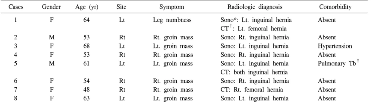
Table 1. Clinical characteristics of patients
Cases Gender Age (yr) Site Symptom Radiologic diagnosis Comorbidity
1 F 64 Lt Leg numbness Sono*: Lt. inguinal hernia Absent
CT†: Lt. femoral hernia
2 M 53 Rt Rt. groin mass Sono: Rt. inguinal hernia Absent
3 F 68 Lt Lt. groin mass Sono: Lt. inguinal hernia Hypertension
4 F 53 Rt Rt. groin mass Sono: Rt. inguinal hernia Absent
5 M 61 Lt Lt. groin mass Sono: Lt. inguinal hernia Pulmonary Tb‡
CT: both inguinal hernia
6 F 54 Rt Rt. groin mass Sono: Rt. inguinal hernia Absent
7 F 48 Rt Rt. groin mass CT: Rt. femoral hernia Absent
8 F 63 Lt Lt. groin mass Sono: Lt. inguinal hernia Absent
*Sono = ultrasonography; †CT = computed tomography; ‡Tb = tuberculosis.
Fig. 2. The ultrasonographic finding of right femoral hernia. The Doppler image shows the femoral hernial sac nearby the femoral vessel.
Fig. 3. The CT finding of left femoral hernia. There are typical findings of femoral hernia: venous compression and lo- calized hernial sac in femoral area (arrow).
여자 2예, 다리저림이 여자 4예였다.
2) 진단
수술 전 이학적 검사로는 동반 탈장이 있던 남자 2예 및 여자 6예에서 두덩뼈결절(pubic tubercle)의 하측방에서 탈 장낭을 촉지하여 대퇴탈장으로 진단하였다. 이학적 검사에 서 대퇴탈장이 의심될 경우 수술 당시 반대측 탈장 여부도 확인하였다. 영상의학적 검사는 이학적 검사에서 대퇴탈장 이 의심되는 경우에 시행하였는데 초음파를 시행한 경우는 남자 2예, 여자 5예였고 전산화단층촬영술을 시행한 경우 는 남자 1예, 여자 2예였다(Table 1). 초음파 결과로는 7예 모두에서 서혜탈장으로 진단했으며(Fig. 2) 전산화단층촬영 술에서는 2예에서 대퇴탈장으로 진단하였다(Fig. 3).
3) 수술소견
탈장의 위치는 우측이 4예(남자 1예, 여자 3예), 좌측이 4예(남자 1예, 여자 3예)였고 반대쪽 간접 서혜탈장이 동반 된 예가 남자 1예, 같은 쪽 직접 서혜탈장이 동반된 예가 남자 1예, 이전 서혜탈장으로 수술을 했던 곳과 같은 쪽에 서 대퇴탈장이 발생한 예가 여자 1예 있었다. 대퇴탈장의 특성상 8예 전부가 감돈이 되어 있었고 탈장낭의 내용물에 지방종 같은 것이 포함된 경우는 7예였다. 탈장 결손부로 복막 또는 복막전조직(preperitoneal structure)의 유착이 동반 된 경우는 7예였다. 탈장낭을 복원할 때 3예에서 복막이 찢 어져 2예에서는 Ligamax Endoscopic clip (Johhn & John K.K., Tokyo, Japan)을 이용해 결찰을 시행하였고 1예에서는 EndoknotⓇ (Ethicon, Cincinnati, OH, USA)을 이용하여 연속 봉합하였다(Table 2).
Table 2. Operative findings
Cases CH* RAGI† HC‡ ATAAW§ PTDR∥ TMD¶
1 − − + + −
2 Contralateral indirect − + + +, clipping
3 − − − − −
4 − − + + −
5 Ipsilateral direct − + + −
6 Recurrent − + + +, clipping
7 − − + + +, suture
8 − − + − −
*CH = coexisting hernia; †RAGI = reduction after gas insufflations; ‡HC = hernia content (associated lipoma like lesion); §ATAAW
= adhesion to anterior abdominal wall; ∥PTDR = peritoneal tearing during reduction and its treatment; ¶TMD = taking mesh down the Cooper’s ligament.
Table 3. Clinical outcomes of laparoscopic TEP* repair for femo- ral hernia
Cases Pain
(acute/chronic)
Wound
problem Seroma Recurrence
1 +/− − − −
2 +/− − − −
3 +/− − − −
4 +/− − − −
5 +/− − − −
6 +/− − + −
7 +/− − − −
8 +/− − − −
*TEP = totally extraperitoneal.
4) 수술 후 경과
추적관찰(범위 2∼26개월, median 8.6개월) 결과 수술 후 심각한 통증은 없었고 상처에 문제가 발생한 예는 없었으 며 여자 1예에서 장액종(seroma)에 의한 불편감을 호소하였 다(Table 3). 재발 및 만성통증은 없었다.
고 찰
대퇴륜은 외측으로는 대퇴정맥(femoral vein), 위로는 서 혜인대(inguinal ligament), 내측으로는 갈고리인대(lacunar ligament), 뒤로는 치골인대(pectineal ligament)로 경계를 이 루며, 대퇴탈장은 탈장낭이 대퇴륜을 통해 서혜인대 하방 에 있는 대퇴관으로 나온 것이다.(10) Naude 등,(11) Mjåland 등(12)의 연구에 의하면 대퇴탈장은 남자보다 여성에 빈발 하며(1:2∼2.5) Yoo 등(13)의 국내보고에서도 여자에게 호 발하는 것으로 보고하였다. 이는 비교적 넓은 골반을 가진
여성의 해부학적 특징으로 상대적으로 대퇴륜이 넓은 이유 에서 기인한다. 본 연구에서도 남녀비는 1대 4로 여성에서 호발하는 것으로 나타나 다른 연구 결과와 일치하였다.
Mikkelsen 등(14)은 대퇴탈장은 서혜탈장이 약 5∼20%에 서 동반된다고 하였다. 또한 서혜탈장의 수술 과거력이 있 는 환자에서 대퇴탈장이 많이 보고된다고 하였으며, 이러 한 이유로는 서혜탈장 수술 시 이미 동반되어 있던 대퇴탈 장을 간과하였거나 서혜탈장 수술 시 과도한 주위 조직 박 리로 대퇴륜을 느슨하게 한 경우, 서혜탈장 수술로 인해 대 퇴륜막이 과도하게 당겨져 대퇴륜이 확장되기 때문이 다.(15) 대퇴탈장은 서혜탈장과 오인되는 경우가 많기 때문 에 수술 전에 정확히 진단하기가 어렵고(16) 대퇴륜의 좁은 해부학적 특징 때문에 뚜렷한 탈장의 돌출 증후가 없어도 교액이나 감돈이 다른 복벽탈장보다 흔하여 응급수술을 받 게 될 가능성이 높아진다.(17) 대퇴탈장이 있는 남자의 경 우 50%에서, 여자의 경우 10%에서 서혜탈장을 동반하는 것으로 알려져 있다.(18) 본 연구에서는 2예의 남자 환자에 서 동측 직접탈장과 반대측 간접탈장을 각 1예씩 동반하였 고 이전에 서혜탈장으로 수술 받은 경우가 여자 환자에서 1예 있었다. 즉 서혜탈장 수술 시 대퇴탈장을 놓치기 쉽고, 대퇴탈장은 다른 서혜탈장을 동반하는 경우가 있을 수 있 어서 개방성 탈장교정술(open hernia repair)을 시행할 때 염 두에 두고 있어야 할 것으로 사료된다.
수술 전 영상학적 검사는 주로 초음파를 이용하는데 본 연구에서는 7예의 대퇴탈장 환자에서 초음파를 시행한 결 과 모두 대퇴탈장을 서혜탈장으로 진단하였다. 이는 초음 파 검사가 시술자의 주관적인 검사라는 점과 서혜부의 복 잡한 해부학적 구조가 서혜탈장과 대퇴탈장을 감별하는 데 정확성이 떨어지는 원인이라 하겠다.(19) 따라서 초음파검
Fig. 4. Laparoscopic view of right femoral hernia. The lipoma-like mass observed in femoral area defect.
사를 할 때는 이학적 검사소견과의 비교가 반드시 필요할 것으로 판단된다. 한편 전산화단층촬영은 대퇴탈장과 서혜 탈장을 구분하는 기준으로 삼을 수 있는 탈장낭(hernia sac) 과 치골결절(pubic tubercle)과의 위치적 관계 및 대퇴정맥 (femoral vein)의 압박여부를 관찰하기 쉬우며(20) 특히 mul- tidetector-row computed tomography를 통한 수술 전 진단은 대퇴탈장의 진단에 중요한 구조물인, 서혜인대와 하 배벽 동맥을 구분하는데 큰 도움이 되는데 본 연구에서도 전산 화단층촬영에서 대퇴정맥 측면으로 대퇴탈장낭이 있는 경 우를 관찰하기 용이하여 수술 전의 영상학적 진단에는 초 음파보다 전산화단층촬영이 더 효과적이었다.
대퇴탈장의 수술방법으로는 과거에는 McVay 술식을 주 로 이용하다가 각종 인공 보형물을 이용한 무긴장성 술식 이 보편화되는 듯하였으나 상처의 크기가 기존 수술법과 다르지 않아 미용적 효과를 크게 보지 못하는 단점이 부각 되면서 최근에는 복강경을 이용한 탈장교정술이 대두되었 다. 복강경술식으로는 복강경을 복강 내로 접근하는 방식 인 TAPP 술식, 복직근의 후면을 통해 복막전공간에 보형물 을 넣는 TEP 술식, 복막을 절개하여 복막전공간을 박리할 필요 없이 바로 복막에 인공보형물을 자동봉합기, 압정, 봉 합사 등을 이용해 부착하여 복벽을 강화하는 방법인 IPOM 술식이 있는데 본원에서는 TEP 술식을 이용하였다.
수술소견에 대하여 대퇴탈장의 해부학적 양태에 대하여 기술된 경우는 없었다. 본 연구에서는 수술을 녹화한 영상 을 토대로 이에 대한 분석을 시행한 바 8예의 TEP 수술에 있어서 복막전지방(preperitoneal fat)이 지방종(lipoma)같은 형태로 관찰되는 경우가 대부분이었다(Fig. 4). 이러한 탈장
결손부에 대한 복막전지방의 유착은 비교적 단단하여 초기 에 시행한 3예에서 탈장낭 복원 도중 복막이 찢어졌다. 이 후 탈장낭 복원을 탈장 결손부의 뒤쪽에서 시도하여 복막 이 찢어지지 않아 탈장낭 복원은 탈장 결손부의 뒤쪽에서 시행하는 것이 좀 더 용이한 접근방식일 것으로 사료된다.
본 연구에서는 8예 전예에서 재발이나 만성통증은 없었 으나 여자 1예에서 장액종이 발생하였는데 이를 방지하기 위한 추후 방법에 관하여 고찰이 필요할 것으로 사료된다.
그물망은 TackerⓇ 및 TisselⓇ을 사용하여 고정하였는데 Lovisetto 등(21)의 연구와 마찬가지로 Tissel이 수술 후 불편 감을 감소시키는 데 역할을 했을 것으로 사료된다.
대퇴탈장은 다른 탈장과의 동반 가능성, 탈장의 뒤쪽 결 손부로의 접근과 탈장낭 교정의 용이함, 수술 후 불편감 및 재발, 그리고 미용적인 측면 등을 고려해 봤을 때 TEP 술식 이 좋은 수술방법이 될 것으로 생각한다.
결 론
저자들은 8예의 환자에서 복강경 TEP 술식으로 대퇴탈 장의 수술을 시도하였다. 수술 후 합병증 및 재발에 대하여 향후 더 많은 증례와 장기간의 추적관찰 기간이 필요하겠 으나 대퇴탈장에 대한 치료방법으로 복강경 TEP 술식은 안 전하고 효과적인 수술방법이라고 여겨진다. 또한 수술 전 영상학적 검사로는 초음파보다 전산화단층촬영술이 더 유 의하다고 사료된다.
REFERENCES
1) Ramshaw B. Surgical anatomy of the inguinal region and ab- dominal wall: clinical application of the total extraperitoneal approach for laparoscopic inguinal hernia repair. In: Soper NJ, editor. Problems in General Surgery. Abdominal Hernias. Vol.
19. Philadelphia: Lippincott Williams & Wilkins; 2002.
p.14-26.
2) Amid PK, Shulman AG, Lichtenstein IL. The femoral canal:
the key to femoral herniorrhaphy. Int Surg 1990;75:69-72.
3) McVay CB, Savage LE. Etiology of femoral hernia. Ann Surg 1961;154:25-32.
4) Rahaman QM, Goswami B, Gumta MK, Samanta S, Mukhopadhyay S, Chattopadhyay M. Bilateral femoral hernia in male--a case report. J Indian Med Assoc 2005;103:237, 42.
5) Lichtenstein IL. Herniorrhaphy. A personal experience with 6,321 cases. Am J Surg 1987;153:553-9.
6) Bátorfi J. The treatment of inguinofemoral hernias with lapa-
roscopic herniorraphy. Our experience of 1210 transabdominal preperitoneal (TAPP) reconstructions. Magy Seb 2005;58:385- 97.
7) Yalamarthi S, Kumar S, Stapleton E, Nixon SJ. Laparoscopic totally extraperitoneal mesh repair for femoral hernia. J Laparoendosc Adv Surg Tech A 2004;14:358-61.
8) Fitzgibbons RJ Jr, Camps J, Cornet DA, Nguyen NX, Litke BS, Annibali R, et al. Laparoscopic inguinal herniorrhaphy.
Results of a multicenter trial. Ann Surg 1995;221:3-13.
9) Napier T, Olson JT, Windmiller J, Treat J. A long-term fol- low-up of a single rural surgeon's experience with laparo- scopic inguinal hernia repair. WMJ 2008;107:136-9.
10) Sandblom G, Haapaniemi S, Nilsson E. Femoral hernias: a register analysis of 588 repairs. Hernia 1999;3:131-4.
11) Naude GP, Ocon S, Bongard F. Femoral hernia: the dire con- sequences of a missed diagnosis. Am J Emerg Med 1997;15:
680-2.
12) Mjåland O, Bakken IJ, Skjeldestad FE, Johnson E. Inguinal and femoral hernia repair in Norway 1990-2003. Tidsskr Nor Laegeforen 2005;125:1338-40.
13) Yoo H, Lee K, Choi U. The diagnostic concordance of femo- ral hernia and the factors influencing diagnosis. J Korean Surg Soc 2009;76:179-86.
14) Mikkelsen T, Bay-Nielsen M, Kehlet H. Risk of femoral her- nia after inguinal herniorrhaphy. Br J Surg 2002;89:486-8.
15) Bendavid R. Femoral hernias: primary versus recurrence. Int Surg 1989;74:99-100.
16) Rhind JR. Lateral femoral hernia. J R Coll Surg Edinb 1971;
16:299-300.
17) Chan G, Chan CK. Longterm results of a prospective study of 225 femoral hernia repairs: indications for tissue and mesh repair. J Am Coll Surg 2008;207:360-7.
18) Malangoni MA, Rosen MJ. Hernias. In: Townsend CM Jr, Beauchamp RD, Evers BM, Mattox KL, editors. Sabiston Textbook of Surgery: The Biological Basis of Modern Surgical Practice. 18th ed. Philadelphia: W.B. Saunders; 2008. p.1155- 79.
19) Zhang GQ, Sugiyama M, Hagi H, Urata T, Shimamori N, Atomi Y. Groin hernias in adults: value of color Doppler so- nography in their classification. J Clin Ultrasound 2001;29:
429-34.
20) Suzuki S, Furui S, Okinaga K, Sakamoto T, Murata J, Furukawa A, et al. Differentiation of femoral versus inguinal hernia: CT findings. AJR Am J Roentgenol 2007;189:W78-83.
21) Lovisetto F, Zonta S, Rota E, Mazzilli M, Bardone M, Bottero L, et al. Use of human fibrin glue (Tissucol) versus staples for mesh fixation in laparoscopic transabdominal preperitoneal hernioplasty: a prospective, randomized study. Ann Surg 2007;
245:222-31.