Received April 28, 2020, Revised May 27, 2020, Accepted May 28, 2020 Corresponding author: Sujung Yeo
Department of Meridian & Acupoint, College of Korean Medicine, Sang Ji University, 83 Sangjidae-gil, Wonju 26339, Korea Tel: +82-33-738-7506, Fax: +82-, E-mail: [email protected]
CCThis is an open access article distributed under the terms of the Creative Commons Attribution Non-Commercial License (http://creativecommons.org/licenses/ by-nc/4.0) which permits unrestricted non-commercial use, distribution, and reproduction in any medium, provided the original work is properly cited.
MPP+로 유도된 신경 독성에 대한 녹각의 보호 효과
여수정
상지대학교 한의과대학 경혈학교실
Neuroprotective Effects of Cervi Cornu in MPP+ Treated SH-SY5Y Cells
Sujung Yeo
Department of Meridian & Acupoint, College of Korean Medicine, Sang Ji University
Objectives : Parkinson’s disease, a progressive neurodegenerative disease, is caused by the loss of dopaminergic neurons in the substantia nigra. There is no clear treatment or remedy for Parkinson’s disease; therefore, the development of novel therapies related to anti-inflammatory and antioxidant effects is required. This study was performed to evaluate the neuroprotective effect of water extracts from Cervi Cornu (CC) in dopaminergic cells. Methods : We studied effects of CC on apoptosis, cell death and inflammation in SH‐SY5Y neuroblastoma cells treated by methylpyridinium ion (MPP+). SH-SY5Y cell line was treated with CC for 24 hours and then 500 μM MPP+ for 18 hours. Results : Cervi Cornu treatment inhibited the decrease in tyrosine hydroxylase (TH) expression and decreased the activation of inflammatory factors mitochondrial cytochrome C oxidase (COX2) and inducible NO synthase (iNOS) against MPP+ neurotoxicity. Apoptosis factors BCL2 associated X, apoptosis regulator (BAX) levels were decreased and B-Cell CLL/Lymphoma 2 (BCL2) levels were increased. Conclusions : These results suggest that CC treatment had neuro- protective effects in the SH‐SY5Y neuroblastoma cells against toxicity induced by MPP+. The results suggest new possibilities of CC for the treatment of Parkinson’s disease.
Key words : Cervi Cornu, Parkinson’s disease, neuroprotection, MPP+, SH‐SY5Y
서 론
1817년에 제임스 파킨슨(James Parkinson)은 손 떨림, 근육 경 직, 자세 불안정 등의 증상을 보이는 환자들에게 ‘떨림 마비’
(paralysis agitans, shaking palsy)라는 이름을 붙였다1). 이후 이 병을 파킨슨병 이라고 명명했다. 파킨슨병은 substantia nigra 부 위에서 도파민성 신경 세포의 감소를 나타내는 진행성 퇴행성 신 경질환이다2,3). 지금까지 국내에서 파킨슨병과 관련된 세포 및 동 물모델에서 신경세포 독성, 염증반응, 활성사노, 세포 사멸 기전
등에 대한 연구들이 진행되었으나 도파민 세포가 감소되는 원인은 아직 명확히 밝혀지지 않았다4-6).
현재 치료법으로는 레보도파(levodopa, L-dopa), 도파민 수용 체 효현제, 항아세틸 콜린성 등의 약물치료 방법이 있으며 약효가 더 이상 듣지 않게 되면 수술요법을 시행한다3,7,8). 레보도파는 도 파민의 전구체로써 도파민으로 대사되어 부족한 뇌내 도파민 농도 를 보충시킴으로써 파킨슨병의 증상을 개선시키려고 사용되고 있 다9). 하지만 3-5년 이상 장기 투여 시 약물효과 시간이 점점 짧아 지는 wearing-off 현상, 약물에 의한 운동조절 기능의 변동이 심해
지는 on-off 현상, 이상운동증 등의 부작용이 나타난다 . 따라 서, 부작용을 줄이면서 약물의 유효성을 지속시키고 파킨슨병의 진행을 늦추거나 막을 수 있는 치료법의 개발이 필요하다.
녹각(old altler, cornu cervi)은 梅花鹿 Cervus nippon Temminck 또는 馬鹿 Cervus elaphus Linnaeus, 수사슴의 골질화된 뿔로 腎 經과 肝經으로 들어가 溫腎, 强筋骨하는 효능을 통해 녹용의 대용 품으로 응용하기도 한다. 그리고 活血, 散瘀, 消腫하는 작용이 우수 한 것으로 알려져 있으며13), 골다공증을 유발한 흰쥐에서 치료효과 가 보고되었다13). 또한 신경세포에서 산화스트레스 개선효과가 있 는 것으로 보고되었으며14) 간세포에서 항염증 효과가 보고되었다15). 산화스트레스와 염증반응은 파킨슨병에서 도파민 세포의 병증화 와 관련이 있으므로 이들 논문을 근거로 녹각이 파킨슨병에서의 치료효과를 나타낼 것으로 생각되었다.
아직까지 파킨슨병 관련 모델에서의 녹각의 효과에 관한 연구는 보고된 바 없다. 본 연구에서는 녹각 물 추출물(CC)의 도파민성 세포에서의 신경보호효과를 측정하고자 하였다. 이에 본 연구에서 는 도파민성 신경세포인 SH-SY5Y 세포에서 MPP+ 유발독성에 대 한 녹각 물 추출물의 apoptosis와 염증성 반응과 관련하여 나타내 는 신경보호효과를 측정하여 파킨슨병 치료를 위한 초석을 다지고 자 하였다.
재료 및 방법
1. 약재
본 실험에 사용한 녹각(CC)은 정도약업사(Seoul, Korea)에서 구입하였다. 잡질을 제거한 후 10배의 증류수에 2시간 동안 환류 추출 한 뒤 −60℃ 이하에서 얼려 동결건조 하였고. −20℃에 보 관 후 매 실험 시 용매에 녹여 사용하였다.
2. 시약
세포배양에 필요한 Dulbeco’s modified Eagle’s medium (DMEM), fetal bovine serum (FBS), minimal essential medium (MEM), penicillin-streptomycin (P/S)은 Gibco사(Auckland, NZ)에서 구입하여 사용하였다. MPP+은 Sigma-aldrich사(St Louis, USA)에서 구입하여 사용하였다. Rabbit anti-tyrosine hydro- xylase (TH) antibody는 Santa Cruz Biotechnology (Texas, USA), cytochrome c와 bax, bcl 2 antibody는 abcam사 (Cambridge, UK), EZ-CYTOX는 두젠바이오(Seoul, Korea), ABC standard kit 는 Vector사(Burlingame, USA)에서 구입하여 사용하였으며, 실험
에 사용된 모든 시약은 분석용 등급 이상으로 사용하였다.
3. 세포배양
본 실험에 사용된 세포주는 human neuroblastoma인 SH- SY5Y 세포이며, 한국세포주은행(Seoul, Korea)에서 분양 받아 사 용하였다. 10% (v/v) FBS, 1% P/S을 포함하는 high-glucose DMEM 배지를 사용하였으며 37℃, 5% CO2 조건에서 배양하였다.
4. Cell viability 측정
96 well plate에 2.0×10⁴/well의 SH-SY5Y 세포를 분주하고 48시간 후, CC 1, 25, 50, 75 μg/ml 농도로 24시간 동안 처리하 였다. 100 μl/ml의 EZ-CYTOX 를 처리하여 3시간 배양 후, spectrophotometer로 450 nm에서 흡광도를 측정하였다.
MPP+ 용액에 대한 Cell viability를 측정하기 위하여 96 well plate에 2.0×10⁴/well의 SH-SY5Y 세포를 분주하고 48시간 후, NSW 1, 25, 50, 75 μg/ml 농도로 처리하였다. 6시간 후 MPP+
용액 500 mg/ml를 처리 하여 18 시간 배양 후, 100 μl/ml의 EZ- CYTOX를 처리 하여 3시간 배양 후, spectrophotometer로 450 nm에서 흡광도를 측정하였다. 세포 생존율을 대조군에 대한 백분 율로 표시하였다.
5. Western blot 측정
SH-SY5Y 세포를 분주하고 48시간 후, CC 1, 25, 50, 75 μg/
ml 농도로 처리하였다. 그 후 MPP+ 용액 500 mg/ml를 처리 하여 18 시간 배양 후, cold PBS 로 세척 후 lysis buffer 로 처리한 후 4℃에서 20분간 원심분리 한 후 (12,000 rpm) 상층액을 12% sodium dodecyl sulfate-polyacrylamide gel electrophoresis (SDS- PAGE)를 이용하여 단백질 농도별로 분리한 후 polyvinylidene difluoride (PVDF) membrane (Bio-Rad, USA)으로 옮겼다. 실온 에서 1시간 동안 0.1% Tris 완충 식염수 (TBS, 20 mM Tris-HCl (pH 7.5)) 및 0.1% Tween-20을 함유한 150 mM NaCl (TBST) 에 5% 탈지유를 첨가한 용액으로 차단하고, mouse anti-tyrosine hyroxylase (TH, 1:2000, Santa Cruz Biotechnology), rabbit anti-Bax (1:2000, abcam) 및 mouse anti-β-actin (1:5000, Santa Cruz Biotechnology) 항체와 함께 배양하였다. 0.1% TBST 로 세척 한 후, 막을 anti-mouse IgG- peroxidase antibody (1:
2000, Bio-Rad)와 항온 배양하고, 항원 - 항체 복합체를 Pierce ECL western blotting substrate (Thermo Scientific, USA)를 사 용하여 현상하였다.
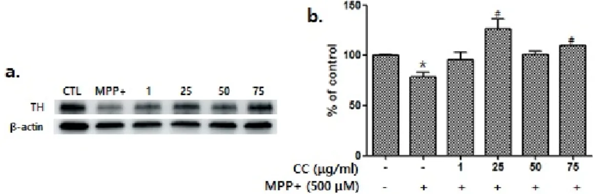
Fig. 3. Western blot to detect dopaminergic cells using TH abtibody in various concentrations of Cervi Cornu treatment (a). SH-SY5Y cell line was treated with Cervi Cornu treatment for 24 hr and 500 μM MPP+ treatment for 18 hrs. Values are expressed as averages±SEM (b).
CTL, control group, MPP+, 500 μM MPP+ treatment, 1, 1 μg/ml Cervi Cornu treatment and 500 μM MPP+ treatment, 25, 25 μg/ml Cervi Cornu treatment and 500 μM MPP+ treatment, 50, 50 μg/ml Cervi Cornu treatment and 500 μM MPP+ treatment, and 75, 75 μg/ml Cervi Cornu treatment and 500 μM MPP+ treatment. *p<0.05 vs. the control and #p<0.05 vs. the MPP+ group.
Fig. 1. Cytotoxicity was analyzed in various concentrations (0, 1, 25, 50, and 75 μg/ml) of Cervi Cornu treatment.
SH-SY5Y cell line was treated with Cervi Cornu treatment for 24 hr.
Fig. 2. Cytotoxicity was analyzed in various concentrations of Cervi Cornu treatment.
SH-SY5Y cell line was treated with Cervi Cornu treatment for 24 hr and 500 μM MPP+ treatment for 18 hrs. *p<0.05 vs. the control and #p<0.05 vs. the MPP+ group.
6. 통계분석
통계분석의 측정값은 평균±표준오차 (mean±standard error) 로 표기하였다. 통계방법은 student t-test 및 ANOVA를 시행했으 며 유의수준은 p-value<0.05로 지정하였다.
결 과
1. CC에 대한 Cell viability 측정
CC에 대한 Cell viability를 측정하기 위하여 SH-SY5Y 세포에 0, 1, 25, 50, and 75 μg/ml 농도의 약물을 처리한 결과 모든 농도에서 세포 생존율이 통계적으로 유의미한 차이가 없었다 (Fig. 1).
2. SH-SY5Y에서 MPP+에 대한 CC의 Cell viability 측정 MPP+ 용액에 대한 CC의 세포보호효과를 확인하기 위하여 0,
1, 25, 50, and 75 μg/ml 농도의 약물을 처리하고 6시간 후 MPP+ 용액을 처리하여 결과를 확인하였다. MPP+만을 처리한 그 룹에서만 대조군과 유의한 차이를 보였으며 (p<0.05), MPP+ 용 액만을 처리한 그룹에 대하여 75 μg/ml의 CC 를 처리한 그룹에 서 유의한 차이를 보였다 (p<0.05, Fig. 2).
3. SH-SY5Y에서 MPP+에 대한 CC의 세포 보호효과 MPP+ 용액의 신경독성에 대한 도파민성 세포에서의 CC의 신 경보호 효과를 관찰하기 위하여 SH-SY5Y세포에 MPP+를 처리하 여 TH 발현을 확인하였다. 대조군에 비하여 MPP+만을 처리한 그 룹에서 통계적으로 유의한 차이를 보였다 (p<0.05). CC 처리 그 룹은 농도마다 차이는 있으나 MPP+ 용액만을 처리한 그룹과의 비교에서 25, 75 μg/ml의 농도에서 유의하게 TH의 발현 감소가 억제 되었다 (p<0.05, Fig. 3).
Fig. 4. Western blot in various concentrations of Cervi Cornu treatment (a). SH-SY5Y cell line was treated with Cervi Cornu treatment for 24 hr and 500 μM MPP+ treatment for 18 hrs. Values are expressed as averages±SEM (b).
CTL, control group, MPP+, 500 μM MPP+ treatment, 1, 1 μg/ml Cervi Cornu treatment and 500 μM MPP+ treatment, 25, 25 μg/ml Cervi Cornu treatment and 500 μM MPP+ treatment, 50, 50 μg/ml Cervi Cornu treatment and 500 μM MPP+ treatment, and 75, 75 μg/ml Cervi Cornu treatment and 500 μM MPP+ treatment. *p<0.05 vs. the control, #p<0.05 vs. the MPP+, and ##p<0.01 vs.
the MPP+ group.
Fig. 5. Immunofluorescence image of SH-SY5Y cells treated with 75 μg/
ml Cervi Cornu and 500 μM MPP+.
(a, e, i) were stained with DAPI to visualize existing cells. (b, f, j) Dopa- minergic cells detected using anti- TH antibodies. (c, g, k) Cox2 expres- sions detected using anti-Cox2 anti- bodies. (d, h, i) show merged TH and Cox2 images.
4. CC의 염증 조절을 통한 MPP+ 용액에 대한 신경보호 효과
MPP+ 용액의 신경독성에 대한 도파민성 세포에서의 CC의 신 경보호 효과를 관찰하기 위하여 SH-SY5Y세포에 MPP+를 처리하 여 cox2와 iNOS 의 발현을 확인하였다. 대조군에 비하여 MPP+만 을 처리한 그룹에서 cox2와 iNOS 가 통계적으로 유의하게 증가하 였다 (p<0.05). CC 처리 그룹은 농도마다 차이는 있으나 MPP+
용액만을 처리한 그룹과의 비교에서 75 μg/ml의 농도에서 유의 하게 cox2의 발현이 감소되었다 (p<0.05). iNOS 의 발현은 MPP+
용액만을 처리한 그룹과의 비교에서 25, 50, 75 μg/ml의 농도에 서 유의하게 감소되었다(p<0.01, Fig. 4).
5. CC의 cox2 조절을 통한 MPP+ 용액의 신경독성에 신경보호효과
MPP+ 용액의 신경독성에 대한 도파민성 세포에서의 CC의 신 경보호 효과를 살펴보기 위하여 cox2 를 이용하여 형광염색을 한 결과 MPP+만을 처리한 그룹에서는 TH와 cox2의 merge 가 더 강하게 나타나고 75 μg/ml CC를 처리한 군에서는 control 과 비슷한 수준으로 나타났다(Fig. 5).
6. CC의 apoptosis 조절을 통한 MPP+ 용액의 신경독 성에 대한 신경보호효과
MPP+ 용액의 신경독성에 대한 도파민성 세포에서의 CC의 신 경보호 효과를 관찰하기 위하여 SH-SY5Y세포에 500 μM MPP+
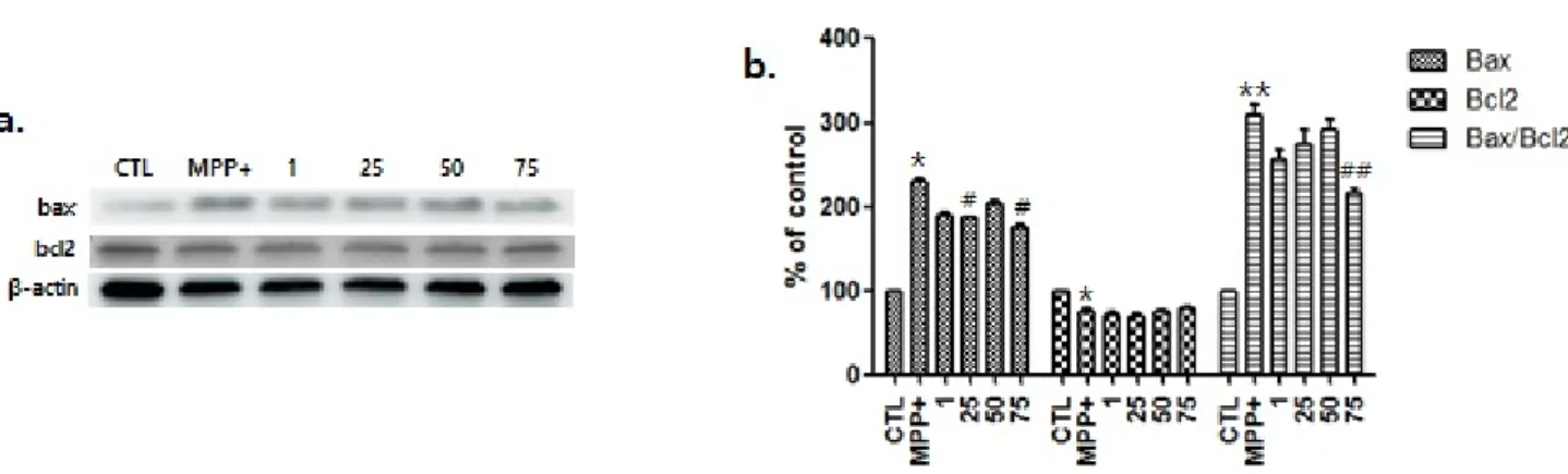
Fig. 6. Western blot to detect the levels of Bax and, Bcl2 in various concentrations of Cervi Cornu treatment (1, 25, 50 and 75 μg/ml) and 500 μM MPP+ in the SH-SY5Y cells.
(a) SH-SY5Y cell line was treated with Cervi Cornu treatment for 24 hr and 500 μM MPP+ treatment for 18 hrs. Values are expressed as averages±SEM. (b) CTL, control group, MPP+, 500 μM MPP+ treatment, 1, 1 μg/ml Cervi Cornu treatment and 500 μM MPP+
treatment, 25, 25 μg/ml Cervi Cornu treatment and 500 μM MPP+ treatment, 50, 50 μg/ml Cervi Cornu treatment and 500 μM MPP+ treatment, and 75, 75 μg/ml Cervi Cornu treatment and 500 μM MPP+ treatment. *p<0.05 vs. the control, **p<0.01 vs. the control, #p<0.05 vs. the MPP+, and ##p<0.01 vs. the MPP+ group.
를 처리하여 cox2와 iNOS 의 발현을 확인하였다. 대조군에 비하 여 MPP+만을 처리한 그룹에서 bax는 유의하게 증가하고 (p<
0.05), bcl2 는 유의하게 감소하였다 (p<0.05). CC 처리 그룹은 농도마다 차이는 있으나 MPP+ 용액만을 처리한 그룹과의 비교에 서 25, 75 μg/ml의 농도에서 유의하게 bax의 발현이 감소되었고 (p<0.05), 75 μg/ml의 농도에서 bax/bcl2 비율이 유의하게 감 소하였다(p<0.01, Fig. 6).
고 찰
SH-SY5Y 세포에 0, 1, 25, 50, and 75 μg/ml 농도의 약물을 처리한 결과 모든 농도에서 세포 생존율이 크게 차이가 없었다.
MPP+의 신경독성에 대한 CC의 세포보호효과를 확인하기 위하여 0, 1, 25, 50, and 75 μg/ml 농도의 약물을 처리하고 6시간 후 MPP+ 용액을 처리한 결과, MPP+만을 처리한 그룹에 비하여 75 μg/ml 의 CC 를 처리한 그룹에서 유의한 신경보호효과를 나타냈다.
CC의 신경보호 효과에 관한 기전을 탐색하기 위하여 산화 스트 레스 및 apoptosis와 관련하여 실험을 진행하였다. 파킨슨병에서 산화 스트레스 및 apoptosis는 뉴런 도파민 세포의 감소를 유발한 다. 파킨슨병을 유도하는 MPP+와 같은 뉴로톡신은 전자 수송 사 슬의 복합체 I에서 과산화물의 생성을 자극하고 미토콘드리아 기 질 탈수소 효소를 포함한 근위 산화 환원 부위에서 자유 라디칼 생성을 유발한다16). 활성 산소 종이 증가 된 미토콘드리아에서 생 성한 산화 스트레스는 미토콘드리아 외막에서 Bax의 발현 및 세포
내 분포를 초래한다. 미토콘드리아의 Ca2+의 축적은 시토크롬 C를 방출하고 Bax 관련 메커니즘 이외의 메커니즘을 통해 과산화물 생성을 촉진한다. 결과적으로 대사 장애, 산화 스트레스 및 apoptosis 가 발생한다. Bax는 미토콘드리아 내 막간 공간으로부터 세포질 내로의 시토크롬 C와 같은 pro-apoptosis 단백질의 방출을 자극 한다. 항- apoptosis 단백질 인 Bcl2는 Ca2+ 또는 Bax에 의해 매개 된 시토크롬 C의 방출을 억제 할 수 있다17,18). 본 실험에서 CC의 처리는 MPP+에 증가된 Bax의 발현 증가를 억제함으로써 Bax/
bcl2의 비율을 낮췄다. 이를 통하여 apoptosis에 의한 도파민성 뉴런의 사멸을 억제하는 기전을 통하여 신경보호효과를 나타낸 것 으로 생각된다.
파킨슨병에서 보여지는 유도성 산화 질소 iNOS는 산화 질소 (NO)를 유도하여 미세 아교 세포를 활성화시킨다. 다양한 염증성 사이토카인과 함께 퇴행성 뇌질환의 과정에서 iNOS가 중요한 역 할을 한다고 보고 되었다6). 또한, NO는 파킨슨병 동물 모델에서 도파민성 세포의 변성을 촉진시키는 역할을 한다4). Cox (Cyclo- oxygenase)는 아라키도네이트로부터 프로스타글란딘의 형성에 관여하는 속도 조절 효소이며, 이의 유도 가능한 형태인 Cox2는 파킨슨병 동물 모델의 SN에서 고도로 발현된다19,20). Cox2의 비정 상적인 증가는 도파민의 산화를 유도하면서 파킨슨병에서와 같이 도파민성 뉴런의 선택적 취약성에 기여한다21,22). 본 실험에서는 MPP+에 의하여 SH-SY5Y 세포에서 iNOS와 Cox2의 증가를 보였 으며, CC를 처리한 그룹에서는 이러한 증가가 유의하게 감소되었 다. 이는 산화스트레스에 의한 도파민성 세포의 선택적 취약성에 대항하는 CC의 신경보호효과의 작용기전을 보여준다.
결 론
이 연구의 주요 목표는 CC의 처리가 MPP+의 신경독성에 신경 보호 효과를 갖는지 여부를 결정하는 것이었다. MPP+의 처리는 SH-SY5Y세포에서 TH 수준을 유의하게 감소시켰으며, CC의 처리 에 의하여 TH감소가 통계적으로 유의하게 억제되었다. 이 실험결 과를 통하여 CC의 물 추출물이 apoptosis와 파킨슨병의 병리기전 으로 알려진 cox2, iNOS를 조절하여 신경보호효과를 나타내는 것 을 확인하였다.
Acknowledgement
None.
Funding
This research was supported by Sangji University Research Fund, 2018
Data availability
The authors can provide upon reasonable request.
Conflicts of interest
저자들은 아무런 이해 상충이 없음을 밝힌다.
References
1. Tolosa E, Wenning G, Poewe W. The diagnosis of Parkinson’s disease. Lancet Neurol. 2006 ; 5(1) : 75-86. https://doi.org/10.
1016/S1474-4422(05)70285-4
2. Albanese A. Diagnostic criteria for Parkinson’s disease. Neurol Sci. 2003 ; 24 Suppl 1 : S23-6. https://doi.org/10.1007/
s100720300032
3. Frank C, Pari G, Rossiter JP. Approach to diagnosis of Parkinson disease. Can Fam Physician. 2006 ; 52 : 862-8.
4. Zheng LT, Ryu GM, Kwon BM, Lee WH, Suk K. Anti-inflammatory effects of catechols in lipopolysaccharide-stimulated microglia cells: inhibition of microglial neurotoxicity. Eur J Pharmacol.
2008 ; 588(1) : 106-13. https://doi.org/10.1016/j.ejphar.2008.
04.035
5. Tasaki Y, Makino Y, Ohta S, Hirobe M. 1-Methyl-1,2,3,4-tetra- hydroisoquinoline, decreasing in 1-methyl-4-phenyl-1,2,3,6- tetrahydropyridine-treated mouse, prevents parkinsonism-like behavior abnormalities. J Neurochem. 1991 ; 57(6) : 1940-3.
https://doi.org/10.1111/j.1471-4159.1991.tb06407.x
6. Salum C, Raisman-Vozari R, Michel PP, Gomes MZ, Mitkovski M, Ferrario JE, et al. Modulation of dopamine uptake by nitric oxide in cultured mesencephalic neurons. Brain Res. 2008 ; 1198 : 27- 33. https://doi.org/10.1016/j.brainres.2007.12.054
7. Hauser RA, Freeman TB, Snow BJ, Nauert M, Gauger L, Kordower JH, et al. Long-term evaluation of bilateral fetal nigral trans- plantation in Parkinson disease. Arch Neurol. 1999 ; 56(2) : 179-87.
https://doi.org/10.1001/archneur.56.2.179
8. Reichmann H. Modern treatment in Parkinson’s disease, a per- sonal approach. J Neural Transm (Vienna). 2016 ; 123(1) : 73-80.
https://doi.org/10.1007/s00702-015-1441-1
9. Fahn S. The history of dopamine and levodopa in the treatment of Parkinson’s disease. Mov Disord. 2008 ; 23 Suppl 3 : S497-508.
https://doi.org/10.1002/mds.22028
10. Cacabelos R. Parkinson’s Disease: From Pathogenesis to Phar- macogenomics. Int J Mol. 2017 ; 18(3) : 551. https://doi.org/10.
3390/ijms18030551
11. Chen H, Fang J, Li F, Gao L, Feng T. Risk factors and safe dosage of levodopa for wearing-off phenomenon in Chinese patients with Parkinson’s disease. Neurol Sci. 2015 ; 36(7) : 1217-23. https://
doi.org/10.1007/s10072-015-2078-4
12. Tolosa E, Marti MJ, Valldeoriola F, Molinuevo JL. History of levo- dopa and dopamine agonists in Parkinson’s disease treatment.
Neurology. 1998 ; 50(6 Suppl 6) : S2-S48. https://doi.org/10.
1212/wnl.50.6_suppl_6.s2
13. Hwang JS, Seo BI, Park JH, Roh SS, Kim MR, Kim SM, et al. Effect of Cervi Cornu On treatment of Osteoporosis in Ovariectomized Rats. Kor J Herbology. 2010 ; 25(2) : 1-10.
14. Kim HD, Lee EY, Park JY, Seo KH, Lee KH, Choi J, et al. Protective effect of ethanolic extract of antler-shaped Ganoderma lingzhi against oxidative stress in PC12 neuronal cell line. J Mushrooms.
2018 ; 16(3) : 213-7.
15. Kim MJ, Cho SY, Park EM, Yoon SH. Effect of Old Antler Extracts on the Benzo(a)pyrene-Induced Hepatotoxicity in Rats. J Korean Soc Food Sci Nutr. 1993 ; 22(4) : 412-7.
16. Gonzalez-Polo RA, Soler G, Fuentes JM. MPP+: mechanism for its toxicity in cerebellar granule cells. Mol Neurobiol. 2004 ; 30(3) : 253-64. https://doi.org/10.1385/MN:30:3:253
17. Fall CP, Bennett JP, Jr. Characterization and time course of MPP+
-induced apoptosis in human SH-SY5Y neuroblastoma cells. J Neurosci Res. 1999 ; 55(5) : 620-8. https://doi.org/10.1002/
(SICI)1097-4547(19990301)55:5<620::AID-JNR9>3.0.CO;2-S 18. Chen J, Sun J, Jiang J, Zhou J. Cyanidin Protects SH-SY5Y Human
Neuroblastoma Cells from 1-Methyl-4-Phenylpyridinium-Induced Neurotoxicity. Pharmacology. 2018 ; 102(3-4) : 126-32. https://
doi.org/10.1159/000489853
19. Wang T, Pei Z, Zhang W, Liu B, Langenbach R, Lee C, et al.
MPP+-induced COX-2 activation and subsequent dopaminergic neurodegeneration. FASEB J. 2005 ; 19(9) : 1134-6. https://doi.
org/10.1096/fj.04-2457fje
20. Teismann P, Vila M, Choi DK, Tieu K, Wu DC, Jackson-Lewis V, et al. COX-2 and neurodegeneration in Parkinson’s disease. Ann N Y Acad Sci. 2003 ; 991 : 272-7. https://doi.org/10.1111/j.
1749-6632.2003.tb07482.x
21. Hsieh YC, Mounsey RB, Teismann P. MPP(+)-induced toxicity in the presence of dopamine is mediated by COX-2 through oxida- tive stress. Naunyn Schmiedebergs Arch Pharmacol. 2011 ; 384(2) : 157-67. https://doi.org/10.1007/s00210-011-0660-8
22. Chae SW, Kang BY, Hwang O, Choi HJ. Cyclooxygenase-2 is in- volved in oxidative damage and alpha-synuclein accumulation in dopaminergic cells. Neurosci Lett. 2008 ; 436(2) : 205-9. https://
doi.org/10.1016/j.neulet.2008.03.031