Introduction
Fractures of condylar process of mandible are common injuries that account for 20-62% of all mandibular frac- tures.1The diagnosis of the mandibular condyle fractures is based on clinical and radiographic examinations. Clini- cal signs such as jaw deviation, mouth opening limitation, malocclusion, and edema of the preauricular region can be indicative of fractures of the condylar process.2The accuracy of physical examination is only 68% of mandi- bular fractures,3therefore radiographic evaluation is essen-
tial to confirm the presence and location of the mandibu- lar fractures.
Computed tomography (CT) is considered as a gold standard for the radiographic evaluation of fractures of the mandibular condyle process.4,5It reveals bony and soft tissue changes at the same time and allows multi-planar evaluation.6,7 However, the routine use of CT for mandi- bular fractures is not justified due to the high cost and increased radiation exposure.
Panoramic radiograph provides a good view of the entire mandible including the condylar region, therefore it is commonly used by many clinicians as an ideal screening view for mandibular fractures.8-10 Condylar region is one of the most difficult areas to detect fractures, especially for many clinicians who do not have much experience of interpreting mandibular fractures.11 The failure to recog- nize the presence of a condylar fracture, especially in child-
Diagnostic performance of dental students in identifying mandibular condyle fractures by panoramic radiography and the usefulness of reference images
Bong-Hae Cho
Department of Oral and Maxillofacial Radiology, School of Dentistry, Pusan National University, Busan, Korea ABSTRACT
Purpose : The purpose of this study was to evaluate the diagnostic performance of dental students in detection of mandibular condyle fractures and the effectiveness of reference panoramic images.
Materials and Methods : Forty-six undergraduates evaluated 25 panoramic radiographs for condylar fractures and the data were analyzed through receiver operating characteristic (ROC) analysis. After a month, they were divided into two homogeneous groups based on the first results and re-evaluated the images with (group A) or without (group B) reference images. Eight reference images included indications showing either typical condylar fractures or ana- tomic structures which could be confused with fractures. Paired t-test was used for statistical analysis of the differ- ence between the first and the second evaluations for each group, and student’s t-test was used between the two groups in the second evaluation. The intra- and inter-observer agreements were evaluated with Kappa statistics.
Results : Intra- and inter-observer agreements were substantial (k==0.66) and moderate (k==0.53), respectively. The area under the ROC curve (Az) in the first evaluation was 0.802. In the second evaluation, it was increased to 0.823 for group A and 0.814 for group B. The difference between the first and second evaluations for group A was statis- tically significant (p⁄0.05), however there was no statistically significant difference between the two groups in the second evaluation.
Conclusion : Providing reference images to less experienced clinicians would be a good way to improve the diag- nostic ability in detecting condylar fracture. (Imaging Sci Dent 2011; 41 : 53-7)
KEY WORDS : Radiography, Panoramic; Mandibular Condyle; Mandibular Fractures
*This study was supported by a grant of Pusan National University Hospital.
Received December 9, 2010; Revised April 30, 2011; Accepted May 6, 2011 Correspondence to : Dr. Bong-Hae Cho
Department of Oral and Maxillofacial Radiology, School of Dentistry, Pusan Nation- al University, Beomeo-ri, Mulgeum-eup, Yangsan, Gyeongsangnam-do 626-870, Korea
Tel) 82-55-360-5255, Fax) 82-55-360-5029, E-mail) [email protected]
Copyright ⓒ 2011 by Korean Academy of Oral and Maxillofacial Radiology
This is an Open Access article distributed under the terms of the Creative Commons Attribution Non-Commercial License (http://creativecommons.org/licenses/by-nc/3.0) which permits unrestricted non-commercial use, distribution, and reproduction in any medium, provided the original work is properly cited.
Imaging Science in Dentistry∙pISSN 2233-7822 eISSN 2233-7830
ren may lead to late complications including facial defor- mity and temporomandibular joint ankylosis.5,12 There were previous studies that the sensitivity of panoramic radiography in diagnosing mandibular condylar fractures ranged from 70 to 90%.5,11,13Lee et al observed that clini- cians with less experience tended to miss more fractures than those with more experience.11
The hypothesis was that providing reference images showing either typical condylar fractures or normal ana- tomic structures mimicking fractures might improve the diagnostic accuracy for the inexperienced.
The purpose of this study was to evaluate the diagnostic performance of dental students in detection of mandibular condyle fractures and the effectiveness of reference pano- ramic images.
Materials and Methods
Materials
From the Oral and Maxillofacial archives of Pusan Na- tional University Hospital, one oral and maxillofacial radiologist and one oral and maxillofacial surgeon with more than 10 years of experience reviewed CT and pano- ramic images of patients suspected of having uncompli- cated mandibular condylar fractures.
A total of 25 panoramic radiographs were selected for this study. The inclusion criteria were as follows: 1. The 2 assessment panels were in complete agreement on the presence or absence of the condylar fracture. 2. All pano- ramic radiographs were of acceptable quality and had no other jaw disease.
The study population comprised 18 males and 7 females and the mean age was 35.2 (9-74 years old). Out of 25 panoramic radiographs, 16 radiographs showed unilateral condylar fracture, 6 bilateral fractures, and 3 no fracture.
The data of the patients’ gender, age, diagnosis, type of fracture, and displacement of the fracture were recorded.
The classification of the fractures was determined using panoramic and CT images. The condylar fractures were grouped into head, neck, and subcondylar fractures by the location and also classified by the type of displacement as suggested by Yamaoka et al.14 The assessment panels’
diagnosis was regarded as a gold standard for confirming condylar fractures.
Image acquisition
Digital panoramic radiographs were taken using PM 2002 CC Proline (Planmeca Oy, Helsinki, Finland) with
the standard panoramic program. The panoramic equip- ment was set at 62-64 kVp, 5-6 mA, and 18 seconds of exposure time.
All CT scans were performed with a multi-detector CT (Somatom Definition AS++, Siemens Medical Systems, Erlangen, Germany) and imaging parameters were as fol- lows: 120 kVp, 230-250 mAs, 3 mm pitch, 0.5 mm inter- val and slice thickness 1.3-2.5 mm.
Image assessment First assessment
The panoramic radiographs were shown to 50 dental students in their final year. All images were directly inter- faced onto a PACS system (M-view, Infinitt Healthcare, Seoul, Korea) on monitors (MFGD 5421, Barco, Kortrijk, Belgium) of 2,048×2,560 image matrices and 145.9-ft- lambert luminescence. All students were blind to the clini- cal information of the subjects imaged. Each assessor was asked to record the presence or absence of fracture by both a simple ‘yes’ or ‘no’ for sensitivity analysis and a 5-point scale for Receiver Operating Characteristic (ROC) analy- sis: 1-fracture definitely absent; 2-fracture probably absent;
3-unsure whether fracture is present; 4-fracture probably present; 5-fracture definitely present.
After the first assessment, the sensitivity and ROC area were calculated. ROC analysis was performed by MedCalc version 6.0 (MedCalc Software, Mariakerke, Belgium).
Four students showing markedly deviated ROC area (¤mean±2 SD) from other students were eliminated as outliers. Using the first assessment results, the assessors were divided into two homogenous groups having the same mean and SD, and each group was composed of 23 students.
Second assessment
A minimum of 4 weeks later, the students performed the second viewing session. For group A (reference group), Eight panoramic radiographs were provided as reference images during the second assessment. The images were indicated with arrows noting either typical condylar frac- ture or normal structures which could be confused with fractures. Figs. 1 and 2 show examples of reference images.
Group B (no reference group) evaluated the images in ran- dom order in the same way as the first session. There was no additional training or education for both groups.
Statistical analysis
The difference between the first and the second evalua- tions within each group was analyzed by means of a paired
t-test and the difference between the two groups in the sec- ond evaluation by student’s t-test using SPSS 11.0 for Win- dows software (SPSS Inc, Chicago, IL). P-values less than 0.05 were considered statistically significant.
Kappa statistics were computed to assess the intra- and inter-observer agreement.15For agreement within the ob- servers, the data from the first and second assessments of group B were used, and for the agreement between the ob- servers, the data from the first assessment of group A and B were used. Kappa statistics are commonly interpreted as
⁄0.00, poor agreement; 0.00 to 0.20, slight agreement;
0.21 to 0.40, fair agreement; 0.41 to 0.60, moderate agree- ment; 0.61 to 0.80, substantial agreement; and 0.81 to 1.00, almost perfect agreement.
Results
The distribution of the condylar fractures is listed in Table 1. There were 5 condylar head fractures, 14 neck fractures, and 9 subcondylar fractures. The overall sensi-
Fig. 1. Panoramic (A) and cone beam CT (B) images show sagittal splitting (white arrow) and condylar neck (black arrow) fracture.
A
B
Fig. 2.Panoramic image shows soft palate (thick white arrow), subcon- dylar fracture (thick black arrow), spine of sphenoid bone (thin black arrow), and pharyngeal wall (thin white arrow).
tivity was 0.81. The sagittal splitting fracture of condylar head showed the lowest sensitivity. Kappa analysis show- ed substantial (k==0.66) agreement within observers and moderate (k==0.53) agreement among observers.
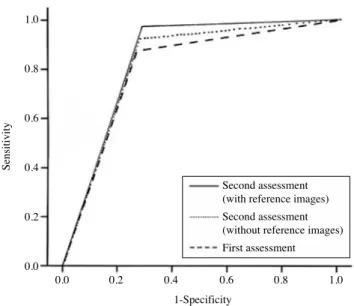
Table 2 and Fig. 3 show the area under ROC curve. Both groups showed higher diagnostic performance in the sec- ond assessment than in the first assessment. Statistically significant difference was shown in group A (reference group) and total assessors (p⁄0.05), however in group B (no reference group), there was no statistically significant difference. Also, the difference of the diagnostic perfor- mance between the groups in the second evaluation was not statistically significant.
Discussion
Panoramic radiography is superior to other conventional radiography and it is adequate in evaluating mandibular fractures.8,9,16With the growing use of the panoramic radio- graphy, the occasions for general dentists to diagnose man- dibular fractures have been increasing. It was reported that clinicians with no experience in the field of fracture show- ed a relatively low diagnostic performance in the detec- tion of condylar fracture.11 This study was performed to explore the possibility of the use of reference images to increase the diagnostic ability.
In this study, the students in their last year of dental school, who have limited experience in this field partici- pated as assessors. They had finished two hours of the lec- ture on facial trauma 4 months before, and no other addi- tional education was given. Having been in the course of clinical patient care program over a year, they were famil- iar with panoramic radiograph.
The students showed the overall sensitivity of 80.9%. It was fairly good, considering the previous report that the sensitivity of the general dentists was 80% or under. It means that they had a considerable knowledge on the frac- ture interpretation. The mandibular condyle is considered as one of the most difficult regions to detect fractures,5,9,17,18 and this is especially true for the sagittal splitting fracture of the condylar head. This study showed that the sensiti- vity of the condylar fractures was different according to the location of the fractures. The sensitivity of the sagittal splitting fracture was only 50%, while that of condylar neck was over 90%. For the subcondyle, the shadow of the soft palate and pharyngeal wall was often mistaken as frac- tures, resulting in relatively low sensitivity. Lee et al stated that condylar fracture was the most common facial fracture in children, and if undiagnosed in a child, it might not be- come apparent until further growth.19The undiagnosed and untreated condylar fractures in children might show facial growth disturbance and asymmetry.12,20Temporomandibu- lar joint disorders such as ankylosis and dysfunction and malocclusion might also occur.19,21 Therefore, Chacon et al recommended additional CT examination in the assess- ment of children suspected to have fractures of the mandi- bular condyle.5
The diagnostic accuracy of condylar fractures was mea- sured by the area under the ROC curve. With or without the reference images, both groups showed improved the diagnostic performance in the second assessment. Statis- tically significant difference was shown in the reference group and in total assessors, however not in the group with-
Table 1.The number of condylar fracture and sensitivity (in paren- theses) according to the fracture classification
Number of fracture (sensitivity) Condylar head Condylar neck Subcondyle
No displacement - 3 (0.97) 7 (0.67)
Deviation and
displacement - 4 (0.92) 2 (0.78)
Dislocation - 7 (0.93) -
Sagittal splitting 5 (0.50) - -
Total 5 (0.50) 14 (0.96) 9 (0.72)
Table 2. Area under ROC curve
1st (Mean±SD) 2nd (Mean±SD) P-value
Reference 0.823±0.04 0.013*
No reference 0.802±0.03 0.814±0.04 0.107
Total 0.819±0.04 0.003*
* statistically significant (p⁄0.05)
Fig. 3.Area under ROC curve.
1.0
0.8
0.6
0.4
0.2
0.0
0.0 0.2 0.4 0.6 0.8 1.0
1-Specificity
Sensitivity
Second assessment (with reference images) Second assessment (without reference images) First assessment
out the reference images. It is understandable that the sup- plementary reference images would contribute to identifi- cation of condylar fractures. However, contrary to expec- tations, there was no statistically significant difference bet- ween the groups in the second evaluation. Even though it was not high enough to show the statistically significant difference, no reference group also showed the increased diagnostic performance in the second evaluation compar- ed with the first evaluation, and the difference between the groups became indistinct. The diagnostic outcome can be improved by training and education. It can be postulat- ed that the first assessment session played as a self-educa- tion, and the diagnostic performance of the no reference group in the second assessment session showed improve- ment, however still not getting higher sensitivity than the reference group.
In conclusion, this study suggests that providing refer- ence images to less experienced clinicians could improve the diagnostic ability in detecting condylar fracture, and along with it, it might be helpful to give a chance of addi- tional education.
References
1. Buchbinder D. Treatment of fractures of the edentulous man- dible, 1943 to 1993: a review of the literature. J Oral Maxillo- fac Surg 1993; 51 : 1174-80.
2. MacLennan WD. Fractures of the mandibular condylar process.
Br J Oral Surg 1969; 7 : 31-9.
3. Thai KN, Hummel RP 3rd, Kitzmiller WJ, Luchette FA. The role of computed tomographic scanning in the management of facial trauma. J Trauma 1997; 43 : 214-8.
4. Roth FS, Kokoska MS, Awwad EE, Martin DS, Olson GT, Hol- lier LH, et al. The identification of mandible fractures by heli- cal computed tomography and panorex tomography. J Cranio- fac Surg 2005; 16 : 394-9.
5. Chacon GE, Dawson KH, Myall RW, Beirne OR. A compara- tive study of 2 imaging techniques for the diagnosis of condy- lar fractures in children. J Oral Maxillofac Surg 2003; 61 : 668- 73.
6. Raustia AM, Pyhtinen J, Virtanen KK. Examination of the temporomandibular joint by direct sagittal computed tomo- graphy. Clin Radiol 1985; 36 : 291-6.
7. Christiansen EL, Thompson JR, Hasso AN. CT evaluation of trauma to the temporomandibular joint. J Oral Maxillofac Surg 1987; 45 : 920-3.
8. Chayra GA, Meador LR, Laskin DM. Comparison of panora- mic and standard radiographs for the diagnosis of mandibular fractures. J Oral Maxillofac Surg 1986; 44 : 677-9.
9. Nair MK, Nair UP. Imaging of mandibular trauma: ROC ana- lysis. Acad Emerg Med 2001; 8 : 689-95.
10. Reiner SA, Schwartz DL, Clark KF, Markowitz NR. Accurate radiographic evaluation of mandibular fractures. Arch Otolaryn- gol Head Neck Surg 1989; 115 : 1083-5.
11. Lee JH, Jung YH, Cho BH, Hwang DS. Diagnostic ability of panoramic radiography for mandibular fractures. Korean J Oral Maxillofac Radiol 2010; 40 : 33-8.
12. Zachariades N, Mezitis M, Mourouzis C, Papadakis D, Spanou A. Fractures of the mandibular condyle: a review of 466 cases.
Literature review, reflections on treatment and proposals. J Craniomaxillofac Surg 2006; 34 : 421-32.
13. Wilson IF, Lokeh A, Benjamin CI, Hilger PA, Hamlar DD, Ondrey FG, et al. Prospective comparison of panoramic tomo- graphy (zonography) and helical computed tomography in the diagnosis and operative management of mandibular fractures.
Plast Reconstr Surg 2001; 107 : 1369-75.
14. Yamaoka M, Furusawa K, Iguchi K, Tanaka M, Okuda D.
The assessment of fracture of the mandibular condyle by use of computerized tomography. Incidence of sagittal split frac- ture. Br J Oral Maxillofac Surg 1994; 32 : 77-9.
15. Cohen J. A coefficient of agreement for nominal scales. Educ Psychol Meas 1960; 20 : 37-46.
16. Moilanen A. Primary radiographic diagnosis of fractures in the mandible. Int J Oral Surg 1982; 11 : 299-303.
17. Wilson IF, Lokeh A, Benjamin CI, Hilger PA, Hamlar DD, Ondrey FG, et al. Contribution of conventional axial comput- ed tomography (nonhelical), in conjunction with panoramic tomography (zonography), in evaluating mandibular fractures.
Ann Plast Surg 2000; 45 : 415-21.
18. Assael LA. Clinical aspects of imaging in maxillofacial trau- ma. Radiol Clin North Am 1993; 31 : 209-20.
19. Lee CY, McCullon C 3rd, Blaustein DI, Mohammadi H. Seque- lae of unrecognized, untreated mandibular condylar fractures in the pediatric patient. Ann Dent 1993; 52 : 5-8.
20. Rowe NL. Fractures of the facial skeleton in children. J Oral Surg 1968; 26 : 505-15.
21. Hovinga J, Boering G, Stegenga B. Long-term results of non- surgical management of condylar fractures in children. Int J Oral Maxillofac Surg 1999; 28 : 429-40.