Photocatalytic activity of Fe treated AC/TiO
2composites between visible light and UV light irradiation
Ze-Da Meng
1, Kan Zhang
1and Won-Chun Oh
1*1Department of Advanced Materials & Science Engineering, Hanseo University
가시광선과 UV광선에 의한 Fe 처리된 AC/TiO
2복합체의 광분해 활성
맹칙달
1, 장간
1, 오원춘
1*1한서대학교 신소재공학과
Abstract FAT compounds photocatalysts were prepared with TiOSO4·xH2O (TOS) by a sol‐gel method. The samples were characterized by scanning electron microscopy (SEM), BET specific surface area, X‐ray diffraction analysis (XRD) and energy dispersive X‐ray spectroscopy (EDX). The SEM results showed that ferric compounds and titanium dioxide were fixed onto the AC surfaces. The XRD results showed that Fe‐AC/TiO2
composites mostly contained anatase phase. EDX showed the presence of C, O, and Ti with Fe peaks in all samples. The photocatalytic activities were evaluated by the photocatalytic oxidation of methylene blue (MB) solution, via compare photodegradation of MB solution under visible light and UV light separately. Fe‐AC/TiO2
composites had an excellent photocatalytic under strong visible light irradiation. A small amount of Fe ions in AC/TiO2 particles could obviously enhance their photocatalytic activity.
요 약 FAT복합체는 TOS (TiOSO4·xH2O)를 사용하여 솔-겔 방법으로 제조한다. 시료들은 SEM, 비표면적, XRD 및 EDX를 사용하여 분석하였다. SEM결과로서 Fe 와 TiO2 입자가 AC 표면에 분포하고 있다. XRD 결과에서 Fe‐ AC/TiO2 복합체는 다 anatase결정상을 하고 있다. 그리고 EDX 결과에서 C, O, Ti 및 Fe 원소가 Fe‐AC/TiO2 복합체 에 다 존재하고 있다. 복합체의 광촉매 활성은 가시광선과 UV광선을 사용하고 메틸렌불루(MB)의 분해 효과에 의해 측정한다. Fe-AC/TiO2 복합체는 강한 가시광선 조사에서 우수한 광촉매 효과가 나타난다. AC/TiO2 복합체에 있는 소 량 존재하는 Fe 원소가 그들의 광촉매 활성을 강화할 수 있다.
Key Words : Activated Carbon, TiO2, Visible Light, SEM, Methylene Blue
*Corresponding Author : Oh, Won-Chun ([email protected])
Received December 17, 2009 Revised (1st March 8, 2010, 2nd May 3, 2010) Accepted May 13, 2010
1. Introduction
In order to obtain drinking water from polluted groundwater, persistent organic compounds have to be removed. Their low concentration and stability pose special demands on the purification procedures. Carey et al. were the first to report the destruction of such organic trace pollutants by means of photocatalysis, the irradiation of a semiconductor with UV light in the presence of oxygen [1, 2].
In recent years, there has been amount of research and development in the area of photocatalytic degradation and heterogeneous photocatalysis. Photo catalysis has been recognised as a promising candidate for purification of water and air, and has since been studied extensively as an alternative to currently used technologies, such as chlorination, ozonation and adsorption on activated carbon [3, 4]. The principles and applications of photocatalysis have been excellently reviewed, especially its utilization for water and air detoxification [5, 6]. Due to its good
photostability and non‐toxicity, titanium dioxide has by far most often been used and investigated as a photocatalyst. A major drawback of TiO2 is the large bandgap of 3.2 eV, so wavelengths below 400 nm are necessary for excitation, which limits the photosensitivity to the UV part of the solar spectrum. Since the concentration of UV radiation in the actual daylight is strongly affected by clouds, day time and annual seasons, efficient use of the visible part of the spectrum would greatly enhance the usefulness of such photocatalysts [7, 8].
Another problem is the high recombination rate of photo‐generated electron–hole pairs which can be limited by introducing charge traps for electrons and/or holes, thus prolonging the recombination time [9]. Many methods have been proposed to solve these problems, but doping TiO2 with foreign ions is one of the most promising strategies for sensitizing TiO2 to visible light and also for forming charge traps to keep electron–hole pairs separate [10].
Two approaches have been applied to extend the shift of photoresponse towards visible range of the titania materials: one direction is doping of metal ions, anions and synthesis of reduced form of TiOx photocatalysts [11]; and the other is ion implantation [12, 13]. However, the ion implantation is quite expensive and possible only in the high crystalline TiO2. Metal elements doping is one of the typical approaches to extend the spectral response of the titanium dioxide to visible‐light region by providing defect states in the band gap [14]. Some metal elements such as Fe, Cu, Mn, Cr and Ni have been employed to tune the electronic structure and enhance the photocatalytic activity of the titanium dioxide [15]. Doping with metal ions may extend the photo‐response of TiO2 into the visible spectrum by introducing additional energy levels in the band gap of TiO2. Among these transition metals, Fe, Cu, and Mn are able to trap both electrons and holes, while Cr and Ni are capable of trapping only single charge carriers [16]. Fe3+ ion with the band gap of 2.6 eV seems to be an interesting dopant in order to extend the absorption threshold towards visible range [17]. Prepared Fe3+ ion doped titania showed the high activity for the photocatalytic under visible light due to the red shift towards visible range.
AC was made excellent alternative because it could concentrate pollutants through adsorption around the
loaded TiO2 leading to an increase in the degradation of the pollutants [18]. AC is cheap and can debase the cost of photocatalyst. In addition, the interaction between pollutants and the surface of AC/TiO2 was also enhanced to further promote the degradation [19]. Consequently, AC/TiO2 is considered to be a promising photocatalyst with an industrial application prospect [20].
In this present work, we reported a new type of photocatalyst based on TiO2, which was prepared by sol‐
gel process using Fe(NO3)3 as a dopant. The catalyst characterizations were determined by employing BET, SEM, XRD and EDX instruments. Fe-AC/TiO2
compounds were irradiation with visible light and UV light to compare the photocatalytic activity. An effective photodegradation of the methylene blue aqueous solution was achieved under visible light (λ>420nm) irradiation.
2. Experimental Procedure
2.1. Materials
All chemicals were used as received without further purification. Active carbon was prepared from coconut.
Fe(NO3)3 and TiOSO4·xH2O (TOS) were purchased from Solvachim and Merck, respectively. H2O2 was purchased from Daejung chemicals metals Co., Ltd which was used to dissolve TOS. Methylene blue was supplied by Duksan pure chemical Co., Ltd.
2.2. Preparation of Fe-AC compounds
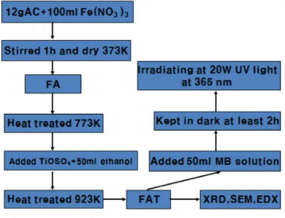
Activated carbon was mulled for 5 hours, and treated with phosphoric acid (0.1M), drying at 373 K for 5 hours, oxidation carbon powders were prepared. 12 g oxidation carbon powders were mixed with 50 ml Fe(NO3)3 solution (0.1M). After churn 1 hour and drying at 373 K, the mixture was heat treatment at 773 K. And then Fe treated AC compounds (Fe-AC) were formed.2.3. Preparation of Fe-AC/TiO
2photocatalysts
A typical procedure is as follows: Due to TOS was easily dissolved by oxydol (H2O2) solution, 3 g, 4 g and 5 g TOS were added to 50 ml of oxydol (H2O2), respectively.
After stirring for 1 hour, TOS‐H2O2 solution was obtained.
And then 3 g Fe-AC compounds were added to each TOS-H2O2 solution, respectively. Churn the mixture for 1h. Then the supported TiO2 particles were first dried at 323 K for 6 hours and calcined at 773 K for 2 hours each. Fe-AC/TiO2 photocatalyst composites were obtained.
The designations for different prepared materials are summarized in Table 1. (FAT1, FAT2, FAT3). Fig. 1 is the course for preparation of Fe‐AC/TiO2 photocatalysts.
[Fig.1] The preparation procedure of Fe-AC/TiO2
photocatalysts.
2.4. Characterization of prepared photocatalyst
Several techniques were used for characterization of the Fe-AC/TiO2 composites. Crystalline phase, particle size and morphology of photocatalysts nanocrystals were investigated by specific surface area (BET), X-ray diffraction analysis (XRD), energy dispersive X-ray spectroscopy (EDX), and scanning electron microscopy (SEM), respectively. The specific surface area (BET) was determined by N2 adsorption measurements at 77 K (Monosorb, USA). XRD (Shimatz XD‐D1, Japan) analysis using CuKa (λ=1.5418 Å) radiation was performed to assess the crystalline phases. EDX spectra were used for the elemental analysis of the samples. SEM measurements were performed using a JEOL apparatus (JSM‐5200 JOEL, Japan) operating at 10 kV on specimens upon which a thin layer of gold or carbon had been evaporated. The pH meter (HI8134) was purchased from Hanna instruments of Italy.
2.5. Photocatalytic activity measurements
To characterize the photocatalytic activity of thenanocomposites under visible light and UV light irradiation, experiments on photodegradation of methyl blue, a common contaminant in wastewater, were carried out at room temperature. The photocatalytic test‐reaction chosen to characterize the samples which contained different amount of titania was the total degradation of methylene blue solution selected as a model organic pollutant. The static batch photo-reactor was a cylindrical flask. Photoactivity of the catalysts was measured for the methylene blue solution decomposition in water.
Photocatalysts in the mass of 0.03 g were added to the 50 ml of methylene blue solution with concentration of 1.0×10‐4 M and that mixture was lay for at least 2 hours in the dark until saturation of adsorption. After adsorption, photodecomposition of MB solution was performed under visible light. place the photo-reactor under visible light (35W led lamp) irradiation for 10 min, 30 min, 60 min, 90 min, 120 min and 150 min, respectively to research the degradation of MB solution. The experiment of UV light was processed as described above.
3. Results and discussion
3.1 X-ray diffraction
Fig. 2 exhibits the XRD patterns of Fe treated AC/TiO2
compounds. XRD analysis was carried out to confirm the TiO2 polymorphs and their crystalline phases. A is anatase, R is rutile, F is iron. Fig. 2 shows the X-ray diffraction patterns of AC/TiO2 with different dosage of TiO2. The peaks corresponding to the anatase TiO2 phase appeared at 25.3, 37.5, 48.0, 53.8, 54.9, and 62.5 were diffractions of (101), (004), (200), (105), (211), and (204) planes of anatase, it indicated that the prepared TiO2
existed in an anatase phase [21]. From Fig. 2 there are six typical peaks with 2θ values of 27.52°, 36.20°, 41.44°
and 54.48°, corresponding to (110), (101), (111) and (211) crystal planes of rutile TiO2 [22], respectively.
Further observation shows that with increasing TiO2, XRD peak intensities of anatase steadily become strong and narrow. There is not any peak assigned to the iron oxide nor is FexTiO2y observed. One possible reason for this might be that the amount of Fe is too low to be detected by XRD. On the other hand, the radius of Ti4+ (0.68 Å)
and Fe3+ (0.64 Å) is almost the same; therefore, the Fe3+
ions could enter into the crystal structure of titania and be located at the interstices or occupy some of the lattice sites of the TiO2, forming an iron‐titanium solid solution [23, 24]. The XRD patterns of the Fe-TiO2 show the same feature as anatase. Therefore, the TiO2 units are successfully introduced into the Fe-AC structure rather than existing in separate free solid phase [25]. The molecular distribution of TiO2 units into Fe‐AC compound and forming a stable composite could be occurred during the process of hydrolysis of TOS in the presence of oxydol solution (0.1M).
3.2 Elemental analysis
Energy Dispersive X-ray (EDX) detector observation (Fig. 3) revealed the inclusive element of the prepared samples. FAT3 has the most TiO2 units because the largest TOS was added in the process of sample preparation. The other element like Si, Cu, Zn were found in Fig. 3, maybe these elements are content in phosphoric acid. The spectra show the presence of C, O, and Ti, as major elements, with strong Fe peaks. The peak of Ti was increased with enhance the dosage of TOS during the process of prepare Fe-AC/TiO2 composites.
3.3 Surface characteristics
The surface characteristics of Fe-AC/TiO2 compounds are shown in Fig. 4 It’s the catalyst surface morphology by scanning electron microscopy (SEM) at 1000 magnifications and 3000 magnifications, can be clearly seen that AC was covered with TiO2 particles.
[Fig.2] XRD for Fe‐AC/TiO2 compounds. A: anatase, R:
rutile, F: Fe and titanium.
(a)
(b)
(c)
[Fig.3] EDX elemental microanalysis of Fe‐AC/TiO2
compounds (a) FAT1, (b) FAT2, and (c) FAT3
(a) (b)
(c) (d)
(e) (f)
[Fig.4] SEM images of Fe‐AC/TiO2 compounds:(a), (c), and (e) 1,000 times magnification for FAT1, FAT2, FAT3; (b), (d), and (f) 2000 times magnification for FAT1, FAT2, FAT3
TiO2 particles were regularly dispersed on the AC surfaces and some large clusters were found with an irregular agglomerate dispersion. It is considered that a good dispersion of small particles on the AC surface could provide evidence for the existence of more reactive sites for photodecompositon of the dye. From SEM image we can see FAT3 compounds has the most TiO2 particles.
In Figure (e) and (f) the AC particles were covered with larger TiO2 particles which is more than that of FAT1 and FAT2 compounds.
The surface areas were calculated using N2 adsorption measurements at 77 K. Table 1. shows that increased the mass of TiO2 units which is the inclusion into Fe-AC structure, the surface areas decreased. The smaller crystallite size caused the larger the specific surface areas.
The Fe-doping usually inhibits the growth of TiO2
crystallite and heat‐treatment of the samples at 773K cause the aggregation and sintering of TiO2 particles [26].
[Table 1] BET surface areas for Fe‐AC/TiO2 compounds Sample name Surface area
FAT1 533.333 m2/g
FAT2 456.923 m2/g
FAT3 399.830 m2/g
3.4 Photocatalytic decomposition of MB
Initially, control tests on degradation of MB solution (ca. 1×10‐4 M) were carried out in specified conditions described below: the date are obtained following a two step experiments. The first step is placed in dark to adsorb dye, and the second step is under light to degrade MB in solution. Because the surface area for FAT1 is much lower than FAT2, FAT3, so its adsorptive property is lower than other samples at our experiments condition.From Fig. 5 a mass of the initial amounts of the dye are adsorbed onto all the adsorbents (catalytsts) following a 120 mins adsorption in the dark. Photocatalytic ability of our prepared catalysts in adsorbing and degradation of MB has been shown in Fig. 5.
Fig. 5 shows the time series of MB (methylene blue) photodegradation using Fe-AC/TiO2 compounds under UV light and visible light irradiation, respectively. Image (a) is the curves for MB solution photodegradation under UV light. From Image (a) we can see FAT3 has the best photocatalytic activity, because FAT3 have the most
-150 -100 -50 0 50 100 150
0.0 0.2 0.4 0.6 0.8 1.0
MB solution(A)
Time(min)
FAT1 FAT2 FAT3
Adsorbtion of MB
Degradation of MB
(a)
-150 -100 -50 0 50 100 150
0.0 0.2 0.4 0.6 0.8 1.0
Time(min)
MB solution(A)
Degradation of MB Adsorbtion of MB
FAT1 FAT2 FAT3
(b)
[Fig.5] MB solution on time of adsorption and light irradiation for Fe‐AC/TiO2 compounds. (a) is irradiation under UV light, (b) is irradiation under visible light.
content of TiO2, which can degradation the dye as a photocatalyst. It is clearly show a sharp decrease in concentration of MB in the initial 120 mins for Fe-AC/TiO2 compounds which is due to adsorption. Then the reduction of the dye concentration is continued with an oppositely gentle slope which is due to photodereadation. (b) is the curves for MB solution photodegradtion under visible light. We found that Fe-AC/TiO2 compounds had a good photocatalytic activity under visible light irradiation, although TiO2 is only active under UV light, because of its relatively large band gap energy of TiO2 (3.2 eV) [27]. Prepared Fe3+ ion treated AC/TiO2 compounds showed a high activity for the photocatalytic under visible light due to the red shift toward the visible range in this present work [28-31]. In
the first case, it was seen that the electronic properties of the photocatalyst gets modified to a large extent and the photocatalyst starts showing absorption of light in the visible region of the spectrum. The doped TiO2
semiconductor with metal ions from the transition series such as V, Cr, Mn and Fe brings about a red shift in the absorption pattern of the TiO2 semiconduction catalyst.
Such red shifts were observed only after calcination of the metal ion implanted in the TiO2 semiconduction [32, 33].
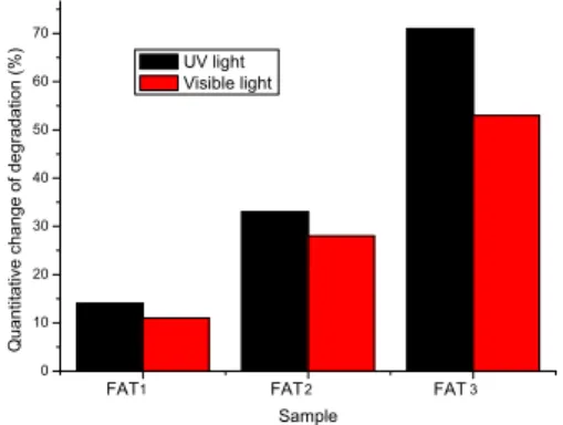
Fig. 5 can not clearly explain the contrast between two kind of methods, so we made the Fig. 6. In Fig. 6, the column is express the quantitative change of MB solution during under the light irradiation for 150 minutes. Fig. 6 is the quantitative change of the MB solution degradation, using Fe-AC/TiO2 compounds under both UV light and visible light irradiation. Fig. 6 shows the Fe-AC/TiO2
compounds have a high activity for the photocatalytic under UV light and visible light, but the degradation of MB under the UV light is batter then under visible light.
Visible light is between 390nm to 780nm, UV light is below 400nm. The onset of the absorption edge for pure TiO2 is ca. 390 nm, which is consistent with the intrinsic bandgap absorption of pure anatase TiO2 (~3.2 eV) [34].
It is apparent that the diffuse reflectance spectra of all Fe-TiO2 grains exhibit a red shift ankelid increased absorption in the visible‐light range [35]. On the inner of Fe-AC/TiO2 compounds the anatase phase is much more than Fe-TiO2 grains, we can find from Fig. 2. So Fe-AC/TiO2 compounds have a batter photocatalytic activity under UV light.
1 2 3
0 10 20 30 40 50 60 70
FAT FAT
FAT
Quantitative change of degradation (%)
Sample UV light Visible light
[Fig.6] Quantitative change of the MB solution degradation, using Fe‐AC/TiO2 compounds under both UV light and visible light irradiation.
4. Conclusions
The Fe ion and TiO2 units were covered on the AC surface and were developed through a deposition process of a sol‐gel method in this paper. SEM shows that Fe-AC/TiO2 composites were synthesized by immobilizing TiO2 particles on the surface of the AC and Fe studded on the composites. The XRD data shows that the Fe-AC/TiO2 composites contained a mixture of anatase and rutile phase forms. C and O with Ti, and Fe peaks were found from the EDX results. The MB decomposition processes confirmed the adsorption and photocatalytic reaction on the composites. The Fe-AC/TiO2 samples show a strong adsorption and can efficiently decompose the methylene blue (MB) solution under visible light. The iron ion acceded to photoactivator can enhance the degradation reaction rate for the MB solution. And the iron ion can reduce the band gap energy of TiO2, which made TiO2 particles active under visible light.
Fe-AC/TiO2 composites can decomposition MB molecule under UV light and visible light, and also have a good photodegradation activity.
참고문헌
[1] E. Piera, M. I. Tejedor, M. E. Zorn, et al.,
“Degradation of chlorophenols by means of advanced oxidation processes: a general review”, Appl. Catal.
B: Environ., 47, 219, 2004.
[2] A. Fujishima, K. Hashimoto, T. Watanabe, “TiO2
Photocatalysis Fundaments and Applications”, BKC, Tokyo, Japan., May 1999.
[3] V. Shah, P. Verma, P. Stopka, et al., “Decolorization of dyes with copper (II)/organic acid/hydrogen peroxide systems”, Appl, Catal, B: Environ., 46, 287, 2003.
[4] I. K. Konstantinou, T. A. Albanis, “Photocatalytic transformation of pesticides in aqueous titanium dioxide suspensions using artificial and solar light:
intermediates and degradation pathways”, Appl, Catal, B: Environ., 42, 319, 2003.
[5] T. Sauer, G. Cesconeto Neto, H. J. Jose, et al.,
“Kinetics of photocatalytic degradation of reactive dyes in a TiO2 slurry reactor”, J, Photochem, Photobiol, A: Chem., 149, 147, 2002.
[6] J. Wiszniowski, D. Robert, J. Surmacz‐Gorska, et al.,
“Solar photocatalytic degradation of humic acids as a model of organic compounds of landfill leachate in pilot‐plant experiments; influence of inorganic salts”, Appl, Catal, B: Environ., 53, 127, 2004.
[7] M. L. Cheng, W. C. Oh, “Preparation of AC/TiO2
Composites from Activated Carbon Codified by HNO3 and their Photocatalytic Activity”, Carbon Science., 8, 108, 2007.
[8] W. C. Oh, J. H. Son, F. J. Zhang, et al.,
“Fabrication of Ni‐AC/TiO2 composites and their photocatalytic activity for degradation of methylene blue”, J, Kor, Ceram, Soc., 46,1, 2009.
[9] M. Ni, M. K. H. Leung, D. Y. C. Leung, et al., “A Review and Recent Developments in Photocatalytic Water‐splitting using TiO2 for Hydrogen Production,”
Renew Sustain Energy Rev., 11, 401, 2007.
[10] W. Choi, A. Termin, M. R. Hoffmann, “The Role of Metal‐ion Dopants in Quantum‐sizen TiO2: Correlation Between Photoreactivity and Charge‐carrier Recombination Dynamics,” J, Phys, Chem., 98, 13669, 1994.
[11] I. Justicia, P. Ordejon, G. Canto, et al., “Designed Self‐doped Titanium Oxide Thin Films for Efficient Visible‐light Photocatalysis,” Adv, Mater., 14 1399, 2002.
[12] M. Anpo, “Utilization of TiO2 Photocatalysts in Green Chemistry”, Pure, Appl, Chem., 72 1265, 2000.
[13] H. Yamashita, M. Hatada, J. Misaka, et al.,
“Application of Ion Beam Techniques for Preparation of Metal Ion‐implanted TiO2 Thin Film Photocatalyst Available under Visible Light Irradiation: Metal Ion‐
implantation and Ionized Cluster Beam Method”, J, Synchrotron, Radiat., 8 569, 2001.
[14] F. J. Zhang, M. L. Chen, W. C. Oh, “Synthesis and Characterization of CNT/TiO2 Photoelectrocatalytic Electrodes for Methlene Blue Degradation”, Kor, J, Mater, Res., 18, 583, 2008.
[15] J. C. Yu, J. G. Yu, W. K. Ho, et al., “Effects of Fe‐ Doping on the Photocatalytic Activity and Microstructures of Nanocrystalline TiO2 Powders”, Chem, Mater., 14, 3808, 2002.
[16] R. S. Sonawane, B. B. Kale, M. K. Dongare,
“Preparation and Photo‐catalytic Activity of Fe–TiO2
Thin Films Prepared by Sol–gel Dip Coating”, Mater, Chem, Phys., 85, 52, 2004.
[17] F. S. Zhu, J. Zhu, W. Zheng, et al., “Characterization
of Fe–TiO2 Photocatalysts Synthesized by Hydrothermal Method and Their Photocatalytic Reactivity for Photodegradation of XRG Dye Diluted in Water”, J. Mol. Catal. A., 216, 35, 2004.
[18] J. Arãna, J. M. Dõna‐Rodr´ıguez, E. Tello Renďon, et al., “TiO2 Activation by Using Activated Carbon as a Support. Part II. Photoreactivity and FTIR Study”, Appl, Catal, B: Environ., 44, 153, 2003.
[19] M. H. Zhang, J. G. Yu, “Effect of Preparation Methods on the Structure and Catalytic Performance of TiO2/AC Photocatalysts”, J, Hazard, Mater., 153, 827, 2008.
[20] J. G. Yu, J. C. Yu, M. K. P. Leung, et al.,
“Preparation and Photocatalytic activity of Fe‐doped Mesoporous Titanium Dioxide Nanocrystalline Photocatalysts”, Mater, Chem, Phys., 93, 159, 2005.
[21] W. C. Oh, A. R. Jung, W. B. Ko, “Characterization and Relative Photonic Efficiencies of a New Nanocarbon/TiO2 Composite Photocatalyst Designed for Organic Dye Decomposition and Bactericidal Activity”, Mater, Sci, Eng., C 29 1338, 2009.
[22] L. Bing, M. X. Song, T. L. Zhou, et al., “Band gap calculation and photo catalytic acticity of rare earths doped rutile TiO2”, Jour, Rare, Earths., 27, 461, 2009.
[23] M. L. Chen, F. J. Zhang W. C. Oh, “Photocatalytic Degradation of Methylene Blue by CNT/TiO2
Composites Prepared from MWCNT and Titanium n‐
butoxide with Benzene”, J, Kor, Ceram, Soc., 45, 651, 2008.
[24] C. Y. Wang, C. Bottcher, D. W. Bahnemann, et al.,
“A Comparative Study of Nanometer Sized Fe(III)‐
doped TiO2 Photocatalysts: Synthesis, Characterization and Activity”, J. Mater. Chem., 13, 2322, 2003.
[25] M. H. Zhou, J. G. Yu, B. Cheng, “Effects of Fe‐
doping on the Photocatalytic Activity of Mesoporous TiO2 Powders Prepared by an Ultrasonic Method”, J, Hazard, Mater., B 137, 1838, 2006.
[26] J. G. Yu, J. C. Yu, M. K. P. Leung, et al.,
“Effects of acidic and basic hydrolysis catalysts on the photocatalytic activity and microstructures of bimodal mesoporous titania”, J, Catal., 217, 69, 2003.
[27] J. Liqianga, S. C. Xiaojuna, X. Weimina, et al.,
“The Preparation and Characterization of Nanoparticle TiO2/Ti Films and their Photocatalytic Activity”, J, Phys, Chem, Solids., 64, 615, 2003.
[28] Y. G. Go, M. L. Chen, F. J. Zhang et al.,
“Fabrication of Zn‐treated ACF/TiO2 Composites and
Their Photocataytic Activity for Degradation of Methylene Blue”, Kor, J, Mater, Res., 19, 142, 1994.
[29] N. Murakami, T. Chiyoya, T. Tsubota, et al,
“Switching Redox Site of Photocatalytic Reaction on Titanium(IV) Oxide Particles Modified with Transition
‐metal Ion Controlled by Irradiation Wavelength”, Appl, Catal, A, Gen., 348, 148, 2008.
[30] W. Huang, X. Tang, I. Felner, et al., “Preparation and Characterization of FexOy‐TiO2 via Sonochemical Synthesis”, Mater, Res, Bull., 37, 1721, 2002.
[31] K. S. Suslick, S. B. Choe, A. A. Cichowlas, et al.,
“Sonochemical Synthesis of Amorphous Iron”, Nature, 353, 414, 1991.
[32] R. Dholam, N. Patel, M. Adami, et al., “Hydrogen Production by Photocatalytic Water‐splitting using Cr or Fe‐doped TiO2 Composite Thin Films Photocatalyst”, J, Hyd, Ene., 34, 5337, 2009.
[33] E. S. Mora, E. G. Barojas, E. R. Rojas, et al.,
“Morphological, Optical and Photocatalytic Properties of TiO2–Fe2O3 Multilayers,” Sola, Ene, Mater. Sol, Cells, 91, 1412, 2007.
[34] B. Xin, Z. Y. Ren, P. Wang, et al., “Study on the Mechanisms of Photoinduced Carriers Separation and Recombination for Fe3+–TiO2 Photocatalysts”, App, Surface Sci., 253, 4390, 2007.
[35] J. G. Yu, H. G. Yu, C. H. Ao, et al, “Preparation, Characterization and Photocatalytic Activity of in Situ Fe‐doped TiO2 Thin Films”, Thin Solid Films., 496, 273, 2006.
Won-Chun Oh
[Regular member]• Feb. 1986 : Dankook Univ., Dept. of Chem., MS
• Feb. 1995 : Dankook Univ., Dept. of Chem., PhD
• Apr. 2008 ~ Current : Anhui Uni. of Arch. in China, Dept.
of New Mat. Sci & Eng, Guest Professor
• Feb. 1998 ~ Current : Hanseo Univ., Dept. of Adv.
Mat. Sci & Eng, Professor
<Research Interests>
Carbon materials, Photocatalysis
Ze-Da Meng
[Associate member]• Feb. 2008 : Hanseo Univ., Dept.
of Adv. Mater. & Sci. Eng., MS
<Research Interests>
Carbon materials, Photocatalysis
Kan Zhang
[Associate member]• Feb. 2008 : Hanseo Univ., Dept.
of Adv. Mater. & Sci. Eng., MS
<Research Interests>
Carbon materials, Photocatalysis