I. 서 론
생명체에 있어 필수 영양소로서 중요한 역할을 하 는 철분 (iron)은 세균의 성장 및 대사에도 영향을 미쳐 감염성 질환의 발병과 진행에 있어 중요한 요 소로작용한다1-3). 일반적으로 숙주 내에는 세포 내 부 및 외부에 철분이 풍부하게 존재하지만 항균기전 의 일환으로 세균에 대해서는 이에 대한 접근이 매 우 제한되어 있다. 예를 들어, 숙주 내 에 존재하는 대부분의 철분은 cytochrome, hemoglobin, my- oglobin, 그리고 hemopexin 같은 heme 함유 단 백질의 형태로 또는 ferritin과 hemosiderin으로 숙주 세포 내에 은폐되어 위치하고 있다1-3). 세포 용 해의 결과 이들이 유리되어 외부로 방출될 수 있으 나, 혈장 내에 존재하는 단백질에 의해 신속히 결합 되어 균주가 이를 용이하게 활용하지는 못하는데, 예를 들어 적혈구의 용해 과정에서 유리되는 hemo- globin과 그것의 분해 과정에서 유리되는 heme (hemin)은 각각 숙주의 haptoglobin과 hemo- pexin 및 albumin에 단단히 결합하여 그들의 제거
와 숙주에의한 재활용에 기여하게 된다4-9).1) 한편, 세포 외부에 존재하는 소량의 철분도 transferrin과 lactoferrin에 결합되어 균주에 의 한 활용이 제한된다1-3). Transferrin과 lacto- ferrin은 혈액 및 세포 간질과 외분비물에 각각 존 재하는 당단백질로 철분의 킬레이션과 이동 및 교환 에 관여한다. 이로 인해 숙주 내에서 실제 활용 가능 한 유리 철분 또는 hemin의 농도는 매우 낮아, 유 리 철분의 경우 그 농도는 단지 10-18M 정도로1), 이 는 대부분의 세균에 있어 성장에 필요한 수준인 10-6M에 크게 미달하여 세균의 성장을뒷받침하는데 있어 일반적으로 불충분하다.
병원성 세균들은 이러한 숙주 내 환경에서 서식하 고 병원성을 발휘하기 위해, 필수 영양소인 철분을 숙주로부터 획득하기 위한 고도의 친화력을 갖는 철 분 획득 기전들을 소유하고 있다. 철분 획득을 위한 세균의 기전들로는 siderophore, transferrin 및 lactoferrin 결합 단백질, 그리고 hemin 결합 단백 질 등이 있으며, 이 과정에서 철분에 의해 조절되는 세포막 단백질 (iron-regulated membrane pro-
* 이 논문은 2002년도 부산대학교병원 의학연구소 연구비 (2002-13)에 의하여 연구되었음.
* 교신저자:김성조, 부산광역시 서구 아미동 1-10 부산대학교 치과대학 치주과학교실, 우편번호:602-739
대한치주과학회지 : Vol. 36, No. 1, 2006
Porphyromonas gingivalis, Prevotella intermedia, 그리고 Prevotella nigrescens에서의 hemin 결합
단백질에 대한 연구
김성조
부산대학교 치과대학 치주과학교실
teins)이 중요한 역할을 한다10-15).
In vitro에서의 세균 성장 과정에서 철분이 억제 되는 경우 새로운 세포막단백질이 발현됨이 몇몇 균 주에서 보고된 바 있는데, 이러한 철분에 의해 조절 되는 세포막 단백질들은 siderophore를 위한 iron transport system으로 작용하거나, transferrin 과 lactoferrin을 결합하는 수용체로 작용할 수도 있고, hemin이나 porphyrin analog를 결합하는 수용체로 작용할 수 있다16-21). 실제로 몇몇 다른 균 주들에서의 연구 보고에 의하면 hemin에 의해 조절 되는 세포막 단백질 (hemin-regulated mem- brane proteins)의 일부가 hemin 획득에 있어 중 요한 역할을 한다20,22-38).
Porphyromonas gingivalis, Prevotella in- termedia, 그리고 Prevotella nigrescens를 포함 하는 black-pigmented Bacteroides는 유리 철분 을 활용하지는 못하고, siderophore를 생성하지 않 으며, hemin 의존성으로 철분 공급원으로써 hemin 이 필수적인데, 이는 hemin과 protoheme의 구성 요소인 porphyrin ring을 생성하는 능력이 이 균주 에 있어 결여되어 있는 것에 기인하는 것으로 여겨 지며, 이들 균주는 electron transport system에 있어 주요한 구성 요소 중의 하나인 cytochrome을 hemin으로부터 형성할 수 있다. Hemin은 P. gin- givalis를 포함한 black-pigmented Bacteroides 에 있어서의 철분요구의 전부를 충족할 수 있는 것 으로 알려져 있다.
P. gingivalis와 P. intermedia는 치주질환 주 요 병인균주 중의 하나로 성인성 치주염 환자의 치 주낭 내에서 우세하게 존재한다39-41). P. inter- media는 또한 급성괴사성 궤양성 치은염과 임신성 치은염과도 연관이 있다42,43). P. nigrescens는 P.
intermedia와 밀접한 연관을 가지고있으며, Shah 와 Gharbia44)에 의해 과거 P. intermedia로 분류 되었던 strain들 중에서, 생화학적 특성 등에 의거 하여, Prevotella genus 내의 별도의 새로운 종 (species)으로 분류된 바 있다. P. intermedia와 P. nigrescens는 용혈 및 혈구응집 활성45) 그리고
beta-lactamase 생성 및 항균제에 대한 감수성 등
46,47)에 있어 상이함이 보고된 바 있고, 이 두가지
균종은 SDS-PAGE와 PCR등의 방법에 의해 구분 될 수 있다48-51).
본 연구는 hemin 의존성을 갖는치주질환 주요 병인균주인 P. gingivalis, P. intermedia, 그리고 P. nigrescens의 hemin 결합 세포막 단백질의 특 성을 비교 분석하기 위해 수행되었다. 본 연구는 이 들 균주에서의 porphyrin 생리 및 hemin 획득 기 전을 밝히는데 있어중요한 의의가 있으리라 사료된 다.
Ⅱ. 연구재료 및 방법
1. 균주 및 배양 조건
사용된 균주는 P. gingivalis 381, P. inter- media ATCC 25611, 그리고 P. nigrescens ATCC 33563이었으며, 이들 균주를 enriched Trypticase soy agar, 또는 2.1% (W/V) Mycoplasma broth base (BBL, Becton Dickinson, Cockeysville, MD)에서, 1 μg/ml의 menadione 과 5 μg/ml의 hemin을 첨가하여, 37℃의 혐기성 조건 하에서 배양하였다. Hemin 제한 조건에서의 배양을 위해서는, hemin을 포함하지 않은 액체 배 지에서 최소 5회 계대 배양하여, 균주 세포 내부에 저장되어 있는 hemin을 고갈시켰다. 세균의 성장은 이중 배양 후 660 nm 파장에서의 흡광도를 측정하 여 결정하였다. 균주 배양의 오염 여부는 Gram 염 색 후 검경하여 판단하였다. 오염되어 있는 철분과 hemin을 제거하기 위해 모든 유리 기구들은 chro- mic acid와 deionized water로 세척한 후 사용하 였다.
2. 세포막의 분리
Early stationary phase의 균주를 원심 분리 (12,000 x g, 20 min at 4℃)하여 회수하여, 냉각
된 phosphate-buffered saline (PBS, pH 7.2) 으로 3회 세척한 후 각각 2 mM의 phenyl- methylsulfonyl fluoride (PMSF), Na-P-Tosyl- L-lysine chloromethyl ketone (TLCK), 그리고 benzamidine을 포함하고 있는 PBS에 분산시켰다.
그 후 sonicator로 세포를 파쇄하고, 저속 (10,000 x g, 30 min) 및 고속 (200,000 x g, 2 hr) 원심 분리를 시행하여 세포막을 분리하였다. 분리된 세포 막은 PBS에 녹여 -20℃에 보관하였다.
3. 단백질 정량
Kennel과Holt52)에 의해 변형된 bicinchoninic acid (BCA) assay (Pierce, Rockford, IL)법에 의거하여 단백질 농도를 결정하였다. 이는 micro- titer plate (flat-bottomed)에서의 단백질 정량을 위해 변형된 방법으로, 이를 간략히 소개하면, 20 μl 의 시료를 microtiter well에 넣고 증류수로 2배 연속 희석한 후, 200 μl의 protein assay reagent 를 각 well에 가하여 30분간 실온에 위치시키고, plate reader (570 nm)로 측정하였다. 표준 단백 질로는 bovine serum albumin이 사용되었다.
4. Polyacrylamide Gel Electrophoresis
단백질 분포의 평가를 위해서는 Laemmli53)의 discontinuous sodium dodecyl polyacrylamide gel electrophoresis (SDS-PAGE)를 실시하였고, 세포막 단백질 중에서 heme-associated perox- idase activity를 갖는 밴드를 확인하기 위해서는 lithium dodecyl sulfate (LDS) PAGE를 시행하 였다. 12% acrylamide separating gel과 4%의 acrylamide stacking gel을 이용하였으며, verti- cal slab gel apparatus (Hoefer Scientific, San Francisco, CA)와 minigel apparatus (Mini PROTEANR II Dual Slab Cell, Bio-Rad, Richmond, CA)를 이용하여 30 mA에 서 전기영동 하였다. 각 lane에는 통상 20 μg 및
60 μg의 단백질을 각각 적용하였으며, low molec- ular weight standards (Bio-Rad, Richmond, CA)를 활용하였다. 전기영동 후 Coomassie bril- liant Blue-R-250 (CBB)으로 염색하고, 각 세포 막 단백질밴드의 분자량 계산은 linear regression analysis에 의거하였다. 세포막 단백질의 hem- in-associated peroxidase activity를 결정하기 위해 LDS-PAGE gel을 tetramethylbenzidine (TMBZ, Sigma Chemical Co., St. Louis, MO) 으로 염색하였다. TMBZ 염색은 Stugard 등31)의 방법에 의거하여 수행하였다. Reducing agent인 2'-mercaptoethanol의 존재가 전기영동에 미치는 영향도 분석하였다.
5. Hemin 결합 단백질의 순수분리
Reference well을 갖는 preparative comb과 12% separating gel을 이용하여, 세포막 단백질 (passage 5)을 전기영동 한 후, gel의 양단을 잘라 내어 CBB로 염색하였다. 이 염색 된 gel을 tem- plate로 활용하여, 분리하고자 하는 hemin 결합 단 백질 밴드 (결과 참조)를 절제한 후, Hager와 Burgess의 방법55)에 의거하여 단백질을 용출하였 는 바 그 방법은 다음과 같다. 잘라낸 gel 밴드를 잘 게 부순 후, 0.1% SDS, 0.05 M Tris-HCl, 0.1 mM EDTA, 5 mM DTT, 그리고 0.2 M NaCl을 포함하고 있는 elution buffer를 가하여, 실온에서 1시간 씩 2차에 걸쳐 단백질을 용출하였다. 그 후 Centricon-10 microconcentrator (10,000 mo- lecular weight cutoff; Amicon Div., Beverly, MA)로 용출액을 농축하였다. 분리된 단백질의 순도 는 SDS-PAGE로 확인하였다.
6. N-terminal 아미노산 서열의 결정
순수분리된 단백질을 SDS-PAGE 후 Polyvinylidine difluoride (PVDF) membrane (ProBlot, Applied Biosystems, Foster City, CA)에 transfer하고
55), Amido black으로 염색한 후 해당 밴드를 잘라 내었으며, Applied Biosystems Model 177A gas-liquid phase sequanator (coupled to an on-line HPLC model 120A analyzer)를 이용하 여 N-terminal 아미노산 서열을 결정하였다.
7. Cyanogen bromide digestion
P. gingivalis의 24 kDa hemin 결합 단백질 (결과 참조)은 N-terminus가 봉쇄되어 cyanogen bromide (CNBr)로 절단한 후 아미노산 서열을 결 정하였다. 간략히 소개하면, acetone으로 침전시킨 24 kDa 단백질에 70% formic acid에 녹인 CNBr 을 가하여 실온에서 16시간 방치한 후, N2와 SpeedVac SC 100 (Savant)을 이용하여 건조하 였다.
Ⅲ. 결 과
각 전기영동 조건에서 정상적으로 7.7 μM의 hemin을 공급하여 배양한 경우와 hemin을 고갈시 킨 경우 (passage 5)에서의 단백질 밴드의 분포와 수는 매우 유사하였다 (Figure 1). 그러나, pas- sage 5의 P. gingivalis에서 약 24 kDa (unheated
30 kDa), 그리고 passage 5의 P. intermedia와 P. nigrescens에서는 약 50 kDa의 단백질 밴드가 현저히 강화되어 발현되었다 (Figure 1). LDS- PAGE gel을 TMBZ로 염색한 결과, passage 5의
(A) (B) (C)
1 2 3 4 5 1 2 3 4 5 1 2 3 4 5
Figure 1. SDS-PAGE of cell envelopes from P. gingivalis (A), P. intermedia (B), and P. ni- grescens (C). Lane 1, M.W. standards; Lane 2 and 3, unheated samples; Lane 4 and 5, samples heated at 100℃ for 5 min; Lane 2 and 4, cells grown in 7.7μM hemin; Lanes 3 and 5, cells grown wit hout hemin (passage 5).
(A) (B)
1 2 3 2 3
Figure 2. Coomassie Brilliant blue stained (A) and tetramethylbenzidine (TMBZ) stained (B) LDS-PAGE of cell envelopes from P.
gingivalis. Protein (70㎍) was applied to each lane following incubation with hemin.
Lane 1, low M.W. standards; Lane 2, cells grownin7.7μM hemin; Lane 3, passage 5.
P. gingivalis 세포막단백질은 30 kD에서 hem- in-associated peroxidase activity를 보였다 (Figure 2).
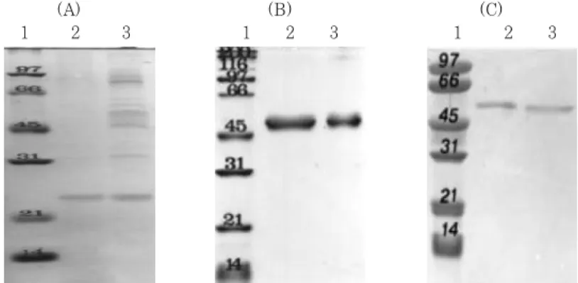
Passage 5의 P. gingivalis에서 약 24 kDa (unheated 30 kDa) hemin 결합 단백질, 그리고 passage 5의 P. intermedia와 P. nigrescens에 서는 약 50 kDa의 hemin 결합 추정 단백질을 각 각 분리하였다. 분리된 단백질은 여타의 단백질에 오염되지 않고 1개의 밴드로 구성되어 있었다 (Figure 3). 분리된 각각의 단백질은 환원재인
2'-mercaptoethanol의 존재 유무와 상관없이 동일 한 전기영동 양상을 보였다 (Figure 4).
P. gingivalis의 24 kDa hemin 결합 단백질을 CNBr로 처리한 결과 최소 3개의 단백질 밴드 (12 kDa, 17 kDa, 그리고 20 kDa)가 확인 되었으며, 이들 밴드의 내부 아미노산 서열이 표 1 (A)에 제시 되어 있다. P. intermedia와 P. nigrescens에서 의 약 50 kDa hemin 결합 추정 단백질의 N-ter- minal 아미노산 서열은 표 1 (B)와 (C)에 제시되 어 있다.
(A) (B) (C)
1 2 3 1 2 3 1 2 3
Figure 4. Electrophoretic mobility of putative hemin-binding proteins from P. gingivalis (A), P.
intermedia (B), and P. nigrescens (C) in the presence and absence of reducing agent, 2-mercaptoethanol. Lane 1, M.W. standards; Lane 2, sample with treatment buffer contain- ing 2-mercaptoethanol; Lane 3, sample without 2-mercaptoethanol.
(A) (B) (C)
1 2 3 4 5 6 1 2 6 1 2 6
Figure 3. SDS-PAGE analysis of the purification of putative hemin-binding proteins from P.
gingivalis (A), P. intermedia (B), and P. nigrescens (C). Lane 1, M.W. standards; Lane 2, cell envelope fract ion; Lane 3, 1% CHAPS-soluble fract ion of P. gingivalis; Lane 4, iso- lation of 30 kDa protein of P. gingivalis; Lane 5, 100℃ heated isolation 30 kDa protein of P. gingivalis; Lane 6, purified putative hemin-binding protein proteins.
Ⅳ. 총괄 및 고찰
병원성 세균들은 숙주 내 환경에서 서식하고 병원 성을 발휘하기 위해, 필수 영양소인 철분을 숙주로 부터 획득하기 위한 고도의 친화력을 갖는 철분 획 득 기전들을 소유하고 있다. 본 연구는 hemin 의존 성을 갖는 치주질환 주요병인균주인 P. gingivalis, P. intermedia, 그리고 P. nigrescens의 hemin 결합 세포막 단백질의 특성을 비교 분석하기 위해수 행되었다.
본 연구에서는 hemin이 고갈된 상태인 passage 5의 P. gingivalis에서 약 24 kDa (unheated 30 kDa), 그리고 passage 5의 P. intermedia와 P.
nigrescens에서는 약 50 kDa의 단백질 밴드가 현 저히 강화되어 발현되었다. Smalley 등30)과 Bramanti와 Holt38)는 몇몇 치주질환 병인 균주에 서의 hemin 조절 세포막 단백질이 hemin 결합 단 백질로서의 역할을 수행할 수 있음을 보고한 바 있 다. 이에 P. gingivalis에서 24 kDa (unheated 30 kDa) hemin 조절 단백질, 그리고 P. inter- media와 P. nigrescens에서의 50 kDa hemin 조 절 단백질은 이들 균주에서의 hemin 결합에 관여하
는 단백질로 추정할 수 있다. 또한, 본 연구에서는 passage 5의 P. gingivalis의 30 kD 세포막 단백 질에서 hemin-associated peroxidase activity를 확인하였다. 이는 P. gingivalis의 경우 30 kD 세 포막 단백질이 hemin 결합에 실질적으로 간여함을 의미한다.
본 연구에서 분리된 P. gingivalis의 24 kDa (unheated 30 kDa) hemin 결합 단백질, 그리고 P. intermedia와 P. nigrescens의 50 kDa hem- in 결합 추정 단백질의 electrophoretic mobility 는 환원제인 2'-mercaptoethanol의 존재 유무에 영향을 받지 않았는 바, 이는 이 단백질에 disulfide bond가 존재하지 않음을 보여준다.
N-terminal 아미노산 서열을 GenBank 자료에 서 분석한 결과에 의하면, P. intermedia의 50 kDa hemin 결합 추정 단백질은 Streptococcus intermedius의 Enolase와 아미노산 서열이 일치 하였으며, P. gingivalis의 24 kDa hemin 결합 단백질과 P. nigrescens의 50 kDa hemin 결합 추정 단백질은 기존의 보고된 단백질과 상동성을 갖 지 않는 새로운 단백질로 추정된다.
본 연구는, hemin 의존성을 갖는 치주질환 주요 Table 1. N-terminal amino acid sequence anaysis of putative hemin-binding proteins from P.
gingivalis (A), P. intermedia (B), and P. nigrescens (C)
(A)
Fragments Amino acid sequence
20 kDa DQATSVPTDG(X)Y(X)TVD(X)KLGRITVK
17 kDa GPDG(H)ZMEYEE
12 kDa EYEEQGFSEVITGKKNAQGFAA(X)G(X)LEF(S)
*(X), unknown; ( ), assume to be correct amino acid.
(B)
Fragment Amino acid sequence
50 kDa SIITDVYAREVLDSRG
(C)
Fragment Amino acid sequence
50 kDa MEVLKIMESLEQKHP
병인균주인 P. gingivalis, P. intermedia, 그리고 P. nigrescens에서의 hemin 조절 세포막 단백질 로, hemin 결합에 관여하여 이들 균주에서의 hem- in 획득에 중요한 역할을하는 것으로 추정되는 단백 질을 확인하여 순수분리하고 몇몇 특성을 비교 분석 한 최초의 보고로 사료된다. 향후, 이 단백질에 대한 보다 심도있는 연구가 수행되어야 할 것으로 사료되 는 바이다.
V. 결론
본 연구는 hemin 의존성을 갖는 치주질환 주요 병인균주인 P. gingivalis, P. intermedia, 그리고 P. nigrescens의 hemin 결합 세포막 단백질의 특 성을 비교 분석하기 위해 수행되었다. 본 연구에서 는 hemin이 고갈된 상태인 passage 5의 P. gin- givalis에서 약 24 kDa (unheated 30 kDa), 그 리고 passage 5의 P. intermedia와 P. ni- grescens에서는 약 50 kDa의 hemin 조절 세포막 단백질이 확인되었으며, 이들은 hemin 결합에 관여 하는 것으로 추정된다. P. gingivalis의 30 kD 세 포막 단백질에서는 hemin-associated peroxidase activity가 확인되었다. N-terminal 아미노산 서열 을 분석한 결과에 의하면, P. intermedia의 50 kDa hemin 결합 추정 단백질은 Streptococcus intermedius의 Enolase와 아미노산 서열이 일치 하였으며, P. gingivalis의 24 kDa hemin 결합 단백질과 P. nigrescens의 50 kDa hemin 결합 추정 단백질은 기존의 보고된 단백질과 상동성을 갖 지않는 새로운 단백질로 추정된다.
참고문헌
1. Bullen JJ. The significance of iron in infection. Rev Infect Dis 1981;3:1127 -1138.
2. Weinberg ED. Iron withholding: a de- fense against infection and neoplasia.
Physiol Rev 1984;64:65-102.
3. Finkelstein RA, Sciortino CV, McIntosh MA. Role of iron in microbe-host interactions. Rev Infect Dis 1983;5:
s759-s777.
4. Koskelo P, Muller Eberhard U.
Interaction of porphyrins with proteins.
Semin Hematol 1977;14:221-226.
5. Laurell CB, Gronvall C. Haptoglobins.
Adv Clin Chem 1962;5:135-172.
6. Martinez JL, Delgado Iribarren A, Baquero F. Mechanisms of iron acquis- ition and bacterial virulence. FEMS Microbiol Rev 1990;75:45-56.
7. Muller Eberhard U, Morgan WT.
Porphyrin-binding protein in serum.
Ann NY Acad Sci 1975;244:624-649.
8. Seery VL, Muller Eberhard U. Binding of porphyrins to rabbit hemopexin and albumin. J Biol Chem 1973;248:3796-3800.
9. Eaton JW, Brandt, JR Mahoney.
Haptoglobin: A natural bacteriostat.
Science 1982;215:691-693.
10. Crosa JH. Genetics and molecular biol- ogy of siderophore-mediated iron trans- port in bacteria. Microbiol Lett 1989;
53:517-530.
11. Schryvers AB, BC Lee. Comparative analysis of the transferrin and lacto- ferrin binding proteins in the family Neisseriaceae. Can J Microbiol 1989;35:
409-415.
12. Gonzalez GC, DI Caamano, AB Schryvers.
Identification and characterization of a porcine-specific transferrin receptor in Actinobacillus pleuropneumoniae. Mol Microbiol 1990;4:1173-1179.
13. Yu R-H, SD Gray-Owen, J Ogunnariwo, AB Schryvers. Interraction of ruminant
transferrin receptors in bovine isolates of Pasteurella haemolytica and Haemophilus somnus. Infect Immun 1992;60:2992- 2994.
14. Schryvers AB, S Gray-Owen. Iron ac- quisition in Haemophilus influenzae re- ceptors for human transferrin. J Infect Dis 1992;165:s103-s104.
15. Ogunnariwo JA, AB Schryvers.
Correlation between the ability of Haemophilus paragallinarum to acquire ovotransferrin-bond iron and the ex- presssion of ovotransferrin-specific receptors.
Avian Dis 1992;36:655-663.
16. Griffith E. The iron uptake systems of pathogenic bacteria. In: Bullen J. J.
and Griffiths, E. eds. Iron and infection.
New York: John Willey & Sons, 1987;
69-137.
17. Neilands JB. Microbial envelope pro- teins related to iron. Ann Rev Microbiol 1982;36:285-309.
18. Schryvers AB. Identification of the transferrin and lactoferrin binding pro- teins in Haemophilus influenzae. J Med Microbiol 1989;29:121-130.
19. Tsai J, DW Dyer, PF Sparling. Loss of transferrin receptor activity in Neisseria meningitidis correlates with inability to use transferrin as an iron source. Infect Immun 1988;56:3132-3138.
20. Bramanti TE, Holt SC. Hemin uptake in Porphyromonas gingivalis: Omp26 is a hemin-binding surface protein. J Bacteriol 1993;175:7413-7420.
21. Coulton JW, Pang JCS. Transport of hemin by Hemophilus influenzae type b.
Curr Microbiol 1983;9:93-98.
22. Chu L, Song M, Holt SC. Effect of iron
regulation on expression and hem- in-binding function of outer-sheath pro- teins from Treponema denticola. Microb Pathog 1994;16:321-335.
23. Fujimura S, Shibata Y, Hirai K, Nakamura T. Some binding properties of the envelope of Porphyromonas gingiva- lis to hemoglobin. FEMS Immunol Med Microbiol 1995;10:109-114.
24. Hanson MS, Hansen EJ. Molecular clon- ing, partial purification, and character- ization of a hemin-binding lipoprotein from Haemophilus influenza type b. Mol Microbiol 1991;5:267-278.
25. Lee BC. Isolation of heamin-binding proteins of Neisseria gonorrheae. J Med Microbiol 1992;36:121-127.
26. Lee BC. Isolation of an outer membrane hemin-binding protein of Hemophilus in- fluenzae type b. Infect Immun 1992;60:
810-816.
27. Morse SA, Chen C-Y, LeFaopu A, Mietzner TA. A potential role for the major iron-regulated protein expressed by pathogenic Neisseria spp. Rev Inf Dis 1988;10:s306-s310.
28. Otto BR, Sparrius M, Verweij-van Vught AMJJ, MacLaren DM. Iron-regu- lated outer membrane protein of Bacteroides fragilis involved in heme uptake. Infect Immun 1990;58:3954-3958.
29. Grenier D. Hemin-binding property of Porphyromonas gingivalis outer membranes.
FEMS Microbiol Lett 1991; 77:45-50.
30. Smalley JW, Birss AJ, McKee AS, Marsh PD. Hemin-binding proteins of Porphyromonas gingivalis W50 grown in a chemostat under haemin-limitation. J Gen Microbiol 1993;139:2145-2150.
31. Stugard CE, Daskaleros PA, Payne SM.
A 101-kilodalton heme-binding protein associated with Congo red binding and virulence of Shigella flexneri and enter- oinvasive Escherichia coli strains. Infect Immun 1989;57:3534-3539.
32. Hanson MS, Slaughter C. Hansen E.
The hbpA gene of Haemophilus influenza type b encodes a heme-binding lip- oprotein conserved among heme-depend- ent Haemophilus species. Infect Immun 1992;60:2257-2266.
33. Scott D, Siboo IR, Chan ECS, Klitorinos A, Siboo R. Binding of hemin and congo red by oral hemolytic spirochetes. Oral Microbiol Immunol 1993;8:245-250.
34. Pendrak ML, RD Perry. Characterization of a haemin-storage reservoirs of hemin and inorganic iron in Yersinia pestis.
Infect Immun 1991;61:32-39.
35. Perry RD, TS Lucier, DJ Sikkema, RR Brubaker. Storage reservoirs of hemin and inorganic iron in Yersinia pestis.
Infect Immun 1993;61:32-39.
36. Maciver I, O'Reilly T, Brown MRW.
Porphyrin ring source can alter the out- er membrane protein profile of non- typeable Hemophilus influenzae. J Med Microbiol 1990;31:163-168.
37. Williams P, Brown MRW. Influence of iron restriction on growth and the ex- pression of outer membrane proteins in Hemophilus influenzae and H. parainfluenzae.
FEMS Microbiol Lett 1985;33:153-157.
38. Bramanti TE, Holt SC. Iron-regulated outer membrane proteins in the perio- dontopathic bacterium Bacteroids gingivalis.
Biochem Biophys Res Commun 1990;166:
1146-1154.
39. Tanner ACR, Haffer C, Bratthall GT, Visconti RA, Socransky SS. A study of the bacteria associated with advancing periodontitis in man. J Clin Periodontol 1979;6:278-307.
40. Slots J, Bragd L, Wikstrom M, Dahlen G. The occurrence of Actinobacillus acti- nomycetemcomitans, Bacteroides gingi- valis and Bacteroides intermedius in de- structive periodontal disease in adults.
J Clin Periodontol 1986;13:570-577.
41. Socransky SS, Haffajee AD. The bacte- rial etiology of destructive periodontal disease: current concepts. J Periodontol 1992;63:322-331.
42. Chung CP, Nisengard RJ, Slots J, Genco RJ. Bacterial IgG and IgM antibody tit- ers in acute necrotizing ulcerative gingivitis. J Periodontol 1983;54:557-562.
43. Kornman KS, Loesche WJ. The sub- gingival microbial flora during pregnancy.
J Periodont Res 1980;15: 111-122.
44. Shah HN, Gharbia SE. Proposal of a new species Prevotella nigrescens sp.
nov. among strains previously classified as P. intermedia. FEMS Immunol Med Microbiol 1993;6:97.
45. Okamoto M, Maeda N, Kondo K, Leung KP. Hemolytic and hemagglutinating ac- tivities of Prevotella intermedia and Prevotella nigrescens. FEMS Microbiol Lett 1999;178:299-304.
46. Matto J, Asikainen S, Vaisanen ML, Von Troil Linden B, Kononen E, Saarela M, Salminen K, Finegold SM, Jousimies Somer H. Beta-lactamase production in Prevotella intermedia, Prevotella ni- grescens, and Prevotella pallens geno- types and in vitro susceptibilities to se-
lected antimicrobial agents. Antimicrob Agents Chemother 1999;43:2383-2388.
47. Andres MT, Chung WO, Roberts MC, Fierro JF. Antimicrobial susceptibilities of Porphyromonas gingivalis, Prevotella intermedia, and Prevotella nigrescens spp. Isolated in Spain. Antimicrob Agents Chemother 1998;42:3022-3023.
48. Baumgartner JC, Bae KS, Xia T, Whitt J, David LL. Sodium dodecyl sulfate polyacrylamide gel electrophoresis and polymerase chain reaction for differ- entiation of Prevotella intermedia and nigrescens. J Endod 1999;25:324-328.
49. Conrads G, Pelz K, Hughes B, Seyfarth I, Devine DA. Optimized oligonucleotides for the differentiation of Prevotella in- termedia and Prevotella nigrescens. Oral Microbiol Immunol 1997;12:117-120.
50. Premaraj T, Kato N, Fukui K, Kato H, Watanabe K. Use of PCR and sodium dodecyl sulfate-polyacrylamide gel elec- trophoresis techniques for differentiation of Prevotella intermedia sensu stricto and Prevotella nigrescens. J Clin Microbiol 1999;37:1057-1061.
51. Guillot E, Mouton C. PCR-DNA probe assays for identification and detection of
Prevotella intermedia sensu stricto and Prevotella nigrescens. J Clin Microbiol 1997;35:1876-1882.
52. Kennel W, Holt SC. Comparative studies of the outer membranes of Bacteroides gingivalis strains ATCC 33277, W50, W83, 381. Oral Microbiol Immunol 1990;5:
121-130.
53. Laemmli UK. Cleavage of structural proteins during the assembly of the head of bacteriophage T4. Nature (London) 1970;227:680-685.
54. Hager DA, Burgess RR. Elution of pro- teins from sodium dodecyl sulfatepolya- crylamide gels, removal of sodium do- decyl sulfate, and renaturation of enzy- matic activity: results with Sigma sub- unit of Escherichia coli RNA polymerase, wheat germ DNA topoisomerase, and other enzymes. Anal Biochem 1980;109:
76-86.
55. Towbin H, Staehelin T, Gordon J.
Electrophoretic transfer of proteins from polyacylamide gels to nitrocellulose sheets:procedure and some applications.
Proc Natl Acad Sci USA 1979;76: 4350- 4354.
-Abstract-
Isolation and Partial Characterization of Hemin-binding Cell Envelope Proteins from Porphyromonas gingivalis,
Prevotella intermedia, and Prevotella nigrescens
Sung-Jo Kim
Department of Periodontology, School of Dentistry, Pusan National University
The results of this study confirm that the availability of hemin influences the expression of selected membrane proteins of Porphyromonas gingivalis, Prevotella intermedia, and Prevotella nigrescens. A 30 kDa (heated 24 kDa) hemin-binding protein whose expression is hemin regulated was identified and purified in P. gingivalis. A strong hemin-binding function was found by LDS-PAGE and TMBZ staining when P. gingivalis cells were grown under hem- in-limited conditions. A 50 kDa cell envelope associated protein, whose expression is hemin regulated, is considered to be a putative hemin binding protein from P. intermedia and P. ni- grescens, respectively. N-terminal amino acid sequence analysis of CNBr-digested 24 kDa hemin binding protein from P. gingivalis revealed that this protein belongs to a new, so far undescribed hemin-binding class of proteins. N-terminal amino acid sequence of a 50 kDa pu- tative hemin binding protein from P. intermedia was identical with Enolase from Streptococcus intermedia. Work is in progress to further characterize the molecular structure of these proteins.2)
Key words:Hemin, Cell envelope proteins, Porphyromonas gingivalis, Prevotella intermedia, Prevotella nigrescens