the advent of distraction osteogenesis (DO), there is another treatment modality available to treat midface hypoplasia. DO has lower relapse rates and better stability2. However, both these methods advance the entire maxilla sagittally, which results in increased velopharyngeal incompetence because of the further separation of the soft palate from the posterior pharyngeal wall3. Increased velopharyngeal incompetence is the biggest disadvantage of performing orthognathic surgery or Le Fort I distraction in cleft patients.
The first successful clinical application of anterior maxil- lary distraction (AMD) using an intraoral tooth-borne dis- tractor was reported by Block and Brister4 on dogs in 1994 and subsequently by Dolanmaz et al.5 on humans in 2003. In AMD, the soft palate is not moved anteriorly and therefore there is no detrimental effect on the velopharyngeal sphincter.
This makes it a suitable treatment option for midfacial hy- poplasia in patients with cleft lip and palate since speech is a major concern in these patients6. AMD can be done near the
I. Introduction
Midface deficiency is a well-known problem in cleft lip and palate patients. The lack of growth of the midfacial structures leads to a flattened appearance of the middle third of the face and a concave profile, all of which have negative psychological impact on the patient. The traditional treat- ment for midfacial hypoplasia associated with cleft lip and palate is orthognathic surgery via Le Fort I osteotomy1. With Sunil Richardson
Richardsons Dental and Craniofacial Hospital, #71 Trivandrum Highway, Parvathipuram, Nagercoil 629003, India
TEL: +91-9944428056
E-mail: sunilrichardson145@gmail.com ORCID: http://orcid.org/0000-0002-2141-9201
This is an open-access article distributed under the terms of the Creative Commons Attribution Non-Commercial License (http://creativecommons.org/
licenses/by-nc/4.0/), which permits unrestricted non-commercial use, distribution, and reproduction in any medium, provided the original work is properly cited.
CC
Use of repeat anterior maxillary distraction to correct residual midface hypoplasia in cleft patients
Sunil Richardson, Shreya Krishna, Avi Bansal Richardsons Dental and Craniofacial Hospital, Nagercoil, India
Abstract(J Korean Assoc Oral Maxillofac Surg 2017;43:407-414)
Objectives: The study was designed to evaluate the efficacy of performing a second, repeat anterior maxillary distraction (AMD) to treat residual cleft maxillary hypoplasia.
Materials and Methods: Five patients between the ages of 12 to 15 years with a history of AMD and with residual cleft maxillary hypoplasia were included in the study. Inclusion was irrespective of gender, type of cleft lip and palate, and the amount of advancement needed. Repeat AMD was executed in these patients 4 to 5 years after the primary AMD procedure to correct the cleft maxillary hypoplasia that had developed since the initial procedure. Orthopantomogram (OPG) and lateral cephalograms were taken for evaluation preoperatively, immediately after distraction, after consoli- dation, and one year postoperatively. The data obtained was tabulated and a Mann Whitney U-test was used for statistical comparisons.
Results: At the time of presentation, a residual maxillary hypoplasia was observed with a well maintained distraction gap on the OPG which ruled out the occurrence of a relapse. Favorable movement of the segments without any resistance was seen in all patients. Mean maxillary advancement of 10.56 mm was achieved at repeat AMD. Statistically significant increases in midfacial length, SNA angle, and nasion perpendicular to point A distance was achieved (P=0.012, P=0.011, and P=0.012, respectively). Good profile was achieved for all patients. Minimal transient complications, for example anterior open bite and bleeding episodes, were managed.
Conclusion: Addressing the problem of cleft maxillary hypoplasia at an early age (12-15 years) is beneficial for the child. Residual hypoplasia may develop in some patients, which may require additional corrective procedures. The results of our study show that AMD can be repeated when residual deformity develops with the previous procedure having no negative impact on the results of the repeat procedure.
Key words: Cleft palate, Osteogenesis, Distraction, Maxilla
[paper submitted 2017. 6. 4 / revised 2017. 8. 8 / accepted 2017. 8. 22]
Copyright © 2017 The Korean Association of Oral and Maxillofacial Surgeons. All rights reserved.
lateral cephalograms (OPG2 and LatCeph2, respectively).
Because the new set of OPGs showed the maintenance of the previously created gap between the premolars and the molar, we determined that the midface hypoplasia was residual and not a relapse. The gap between the premolar and the molar was calculated using the Sidexis 4 software (Sirona Dental Company, Bensheim, Germany) as described here.
A line, termed the ‘O’ line, was made between the right and left orbitales on both the OPGs. The ‘O’ line was con- sidered the stable line8. The cemento-enamel junction of the distal border of the right and left premolars was marked in both OPGs as CPr1, CPl1, CPr2, and CPl2. Similarly, the cemento-enamel junction of the mesial border of the right and left molars was marked on both OPGs as CMr1, CMl1, CMr2, and CMl2. From all these points, perpendicular lines were drawn to the ‘O’ line and measurements between CPr1- CMr1, CPl1-CMl1, CPr2-CMR2 and CPl2-CMl2 were made in millimeters for every patient.(Fig. 1) The means of CPr1- CMr1 and CPl1-CMl1 were calculated for every patient. The same method was repeated for CPr2-CMR2 and CPl2-CMl2, and the results were tabulated. IBM SPSS Statistics 19.0 for Windows (IBM Co., Armonk, NY, USA) was used for statis- tical analysis. A Mann-Whitney U-test was used to compare values, and the significance value was set at P<0.05.
The amount of advancement required for correction of the residual midface hypoplasia was based on the new radio- graphs (OPG2 and LatCeph2) taken at the time of presenta- tion during adulthood. Cephalometric studies using McNa- mara’s analysis9 was performed using the Sidexis 4 software on two radiographs, LatCeph2, and on the one-year follow- up radiograph, LatCeph5. IBM SPSS Statistics 19.0 for Win- dows was used for statistical analysis. A Mann-Whitney U- test was used to compare values, and the significance value was set at P<0.05.
The appliance was fabricated as described previously on a dental model with the hyrax screw (Forestadent Co., Pforzheim, Germany) oriented parallel to the midpalatal end of the mixed dentition period, after the second premolars
erupt. There are no risks of performing AMD at an early age, for instance before the patient reaches skeletal maturity. In fact, it is desirable to correct the facial profile of these chil- dren as early as possible in order to provide these children with normal social lives.
Despite correction at an early age, many patients develop a concave profile again by the time they attain adulthood. The development of hypoplasia is thought to be due to the mandi- ble continuing to grow at its normal rate through the remain- ing growth years while the maxilla ceases growth7. Although overcorrection may be done at this stage, it often leads to an unappealing profile at a very crucial age and is best avoided.
For this reason, there is a need to find a treatment modality that can be performed again if residual deformity develops after the primary AMD had been performed. This study was conducted to understand the efficacy and feasibility of repeat AMD as a treatment option for correcting residual cleft max- illary hypoplasia.
II. Materials and Methods
Richardsons Dental and Craniofacial Hospital’s Institu- tional Ethical Committee approval was procured and an informed consent agreement was signed by all patients. Five patients were included in this retrospective study from June 2010 to March 2016. There were three cases of bilateral cleft palate and two cases of unilateral cleft palate that were previ- ously repaired between 10-13 months of age. All patients had a history of a single AMD procedure done at the age group of 12-15 years. After the initial AMD, an orthopantomogram (OPG1) and a lateral cephalogram (LatCeph1) were taken.
All patients developed residual midface hypoplasia requiring a second AMD by the time they attained 17-20 years of age.
(Table 1)
When patients presented with AMD a second time, they were evaluated with a new set of orthopantomograms and
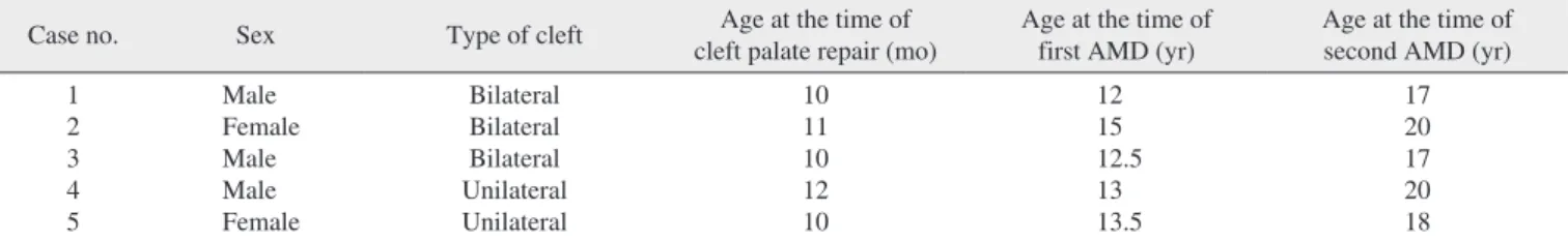
Table 1. Summary of patient details
Case no. Sex Type of cleft Age at the time of
cleft palate repair (mo)
Age at the time of first AMD (yr)
Age at the time of second AMD (yr) 1
2 3 4 5
Male Female Male Male Female
Bilateral Bilateral Bilateral Unilateral Unilateral
10 11 10 12 10
12 15 12.5 13 13.5
17 20 17 20 18 (AMD: anterior maxillary distraction)
Sunil Richardson et al: Use of repeat anterior maxillary distraction to correct residual midface hypoplasia in cleft patients. J Korean Assoc Oral Maxillofac Surg 2017
raphe. The arms of the hyrax were soldered to bands and the appliance was cemented to the premolars anteriorly and molars posteriorly.(Fig. 2. A) In one case, the premolar was missing and the band was cemented to the canine. This ap- pliance was cemented preoperatively in the patient’s mouth.
(Fig. 2. B) Then, acrylic bite blocks were prepared on the molars to open the bite and avoid any occlusal interference during distraction. An acrylic plate was fabricated separately around the anterior teeth, which included the premolars, and encompassed the teeth from the lingual, incisal/occlusal, and buccal sides in order to hold the anterior fragment firmly and distribute the forces of distraction.
1. Surgical technique
Under general anesthesia, a maxillary degloving incision was performed between the molars. Subperiosteal dissection proceeded bilaterally up until the infraorbital foramen. After reflection of the mucoperiosteal flap, the previous osteotomy cuts were faintly visible in three cases and were utilized. In the other two cases, new osteotomy cuts were made hori- zontally, starting from the pyriform rim till a point where the deemed vertical cut would begin. Using a bur, vertical interdental cuts were made between the second premolar and first molar through the buccal cortex.(Fig. 3) Lateral nasal osteotomes were used on both sides to separate the lateral na- sal wall from the pyriform rim at the same level of the buccal cut. An elevator was used to protect the nasal mucosa. The palatal osteotomy was performed using a spatula osteotome, with particular care taken to avoid perforating the palatal mucosa. Gentle luxation was done between the cuts to ensure
A B
Fig. 2. The distractor appliance. A. The distractor appliance after its laboratory fabrication consisting of the hyrax screw and tooth bands. B.
Appliance cemented intra-orally.
Sunil Richardson et al: Use of repeat anterior maxillary distraction to correct residual midface hypoplasia in cleft patients. J Korean Assoc Oral Maxillofac Surg 2017
CMr1 CPr1 CPl1 CMl1
CMr2
CMr2 CPr2CPr2 CPl2CPl2 CMl2CMl2
A
B
'O' line
'O' line
Fig. 1. OPG1 and OPG2 showing maintenance of the bone formed after initial AMD procedure. A. Measuring distance between CMr1-CPr1 and CMl1-CPl1 in OPG1. B. Measur- ing distance between CMr2-CPr2 and CMl2-CPl2 in OPG2.
CPr1/2=cemento-enamel junction of the distal border of the right premolar in OPG1/OPG2, respectively; CMr1/2=cemento-enamel junction of the mesial border of the right molar in OPG1/OPG2, respectively; CPl1/2=cemento-enamel junction of the distal border of the left premolar OPG1/OPG2, respectively; CMl1/2=cemento- enamel junction of the mesial border of the left molar OPG1/
OPG2, respectively. (OPG1: orthopantomogram taken after completion of intital AMD, OPG2: orthopantomogram taken at the second time of presentation)
Sunil Richardson et al: Use of repeat anterior maxillary distraction to correct residual midface hypoplasia in cleft patients. J Korean Assoc Oral Maxillofac Surg 2017
ate. The mean age at the second presentation was 18.4 years.
The minimum follow-up period was one year.
The results of the Mann-Whitney U-test showed no signifi- cant change in the measurements taken on OPG1 and OPG2 mobilization of the segment. The distractor screw was acti-
vated intraoperatively to check the symmetrical movement on both sides. Closure was performed with 3-0 Vicryl sutures (Ethicon Inc., Somerville, NJ, USA).
2. Distraction protocol
We followed mean latency period of 5 to 7 days. Due to an accidental tear of the palatal mucosa intraoperatively in one case, we used an eight-day latency period. Following the latency period, the screw was turned three revolutions, twice daily. The pitch of the screw was 0.18 mm and therefore the amount of distraction achieved per day was 1.08 cm. Distrac- tion proceeded until a positive overjet and the desired facial profile was achieved. Consolidation was carried out for a pe- riod of 8 to 10 weeks by keeping the appliance in situ. After removing the distractor, an acrylic plate was inserted for a pe- riod of 3 to 4 weeks in order for the occlusion to settle. Fixed prosthesis was fabricated in the space created between the distraction segments; crowding, if present, was corrected by orthodontics. Orthopantomograms and lateral cephalograms were taken immediately after distraction (OPG3 and Lat- Ceph3), after the consolidation period (OPG4 and LatCeph4), and before the appliance removal at the one-year follow-up visit (OPG5 and LatCeph5).
III. Results
There was a total of five patients in the study. Three were male and two were female. Three patients had a repaired bi- lateral cleft palate, and two had a unilateral repaired cleft pal-
A B
Fig. 3. The osteotomy cuts. A. The arrows pointing towards the previous osteotomy cuts at the time of second anterior maxillary distrac- tion (AMD). B. Fresh osteotomy cuts to be made for the AMD (dashed line).
Sunil Richardson et al: Use of repeat anterior maxillary distraction to correct residual midface hypoplasia in cleft patients. J Korean Assoc Oral Maxillofac Surg 2017
A B
C D
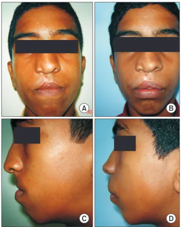
Fig. 4. Clinical photographs of the initial anterior maxillary distrac- tion procedure (at 12 years). A. Frontal view pre-distraction. B.
Frontal view post-distraction. C. Side profile pre-distraction. D.
Side profile post-distraction.
Sunil Richardson et al: Use of repeat anterior maxillary distraction to correct residual midface hypoplasia in cleft patients. J Korean Assoc Oral Maxillofac Surg 2017
gram taken after the completion of the first AMD procedure.
Cephalometric analysis showed significant improvement of most parameters in LatCeph5 when compared to LatCeph2.
(Table 3) The mean increases in the midfacial length (10.56 mm), in the SNA angle (5.09°), and the distance between the nasion perpendicular to point A (5.89 mm) were all statisti- cally significant (P=0.012, P=0.011, and P=0.012, respective- ly). The changes in the SNB angle (1.82°) and lower facial height (3.27 mm) were not statistically significant (P=0.14 and P=0.52, respectively). A straight profile was maintained for all patients, and a positive overjet was achieved.(Fig. 6) There was also evidence of solid bone formation between the osteotomy site on LatCeph5, which was taken after comple- tion of the second AMD.(Fig. 7) Dental rehabilitation for the created gap was conducted on four of the patients by fixed prosthesis. One patient underwent orthodontic treatment to fill the created gap by moving the crowded teeth to an opti- mal position. No complications from the surgical procedures were observed either intraoperatively or postoperatively.
IV. Discussion
Maxillary hypoplasia is a common deformity in cleft lip (P=0.67). The lack of a significant change demonstrated that
the cleft maxillary hypoplasia at presentation was residual since the gap created by the initial distraction procedure was still maintained.(Fig. 4, Table 2) Fig. 5 shows the cephalo-
Table 2. Comparison of the created distraction gap in OPG’s taken after the first distraction (OPG1) and after the subsequent presentation (OPG2)
Case no. CPr1-CMr1 (mm) CPl1-CMl1 (mm) Mean OPG1 (mm) CPr2-CMr2 (mm) CPl2-CMl2 (mm) Mean OPG2 (mm) P-value 1
2 3 4 5
8.30 13.83 12.12 10.52 9.55
8.33 13.83 12.02 10.33 9.47
8.31 13.83 12.07 10.42 9.51
8.25 13.23 12.08 10.48 9.52
8.31 13.92 12.00 10.15 9.39
8.28 13.57 12.04 10.31 9.45
0.67
(OPG: orthopantomogram)
CPr1/2=cemento-enamel junction of the distal border of the right premolar in OPG1/OPG2, respectively; CMr1/2=cemento-enamel junction of the mesial border of the right molar in OPG1/OPG2, respectively; CPl1/2=cemento-enamel junction of the distal border of the left premolar OPG1/
OPG2, respectively; CMl1/2=cementoenamel junction of the mesial border of the left molar OPG1/OPG2, respectively.
Sunil Richardson et al: Use of repeat anterior maxillary distraction to correct residual midface hypoplasia in cleft patients. J Korean Assoc Oral Maxillofac Surg 2017 Fig. 5. Lateral cephalogram taken after the completion of the ini-
tial anterior maxillary distraction procedure (LatCeph1).
Sunil Richardson et al: Use of repeat anterior maxillary distraction to correct residual midface hypoplasia in cleft patients. J Korean Assoc Oral Maxillofac Surg 2017
Table 3. McNamara analysis performed on LatCeph2 and LatCeph5 to assess the skeletal changes after the second anterior maxillary distraction (AMD)
Measurement LatCeph2 LatCeph5 Mean difference P-value
SNA (°) SNB (°)
Nasion perpendicular to point A (mm) Effective midface length (Co-point A) (mm) Lower facial height (ANS-Me) (mm)
76.02±0.80 (74.82-76.82) 75.45±4.24 (71.10-82.47)
7.04±1.82 (5.11-9.01) 85.47±4.84 (79.25-91.10) 74.87±6.61 (66.11-83.02)
81.11±1.07 (79.9-82.62) 77.27±3.25 (74.82-82.92)
1.15±2.60 (–3.20-3.58) 96.03±4.41 (91.28-102.30) 78.14±9.27 (63.50-87.00)
5.09 1.82 5.89 10.56 3.27
0.011 0.14 0.012 0.012 0.52 (LatCeph2: lateral cephalogram taken at the time of presentation, LatCeph5: lateral cephalogram taken 1 year after the second AMD, SNA: angle formed by the sella, nasion, and point A, SNB: angle formed by the sella, nasion, and point B, Co: most postero-superior point on the condyle, ANS: anterior nasal spine, Me: menton)
Values are presented as mean±standard deviation (range).
P-values calculated from Mann-Whitney U-test.
Sunil Richardson et al: Use of repeat anterior maxillary distraction to correct residual midface hypoplasia in cleft patients. J Korean Assoc Oral Maxillofac Surg 2017
the maxilla15. Since the advancement only affects the anterior part of the maxilla, the velopharyngeal muscles are not af- fected and velopharyngeal closure is maximally preserved16. Not only is the velopharyngeal sphincter preserved, it has been found that all parameters of speech, such as velopharyn- geal incompetence, nasal air emission, resonance, articulation errors, and intelligibility, are significantly improved after AMD6. This is the biggest advantage that AMD has over the other conventional treatment modalities. Another advantage is that the new bone deposition between the osteotomy cuts create space in the arch to correct crowding and to assist in the eruption of the canines without the need for tooth extrac- tion10,15.
There are only two treatment options available to treat cleft maxillary hypoplasia for patients 11 to 13 years; total maxil- lary distraction or AMD. Orthognathic surgery can be per- formed only after growth completion and face-mask therapy should be provided for the younger age group16,17. Although total maxillary distraction can be done at any age, its negative impact on velopharyngeal function makes it the least viable of the two alternatives. It may be preferred when advance- ments greater than 13 mm are required since the maximum movement achieved with AMD is 13 mm due to the limita- tion of the screw used.
As seen in our study, all five patients had undergone AMD once before at mean age of 13 years. At the time of the first procedure we had achieved good facial profile and positive overjet. No overcorrection was done. Over time, the rest of and palate patients, and approximately 25% of these patients
require orthognathic surgery to correct10. Sagittal advance- ment of the entire maxilla has a relapse rate of 20% to 60%11. Sagittal advancement of the maxilla also alters the speech of the patient12.
With the application of DO to facial structures in the 1990s, maxillary distraction has become a viable alternative to osteotomy for correcting midfacial hypoplasia. Distraction can be achieved using a cranially attached rigid distractor, an orthodontic facemask, or by using internal distractors13. Usu- ally distraction is performed using a rigid external device or an intraoral distractor at a Le Fort I level. This procedure also has a negative impact on speech14. Since both Le Fort I oste- otomy and a LeFort I DO involve moving the entire maxilla off the posterior pharyngeal wall, the risk of velopharyngeal insufficiency is similar in both procedures14.
AMD is a method of advancing only the anterior part of the maxilla. It relieves dental crowding of the upper arch, al- lows normal eruption of canines, and corrects the retrusion of
A B
C D
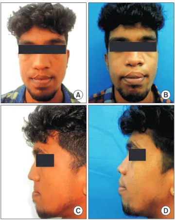
Fig. 6. Clinical photographs of anterior maxillary distraction proce- dure the second time (at 17 years). A. Front view showing residual maxillary hypoplasia (pre-distraction). B. Front profile after distrac- tion. C. Side profile before distraction. D. Side profile after distrac- tion.
Sunil Richardson et al: Use of repeat anterior maxillary distraction to correct residual midface hypoplasia in cleft patients. J Korean Assoc Oral Maxillofac Surg 2017
Fig. 7. Lateral cephalogram taken after the second anterior maxil- lary distraction was completed (LatCeph5).
Sunil Richardson et al: Use of repeat anterior maxillary distraction to correct residual midface hypoplasia in cleft patients. J Korean Assoc Oral Maxillofac Surg 2017
the patient’s facial skeleton (in particular, the mandible) con- tinued to grow normally and the patient developed a retrusive midfacial appearance. Cleft maxillary hypoplasia is thought to occur due to both the inherent developmental deficiency of the cleft structures and due to the ill effects of surgies done in childhood to treat the primary cleft18. Although relapse is also encountered after AMD, those cases were excluded from our study. Radiographic analysis proved that it was a residual deformity, and not a relapse, since the distraction gap was maintained.
At presentation, a negative overjet and a concave facial profile was observed in the five patients. Repeat AMD was planned and we achieved satisfactory results in all cases.
There were no negative effects from the previous AMD in our results. One-year follow-up showed stable results. There were significant improvements in the cephalometric param- eters. The parameters pertaining to the maxilla (midface length), SNA angle, and the nasion perpendicular line dis- tance to point A increased significantly.
There was minor variation in the surgical technique when performing the procedure for the second time. First, nasal septal separation from the maxillary crest was not required.
Second, in most cases the previous osteotomy line, if visible, could be followed. Also, if there was sufficient space cre- ated from the primary AMD between the second premolar and first molars, the buccal vertical osteotomy cuts could be safely used a second time without injuring the teeth. After completion of the final AMD, dental rehabilitation could be performed using a crown or implant.
A few disadvantages of AMD were as follows. 1) The lack of effective vector controls, which caused anterior open bite in a few cases but was corrected with the application of box elastics before the completion of consolidation. 2) Minor bleeding episodes were encountered during the patient recov- ery period, which were managed conservatively.
Our findings are encouraging and suggest that AMD could be a preferred treatment modality to treat cleft maxillary hy- poplasia at our center since it can be performed as early as 11 years of age and can be repeated later without complication.
Treating cleft maxillary hypoplasia at a young age provides the child with an opportunity to live normally and does not jeopardize future treatments that may be done once the child attains skeletal maturity.
V. Conclusion
Children suffering from cleft maxillary hypoplasia do
not have to wait for skeletal maturity to be treated. We have found AMD to be an effective treatment modality that can correct the facial deformities as early as 11 years and can be repeated again at any stage if residual deformity develops.
Conflict of Interest
No potential conflict of interest relevant to this article was reported.
ORCID
Sunil Richardson, http://orcid.org/0000-0002-2141-9201 Shreya Krishna, http://orcid.org/0000-0001-8031-0014 Avi Bansal, http://orcid.org/0000-0003-0407-0076
References
1. Janulewicz J, Costello BJ, Buckley MJ, Ford MD, Close J, Gas- sner R. The effects of Le Fort I osteotomies on velopharyngeal and speech functions in cleft patients. J Oral Maxillofac Surg 2004;62:
308-14.
2. Cheung LK, Chua HD. A meta-analysis of cleft maxillary oste- otomy and distraction osteogenesis. Int J Oral Maxillofac Surg 2006;35:14-24.
3. Bengi O, Karaçay S, Akin E, Okçu KM, Olmez H, Mermut S.
Cephalometric evaluation of patients treated by maxillary anterior segmental distraction: a preliminary report. J Craniomaxillofac Surg 2007;35:302-10.
4. Block MS, Brister GD. Use of distraction osteogenesis for maxil- lary advancement: preliminary results. J Oral Maxillofac Surg 1994;52:282-6.
5. Dolanmaz D, Karaman A, Ozyesil AG. Maxillary anterior segmen- tal advancement by using distraction osteogenesis: a case report.
Angle Orthod 2003;73:201-5.
6. Richardson S, Seelan NS, Selvaraj D, Khandeparker RV, Gnana- mony S. Perceptual speech assessment after anterior maxillary distraction in patients with cleft maxillary hypoplasia. J Oral Max- illofac Surg 2016;74:1239.e1-9.
7. Wolford LM, Karras SC, Mehra P. Considerations for orthognathic surgery during growth, part 2: maxillary deformities. Am J Orthod Dentofacial Orthop 2001;119:102-5.
8. Bansal A, Prakash AT, Deepthi, Naik A. A noble, easy and concep- tual radiographic analysis to assess the type of tooth movement (molar distalization). J Clin Diagn Res 2015;9:ZC22-5.
9. McNamara JA Jr. A method of cephalometric evaluation. Am J Or- thod 1984;86:449-69.
10. Wang XX, Wang X, Li ZL, Yi B, Liang C, Jia YL, et al. Anterior maxillary segmental distraction for correction of maxillary hypo- plasia and dental crowding in cleft palate patients: a preliminary report. Int J Oral Maxillofac Surg 2009;38:1237-43.
11. Posnick JC, Dagys AP. Skeletal stability and relapse patterns after Le Fort I maxillary osteotomy fixed with miniplates: the unilateral cleft lip and palate deformity. Plast Reconstr Surg 1994;94:924-32.
12. Hirano A, Suzuki H. Factors related to relapse after Le Fort I max- illary advancement osteotomy in patients with cleft lip and palate.
Cleft Palate Craniofac J 2001;38:1-10.
13. Precious DS. Treatment of retruded maxilla in cleft lip and palate-- orthognathic surgery versus distraction osteogenesis: the case for orthognathic surgery. J Oral Maxillofac Surg 2007;65:758-61.
14. Guyette TW, Polley JW, Figueroa A, Smith BE. Changes in speech following maxillary distraction osteogenesis. Cleft Palate Cranio- fac J 2001;38:199-205.
15. Gunaseelan R, Cheung LK, Krishnaswamy R, Veerabahu M. An- terior maxillary distraction by tooth-borne palatal distractor. J Oral Maxillofac Surg 2007;65:1044-9.
16. Oluwajana F. Seeking beauty: understanding the psychology behind orthognathic surgery. Br J Oral Maxillofac Surg 2015;53:953-6.
17. Dogan S. The effects of face mask therapy in cleft lip and palate patients. Ann Maxillofac Surg 2012;2:116-20.
18. Ross RB. Treatment variables affecting facial growth in complete unilateral cleft lip and palate. Cleft Palate J 1987;24:5-77.