Korean J Gastroenterol Vol. 57 No. 6, 370-373 DOI: 10.4166/kjg.2011.57.6.370
CASE REPORT
Korean J Gastroenterol, Vol. 57 No. 6, June 2011 www.kjg.or.kr
복부 둔상 3개월 후에 발생한 회장 협착
강건희, 전태주, 서동대, 오태훈, 김수현1, 조현선1, 배병노2, 김정연3
인제대학교 의과대학 상계백병원 내과학교실, 영상의학교실1, 외과학교실2, 병리학교실3
Ileal Stenosis Occurred 3 Months after Blunt Abdominal Trauma
Gun-Hi Kang, Tae Joo Jeon, Dong Dae Seo, Tae-Hoon Oh, Soohyun Kim1, Hyun Sun Cho1, Byung Noe Bae2 and Jung Yeon Kim3 Departments of Internal Medicine, Radiology1, Surgery2 and Pathology3, Sanggye Paik Hospital, Inje University College of Medicine, Seoul, Korea
We present a case of ileal stenosis with delayed presentation 3 months after car accident. Ileal stenosis after blunt abdominal trauma is a rare clinical entity. We present CT and small bowel series 3 months after trauma. Image showed segmental thickening of intestinal wall and proximal bowel dilation. At surgery, a stenotic bowel loop was adjacent to a fibrotic mesentery.
Histological examination showed ulcers, inflammatory cells and fibroblasts infiltrated to the muscularis mucosae, submucosa, and mesentery. The most likely cause, supported by most authors, implicates an injury to the mesentery. Post-traumatic ischemic bowel stenosis may result from even small tears and contusions of mesentery. Posttraumatic intestinal stenosis should be included in the differential diagnosis in a patient with a history of blunt abdominal trauma and signs of intestinal obstruction.
(Korean J Gastroenterol 2011;57:370-373) Key Words: Blunt abdominal trauma; Stenosis
Received March 30, 2010. Revised June 5, 2010. Accepted June 8, 2010.
CC This is an open access article distributed under the terms of the Creative Commons Attribution Non-Commercial License (http://creativecommons.org/licenses/
by-nc/3.0) which permits unrestricted non-commercial use, distribution, and reproduction in any medium, provided the original work is properly cited.
교신저자: 전태주, 139-707, 서울시 노원구 상계 7동 761-1, 인제대학교 의과대학 상계백병원 내과학교실 소화기내과
Correspondence to: Tae Joo Jeon, Division of Gastroenterology, Department of Internal Medicine, Sanggye Paik Hospital, Inje University College of Medicine, 761-1, Sanggye 7-dong, Nowon-gu, Seoul 139-707, Korea. Tel: +82-2-950-8889, Fax: +82-2-950-1955, E-mail: [email protected]
Financial support: None. Conflict of interest: None.
INTRODUCTION
Post-traumatic intestinal stenosis is a rare complication of blunt abdominal trauma and a disease causing local ische- mic stenosis by damaging mesentery or intestinal wall after blunt abdominal trauma. The authors experienced a case of 42 year-old man who had shown incomplete small bowel ob- struction 3 months after car accident. He was diagnosed with ileal stenosis and had successful surgical treatment. Herein we present our clinical experience with ileal stenosis oc- curred 3 months after blunt abdominal trauma.
CASE REPORT
A 42 year-old man who had lived healthily without any par- ticular symptoms had multiple fractures of extremities be- cause of car accident. He was driving with the seat belt buck- led up. After 3 months, he had postprandial abdominal dis- tension and intermittent colicky pain in his right lower abdomen. Only constipation was accompanied. At admis- sion, vital sign was stable. On physical examination, abdomi- nal distension was found, but there were no tenderness and palpable mass in the abdomen. Laboratory values were as follows: hemoglobin 13.1 g/dL, white blood cell 7,710/mm3, platelet 413,000/mm3, total bilirubin 0.5 mg/dL, aspartate aminotransferase 11 U/L, alanine aminotransferase 4 U/L,
Kang GH, et al. Ileal Stenosis Occurred 3 Months after Blunt Abdominal Trauma 371
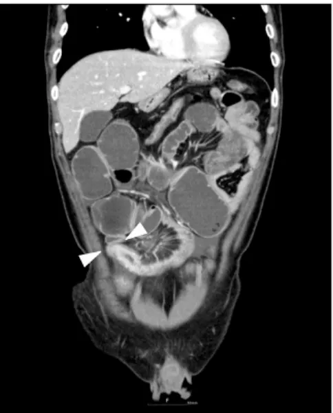
Vol. 57 No. 6, June 2011 Fig. 1. Coronal abdominal computed tomography. It demonstrated
segmental luminal narrowing and stricture in mid ileum (arrow heads).
Fig. 2. Small bowel series. It showed segmental luminal narrowing and stricture with ulceronodular mucosa (arrow heads).
Fig. 3. Gross findings of resected specimen. (A) In gross finding, the ileum was stenosed, the surrounding mesentery was fibrosed hardly, adhesion was serious, and the mesenteric root was shortened. (B) The mucosa showed ulcerative change with necrotic exudates.
Fig. 4. Histopathological finding. Ulcers were observed and in- flammatory cells and fibroblasts infiltrated to the whole layers (H&E,
×40).
alkaline phosphatase 75 U/L, creatinine 0.7 mg/dL, amy- lase 46 U/L, lipase 56 U/L. Simple X-ray was unremarkable.
Abdominal pelvic CT revealed segmental thickened in- testinal wall in the ileum of the right lower abdomen and the expanded proximal small bowel due to the stenosis (Fig. 1).
CT also showed mesenteric haziness near stenotic area. In small bowel series, stenosis in the ileum was 10 cm in length, and the small bowel proximal to the stenosis was dilated (Fig.
2).
In the operation, segmental resection and reanastomosis was done for the stenotic lesion followed by adhesiolysis.
Operative findings showed that midileum was stenosed, the surrounding mesentery was fibrosed hardly, adhesion was serious, and the mesenteric root was shortened (Fig. 3A).
And, the mucosa showed ulcerative change with necrotic exu- dates (Fig. 3B). In histopathological examination, ulcers were observed and inflammatory cells and fibroblasts infiltrated to the whole layers. The vessel wall was thickened and filled with thrombus (Fig. 4). The patient has been well without any problems after discharge.
372 강건희 등. 복부 둔상 3개월 후에 발생한 회장 협착
The Korean Journal of Gastroenterology
DISCUSSION
Delayed small bowel obstruction after blunt abdominal trauma is a rare clinical entity, comprising fewer than 1% of non-penetrating trauma admissions in some institutions.1
Symptoms of posttraumatic intestinal stenosis are nau- sea, vomiting, and abdominal distension. According to pre- vious reports, posttraumatic intestinal stenosis is charac- terized by delayed appearance of the symptoms. However, the period from the occurrence of the trauma to the onset of the symptom varies from a week to 18 weeks.2,3 The length of stenosis also varies from 1 cm to 28 cm, and the site is usu- ally limited to single region but sometimes multiple.2,3 The disease usually induces the stenosis of the small bowel, but it may also cause the stenosis of the large bowel.3
The thickening of the small bowel wall and the loss of the layer in ultrasonography are not specific findings, and they can be observed in intestinal ischemia.4 The uniformly in- creased echo of the mesentery indicates fat necrosis caused by hemorrhage resulting from the injury of the mesentery.
Such a change in the mesentery suggests that damage was inflicted not on the intestinal wall but on the mesentery.5 CT showed concentric thickened intestinal wall of various lengths and the expansion of the proximal bowel and the de- crease of the inner diameter of the distal bowel.6 Barium ser- ies showed various narrowed concentric inner diameters of the bowel and ulcers in the mucous layer.7 In addition, the ex- pansion of the proximal bowel and the collapse of the distal bowel observed. In angiography, some branches of the supe- rior mesentric artery were blocked and ischemic intestinal wall was observed.7
Diagnostic criteria includes the obvious history of blunt ab- dominal trauma, the absence of disease before the trauma, the occurrence of intestinal symptoms after the trauma, the confirmation of intestinal stenosis in radiological examina- tion, and no inflammatory or neoplastic change in the site re- sected through operation.3 In radiological examination, it may be difficult to distinguish the disease from Crohn's dis- ease,8 intestinal tuberculosis, radiation enteritis, and small intestine cancer. The histological examination should be done to confirm the diagnosis.
One of the theories suggests that the fixed portions of the small bowel, namely, the terminal ileum and proximal jeju-
num, are prone to perforation during blunt abdominal trauma.9 A small subclinical perforation may seal off sponta- neously producing a stricture due to scar formation. Welch and Anderson presented such a case.10 This explanation is indeed plausible, but is not found in the majority of cases.
Another mechanism for posttraumatic intestinal stenosis is localized bowel ischemia. It is more likely, however, that the focal ischemia of the gut is secondary to a mesenteric blunt trauma rather than direct ischemia of the bowel wall.1
The most likely cause, supported by most authors, im- plicates an injury to mesentery. Breen et al.11 classified mes- enteric injuries as major and minor. Minor mesenteric in- juries are defined as small hematomas, contusions, or lacer- ations without compromising the bowel circulation. Although small lacerations do not result in significant hemorrhage or devitalization of the bowel, they still present a risk for internal herniation if left unrepaired.11
Because there was no evidence of small bowel perforation in our patient, it is highly likely that the small bowel ob- struction was caused by small bowel ischemia. CT showed mesenteric haziness, operative findings and biopsy showed fibrosis in the mesentery, suggesting the small bowel steno- sis caused by the injury of the mesentery.
Posttraumatic small bowel stenosis is an entity that is not widely known. Patients complaining of abdominal pain with no prior abdominal surgery and with a suspicion of partial small bowel obstruction should be specifically questioned about previous blunt trauma. The work-up for these patients should include an abdominal CT and small bowel series.
Resection of the affected bowel will resolve the symptoms.12
REFERENCES
1. Kaban G, Somani RA, Carter J. Delayed presentation of small bowel injury after blunt abdominal trauma: case report. J Trauma 2004;56:1144-1145.
2. Lynch JM, Albanese CT, Meza MP, Wiener ES. Intestinal stricture following seat belt injury in children. J Pediatr Surg 1996;
31:1354-1357.
3. Hirota C, Iida M, Aoyagi K, Matsumoto T, Yao T, Fujishima M.
Posttraumatic intestinal stenosis: clinical and radiographic fea- tures in four patients. Radiology 1995;194:813-815.
4. Teefey SA, Roarke MC, Brink JA, et al. Bowel wall thickening: dif- ferentiation of inflammation from ischemia with color Doppler and duplex US. Radiology 1996;198:547-551.
5. Loberant N, Szvalb S, Herskovits M, Cohen I, Salamon V.
Kang GH, et al. Ileal Stenosis Occurred 3 Months after Blunt Abdominal Trauma 373
Vol. 57 No. 6, June 2011 Posttraumatic intestinal stenosis: radiographic and sono-
graphic appearance. Eur Radiol 1997;7:524-526.
6. Tsushima Y, Yamada S, Aoki J, Endo K. Ischaemic ileal stenosis following blunt abdominal trauma and demonstrated by CT. Br J Radiol 2001;74:277-279.
7. De Backer AI, De Schepper AM, Vaneerdeweg W, Pelckmans P.
Intestinal stenosis from mesenteric injury after blunt abdominal trauma. Eur Radiol 1999;9:1429-1431.
8. Fanelli C, Bassotti G, Giansanti M, Bartoli A, Bolli GB.
Posttraumatic ileal stenosis mimicking Crohn’s disease. J Clin Gastroenterol 1995;20:338-340.
9. Allen JC. Post-traumatic small bowel obstruction. J R Army Med Corps 1994;140:47-48.
10. Welch GH, Anderson JR. Small bowel stricture following abdomi- nal trauma. Postgrad Med J 1985;61:1087-1088.
11. Breen DJ, Janzen DL, Zwirewich CV, Nagy AG. Blunt bowel and mesenteric injury: diagnostic performance of CT signs. J Comput Assist Tomogr 1997;21:706-712.
12. Bala M, Lebenthal A, Pikarsky E, Faroja M, Rivkind AI, Mintz Y.
Intestinal stenosis causing small bowel obstruction after non- operative management of blunt abdominal trauma. J Trauma 2007;62:1511-1513.