WWW.KJOG.ORG 257
PRENATAL DIAGNOSIS OF CYSTIC PERIVENTRICULAR LEUKOMALACIA IN A FULL TERM FETUS
Heui Jin Joo, MD, Hyun Chul Jeong, MD, Youn Jeong Lee, MD, Ja Young Hwang, MS, Sang Hoon Lee, MD, Dong Ho Kim, MD, Seung Su Han, MD, Gwang Jun Kim, MD
Department of Obstetrics and Gynecology, Chung-Ang University Hospital, Chung-Ang University College of Medicine, Seoul, Korea
Periventricular leukomalacia is a disorder of white matter injury that is frequently found in premature infants and is the major cause of cerebral palsy. However, only a few cases of periventricular leukomalacia have been reported prenatally. Ultrasound investigation of the periventricular region could provide clues for the prenatal diagnosis and prediction of cerebral palsy. Herein, we report a case of cystic periventricular leukomalacia that was diagnosed prenatally and confi rmed postnatally by brain magnetic resonance imaging.
Keywords:
Periventricular leukomalacia; Cerebral palsy; Periventricular echodensities; Prenatal ultrasonogram
CASE REPORT
Received: 2012.1.20. Accepted: 2012.3.4.
Corresponding author: Gwang Jun Kim, MD
Department of Obstetrics and Gynecology, Chung‐Ang University Hospital, 224‐1 Heukseok‐dong, Dongjak‐gu, Seoul 156‐755, Korea
Tel: +82‐2‐6299‐1646, 1656 Fax: +82‐2‐6263‐2187 E‐mail: [email protected]
Th is is an Open Access article distributed under the terms of the Creative Commons Attribution Non-Commercial License (http://creativecommons.org/licenses/
by-nc/3.0/) which permits unrestricted non-commercial use, distribution, and reproduction in any medium, provided the original work is properly cited.
Copyright © 2012. Korean Society of Obstetrics and Gynecology Korean J Obstet Gynecol 2012;55(4):257-260
http://dx.doi.org/10.5468/KJOG.2012.55.4.257 pISSN 2233-5188 · eISSN 2233-5196
Periventricular leukomalacia (PVL) is a typical cerebral lesion in premature infants, largely due to hypoxia-ischemia and reperfu- sion, which may result in cerebral palsy (CP) [1]. The pattern of perinatal brain injury is age-dependent, which in turn results in prognoses of different pathways. Depending on the type of lesion, the suspected incidental time may vary. PVL can be suspected on routine ultrasound screening. Periventricular echodensities (PVE), echolucent cysts, or ventricular dilatations are some of the abnor- mal prenatal ultrasonographic fi ndings suggestive of PVL, which lead to a neurosonography and follow-up throughout the rest of the perinatal period.
Case Report
A 27-year-old, para 1 woman was referred for consultation due to the fi ndings of a mildly dilated lateral ventricle in the fetus at 34 + 6 weeks of gestation. She had no past medical history, no op- eration history, and had not taken any kind of medication, except for oral iron for the current pregnancy. Her family did not have any history of medical illnesses, and she did not smoke nor drink. She had one full-term vaginal delivery two years prior. At the beginning of the current pregnancy, the patient did not have any obstetrical problems. Mid-gestation screening was reported low risk. Mild ventricle dilatation in the fetus was detected on the 2nd trimester
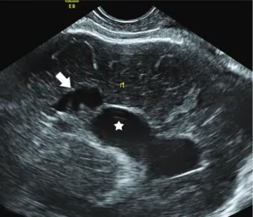
ultrasound examination and was maintained until 35 weeks of gestation. Therefore, she was referred to our hospital at 34 + 6 weeks of gestation due to mild but persistent fetal ventriculo- megaly. The biometry showed normal measures for head circum- ference, femur length, abdominal circumference, and estimated fetal weight. A closer look at the brain revealed the width of the lateral ventricle to be 9.8 mm. Cystic changes in the right frontal periventricular region were detected, along with a prominent lateral ventricle (Fig. 1). At 36 +5 weeks of gestation, the mother visited the delivery fl oor complaining of watery vaginal discharge.
At 36 + 6 weeks of gestation, a baby girl, 3,150 g, was born by
normal vaginal delivery, with an Apgar score of 9 at 1 minute
WWW.KJOG.ORG 258
KJOG Vol. 55, No. 4, 2012
and 9 at 5 minutes. The infant looked healthy without spasm. No hypotonia and spasticity were detected on physical examination done by a pediatric neurologist. On the second day, brain mag- netic resonance imaging (MRI) showed grade 3 PVL with a mildly dilated right lateral ventricle with cystic encephalomalacia near the right lateral ventricle (Fig. 2) and focal chronic old hemorrhage in the inferolateral wall of the right lateral ventricle body. The brain ultrasound revealed grade 2 PVL in the right lateral ventricle and
grade 1 PVL in the left lateral ventricles, with periventricular cysts and echogenicities (Fig. 3), respectively. Despite the severity of the fi ndings, the patient had no severe neurologic complications. The baby was well and discharged at 4 days of age, and outpatient department follow-ups have included physical therapy for hypo- tonicity and weak upper extremities that have developed over the postnatal months.
Discussion
PVL is a cerebral injury that is mainly prevalent in preterm infants but also arises in full-term neonates as well as infants as old as two months postnatally [1]. It is known to be a leading cause of CP in newborns. It is a disorder of diffuse white matter injury characteristic of focal cystic necrotic lesions to diffuse myelination disturbances [2]. The main pathogenesis of this disease is thought to be due to hypoxic-ischemia and maternal-fetal infection. The vasculature of the premature infant has not yet matured, and the walls of the arteries and veins are still fragile and thin. During epi- sodes of decreased oxygenation, it is likely that these vessels are not able to maintain a suffi cient amount of blood fl ow, which in turn results in hypoxic-ischemic events [3]. Therefore, incomplete development of the peripheral cerebral arteries and impaired tis- sue perfusion in periventricular white matter result in white matter damage, which is PVL. The damage may show as a diffuse lesion or as cystic, focal lesions. Another mechanism is infection-infl am- mation, which seems to be an important risk factor for brain in- jury, especially in term infants. Other risk factors for neonatal PVL
Fig. 1. Prenatal sonogram: Enlarged right lateral ventricle (white star)with periventricular cystic changes (white arrow).
Fig. 2. Postnatal brain magnetic resonance imaging: Cystic encephalo- malacia (white arrow) with dilated right lateral ventricle (black star).
Fig. 3. Postnatal brain sonogram: Increased left periventricular echo- genicity (white arrows), suggesting periventricular leukomalacia grade 1.
WWW.KJOG.ORG 259
Heui Jin Joo, et al. Prenatal diagnosis of full term PVL
mentioned in the literature are premature rupture of membrane, preterm birth, multiple pregnancy, intrauterine growth retardation, reduced amniotic fl uid, cord compression, and low Apgar scores [4,5]. The fetal brain has restorative powers, in which intrauterine damage to the brain can be a brief incident where regrowth of the brain tissue occurs; consequently, the neonatal outcome would be normal, and PVE would disappear. The repairing function is thought to be due to a signifi cant increase in insulin-like growth factor 1 (IGF-1) of maternal origin in maternal and cord sera in late gestation, which is supposed to play an essential role in fetal growth. After birth, the newborn is separated from the mother, and the IGF-1 is lost in the neonatal circulation, diminishing re- growth abilities. The PVE repairing function is diminished in the neonatal brain, necrotic changes progress to develop a cyst in PVE, and PVL is formed [6]. Early changes associated with cystic PVL may be apparent histologically within hours of insult, but at least 2 to 6 weeks are required before lesions can be visualized on the sonogram. Therefore, if cystic PVL is observed within 7 days after birth, the origins must be intrauterine [7]. Another big risk factor of term PVL, clinical chorioamnionitis, is strongly connected to CP, as the infectious products sensitize the fetus to secondary insults [8]. A study has suggested that maternal infection can activate the cytokine network, causing interleukin-6 levels to rise, which were found in high levels in the umbilical cord blood of neonates with white matter lesions [9].
Depending on the fi ndings of the ultrasound, PVL is graded into four findings: grade 1 correlates with findings of increased PVE at the external angle of the lateral ventricle, corresponding to the early stages of PVL. Grade 2 is when small cysts develop 2 to 3 weeks after the onset of PVE in the same area as grade 1. Grade 3 shows PVE extending to the parieto-occipital white matter, breaking down into cystic lesions 2 to 4 weeks later. With grades 2 and 3, cysts are usually no longer visible when the infant is 2 to 3 months old. Owing to loss of brain tissue, dilation of the ventricles develops, either uni- or bilateral, as predicted by the early localization of the lesions. Grade 4 mainly affects full-term infants and is often called subcortical leukomalacia. Localization of PVE and cysts is farther away from the lateral ventricles than in premature infants, and the cysts do not resolve [10]. The baby in this case has fi ndings consistent with grades 2 and 3 in the right lateral ventricle and grade 1 in the left lateral ventricle. Identifying PVL during a pregnancy can be difficult, and depending on the type of lesion, PVL of the diffuse, echodense type could be missed if not examined closely with caution. Usually it is diagnosed dur- ing a routine sonogram, and depending on whether the lesion is
cystic or atrophic, the suspected incidental time may be very early in pregnancy or as late as 32 to 35 weeks of gestation. The baby in this case was diagnosed with cystic PVL with periventricular echogenic shadows or echodensities. A study by Yamamoto et al. [4] states that there is a relationship between the gestational age at delivery and the persistence of fetal PVE, which eventually leads to neonatal PVL. White matter damage causes accumulation of astroglia, which may cause high echogenicity on ultrasound examination. Motor nerve conduction is disconnected by the cystic change of the white matter, after which CP appears as a result.
PVE may be a precedent to PVL locally, showing hypoxic-ischemia before motor nerve fiber necrosis occurs. In another study that aimed to fi nd out the relationship between the grade and dura- tion of PVE and its subsequent development of CP, the result was that no matter what the gestational age, the longer the duration of PVE, the worse the CP. However, the authors could not fi nd a correlation between grade of PVE and adverse neurodevelopmen- tal outcome [11]. As for the present case, the baby was evaluated by a pediatrician and a rheumatologist one month post birth. She showed overall hypotonicity and weakness in the upper extremities, especially the axilla. Development of motor movement and muscle control was lagging, so physical therapy was started. It has been a year now, and so far therapy has helped the baby recover some of her muscle strength. There have been no occurrences of spastic- ity, especially in the lower extremities. However, periodic follow-up investigation is mandatory to fi nd a correlation between the grade and duration of PVE and the possible development of CP.
Cerebral palsy is one of the most prevalent conditions associated with obstetric malpractice litigation, and PVL is a major cause of CP. For this reason, precise prenatal diagnosis of PVL and proper counseling about the prognosis are essential. Periventricular cyst and ventricle dilatation with prolonged PVE increase the risk of CP.
When performing the prenatal sonogram, caution must be taken, especially when the baby is full term with risk factors, in which case a detailed examination of the periventricular area would be indispensible. This case suggests the importance of the meticulous surveillance of the fetus with prenatal sonographic features of suspected brain lesions. Close investigation and follow-up of a PVL fetus would prepare us for increasing medical litigations and pre- and postnatal counseling, including possible early treatment.
References
1. Folkerth RD. Periventricular leukomalacia: overview and recent
WWW.KJOG.ORG 260
KJOG Vol. 55, No. 4, 2012
fi ndings. Pediatr Dev Pathol 2006;9:3-13.
2. Back SA. Perinatal white matter injury: the changing spectrum of pathology and emerging insights into pathogenetic mecha- nisms. Ment Retard Dev Disabil Res Rev 2006;12:129-40.
3. Rezaie P, Dean A. Periventricular leukomalacia, infl ammation and white matter lesions within the developing nervous sys- tem. Neuropathology 2002;22:106-32.
4. Yamamoto N, Utsu M, Serizawa M, Ohki S, Murakoshi T, Seguchi M, et al. Neonatal periventricular leukomalacia pre- ceded by fetal periventricular echodensity. Fetal Diagn Ther 2000;15:198-208.
5. Murata Y, Itakura A, Matsuzawa K, Okumura A, Wakai K, Mizutani S. Possible antenatal and perinatal related factor in development of cystic periventricular leukomalacia. Brain Dev 2005;27:17-21.
6. Funakoshi T, Ueda Y, Kobayashi A, Morikawa H, Mochizuki M. Studies on insulin-like growth factors (IGF-I, -II) and there binding proteins in normal human pregnancy. Nihon Naibunpi Gakkai Zasshi 1990;66:688-99.
7. Nitta A, Suzumura H, Kano K, Arisaka O. Congenital cystic periventricular leukomalacia in a small-for-gestational age full-term infant. Pediatr Int 2008;50:696-7.
8. Hagberg H, Peebles D, Mallard C. Models of white matter inju- ry: comparison of infectious, hypoxic-ischemic, and excitotoxic insults. Ment Retard Dev Disabil Res Rev 2002;8:30-8.
9. Yoon BH, Romero R, Yang SH, Jun JK, Kim IO, Choi JH, et al. Interleukin-6 concentrations in umbilical cord plasma are elevated in neonates with white matter lesions associ- ated with periventricular leukomalacia. Am J Obstet Gynecol 1996;174:1433-40.
10. Gilbert-Barness E, Kapur RP, Oligny LL, Siebert JR. Potter’s pathology of the fetus, infant, and child. 2nd ed. Philadelphia (PA): Mosby Elsevier; 2007.
11. Resch B, Jammernegg A, Perl E, Riccabona M, Maurer U, Mül- ler WD. Correlation of grading and duration of periventricular echodensities with neurodevelopmental outcome in preterm infants. Pediatr Radiol 2006;36:810-5.
만삭아에서 산전진단된 낭종성 뇌실주위백질연화증
중앙대학교병원 산부인과
주희진, 정현철, 이윤정, 황자영, 이상훈, 김동호, 한승수, 김광준
뇌실주위백질연화증은 미숙아에서 특징적으로 발견되는 대뇌백질의 질환이며, 뇌성마비의 주요 원인이다. 그러나 산전에 뇌실주위백질 연화증이 진단되어 보고된 경우는 매우 드물다. 산전 초음파검사 시 뇌실 주위를 주의 깊게 관찰을 한다면 뇌성마비를 산전에 진단할 수 있고 산후 상담 및 처치에 도움이 될 수 있다. 저자들은 산전 초음파검사로 낭종성 뇌실주위백질연화증을 진단하고 출산 후 뇌자기공명영 상으로 확진이 된 증례를 보고한다.
중심단어: 뇌실주위백질연화증, 뇌성마비, 뇌실주위 음영증가, 산전 초음파