Orthodontic and surgical management of cleidocranial dysplasia
13
0
0
전체 글
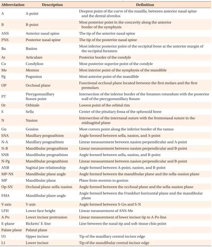
(2) Park et al • Mangement of CCD. INTRODUCTION Cleidocranial dysplasia (CCD), also known as cleido cranial dysostosis or Marie-Sainton syndrome, is an. autosomal dominant disorder with a prevalence of 1 in 1,000,000 individuals. It is characterized by delayed or absent fontanel closure, parietal and frontal bossing, hypoplastic clavicles, and narrow thorax; it may also. Table 1. Abbreviations, descriptions, and definitions of the relevant cephalometric points and measurements Abbreviation. Description. Definition. A. A-point. Deepest point of the curve of the maxilla, between anterior nasal spine and the dental alveolus. B. B-point. Most posterior point in the concavity along the anterior border of the symphysis. ANS. Anterior nasal spine. The tip of the anterior nasal spine. PNS. Posterior nasal spine. The tip of the posterior nasal spine. Ba. Basion. Most inferior posterior point of the occipital bone at the anterior margin of the occipital foramen. Ar. Articulare. Posterior border of the condyle. Co. Condylion. Most posterior superior point of the condyle. Me. Menton. Most interior point of the symphysis of the mandible. Pg. Pogonion. Most anterior point of the mandible. OP. Occlusal plane. Functional occlusal plane located between the first molars and the first premolars. PT. Pterygomaxillary fissure point. Intersection of the inferior border of the foramen rotundum with the posterior wall of the pterygomaxillary fissure. Or. Orbitale. Lowest point of the orbital rim. S. Sella. Center of the pituitary fossa of the sphenoid bone. N. Nasion. Intersection of the internasal suture with the frontonasal suture in the midsagittal plane. Gn. Gonion. Most convex point along the inferior border of the ramus. SNA. Maxillary prognathism. Angle formed between sella, nasion, and A-point. N-A. Maxillary prognathism. Linear measurement between nasion perpendicular and A-point. N-B. Mandibular prognathism. Linear measurement between nasion perpendicular and B-point. SNB. Mandibular prognathism. Angle formed between sella, nasion, and B-point. N-Pg. Mandibular prognathism. Linear measurement between nasion perpendicular and B-point. ANB. Sagittal jaw relationship. Angle formed between A-point, nasion, and B-point. MP-NS. Mandibular plane angle. Angle formed between the mandibular plane and the sella-nasion plane. MP. Mandibular plane. Plane from menton to gonion. Op-SN. Occlusal plane-sella-nasion. Angle formed between the occlusal plane and the sella-nasion plane. FMA. Mandibular plane angle. Angle formed between the Frankfurt horizontal plane and the mandibular plane. Y-axis. Y-axis. Angle formed between S-Gn and S-N. LFH. Lower face height. Linear measurement of ANS-Me. A-Po. Lower incisor protrusion. Linear measurement of lower incisor tip to A-Po line. E-plane. Ricketts’ E-line. Line between the nasal tip and soft-tissue chin point. Palate plane Palatal plane U1. Upper incisor. Tip of the maxillary central incisor edge. L1. Lower incisor. Tip of the mandibular central incisor edge. www.e-kjo.org. http://dx.doi.org/10.4041/kjod.2013.43.5.248. 249.
(3) Park et al • Mangement of CCD. present deafness, hypertelorism, and low nasal bridge. Young individuals with CCD show relatively normal jaw proportions and mandibular morphology, whereas older affected individuals tend to have short lower fa cial height, acute gonial angle, anterior inclination of the mandible, and mandibular prognathism. These diff erences are attributed to pronounced horizontal mandibular growth resulting from impaired vertical maxillary growth and eruption of permanent teeth.1 The dental manifestations include delayed exfoliation of primary teeth, delayed eruption or noneruption of per manent teeth, supernumerary teeth, retention cysts, and enamel hypoplasia (OMIM #119600). Heterogeneous mutations in core-binding factor. subunit alpha-1 (CBFA1) are associated with CCD. 2 CBFA1, also known as runt-related transcription factor 2 (RUNX2), is a transcription factor of the runt family, located on chromosome 6p21. 3 RUNX2 binds to and regulates multiple genes in osteoblasts; it has been identified as an osteoblast-specific transcription factor and is also regulated by bone morphogenetic proteins (BMPs) and vitamin D3.4,5 Along with its role in bone development, RUNX2 participates in tooth development by mediating interactions between epithelial and mesen chymal cells during tooth morphogenesis.6 Studies of the role of RUNX2 in normal tooth develop ment have provided some molecular explanations for the abnormal dental features in CCD. In the Lossdörfer. Figure 1. Initial record of patient 1 at age 10 years and 11 months. A, Frontal facial view; B, lateral facial view; C, frontal intraoral view; D, lateral intraoral view; E, panoramic radiograph; and F, lateral cephalogram.. 250. http://dx.doi.org/10.4041/kjod.2013.43.5.248. www.e-kjo.org.
(4) Park et al • Mangement of CCD. et al.7 study, an individual with CCD having a missense mutation in RUNX2 showed reduced basal expression of mRNA for receptor activator of nuclear factor kappaB ligand (RANKL), which is normally expressed on the cell membrane of osteoblasts. Given that RANKL is responsible for osteoclastic differentiation and activa tion, insufficient resorption of alveolar bone and failure of root resorption of primary teeth could lead to delayed exfoliation of primary teeth in affected individuals with an RUNX2 mutation. One explanation for the noneruption of permanent and supernumerary teeth is the lack of cellular cementum in the apical region of impacted teeth. Manjunath et. al.8 found that all the examined teeth from unaffected individuals had cellular cementum at the apices, whereas only 8% of the teeth from a 60-year-old patient with CCD had cellular cementum at the apices. Because the patient’s teeth had spontaneously erupted, the authors did not correlate the absence of cellular cementum with the failure of tooth eruption. Further, Counts et al.9 studied two teeth from a 22-year-old patient with CCD and found no significant correlation between ce mentum in this patient and that in a patient without CCD. These incongruent results indicate the need for further research on this topic. The issue becomes more perplexing because 40% of the CCD cases do. Figure 2. Progress record of patient 1 at age 17 years. A, Frontal facial view; B, lateral facial view; C, frontal intraoral view; D, lateral intraoral view; E, panoramic radiograph; and F, lateral cephalogram. www.e-kjo.org. http://dx.doi.org/10.4041/kjod.2013.43.5.248. 251.
(5) Park et al • Mangement of CCD. not have any apparent genetic cause.10 A possible role of environmental and other nongenetic factors in the variation of CCD symptoms, especially in the formation of supernumerary teeth, has been suggested by Suda et al.,11 who studied a case of monozygotic twins with an identical mutation in RUNX2 showing different numbers and positions of supernumerary teeth. The most recognizable dental features of CCD are supernumerary and retained primary teeth. The cranial defects may not be noticeable at birth. The physical symptoms are not progressive or debilitating. However, retention of primary teeth, midfacial hypoplasia, over closure, and skeletal class III malocclusion cause both functional and aesthetic problems.12 The dental management of individuals with CCD is. challenging and involves comprehensive orthodontic and surgical treatments. The four main therapeutic ap proaches published in the literature are the TorontoMelbourne, Belfast-Hamburg, Jerusalem, and Bronx methods.13 Here, we report two cases of CCD that had been treated surgically and orthodontically at the Uni versity of California, San Francisco (UCSF) School of Dentistry by using modifications of the Bronx (case 1) and Belfast-Hamburg (case 2) approaches. We also summarize the clinical features of 17 individuals who had been diagnosed with CCD at UCSF. Refer to Table 1 for details of the cephalometric points and measure ments.. Figure 3. Progress record of patient 1 at age 23 years. A, Frontal facial view; B, lateral facial view; C, frontal intraoral view; D, lateral intraoral view; E, panoramic radiograph; and F, lateral cephalogram. 252. http://dx.doi.org/10.4041/kjod.2013.43.5.248. www.e-kjo.org.
(6) Park et al • Mangement of CCD. CASE REPORT Case 1. Features A 12-year-old Asian boy diagnosed with CCD presented with his father, who had the same condition. The pa tient’s chief complaint was described by his father as “wanting normal eruption of his permanent teeth and better alignment of his upper and lower arches.” His medical and surgical histories included recurrent otitis media with tubes placed in the tympanic membranes and bilateral femoral osteotomies to correct coxa vara. The boy showed the classical craniofacial features of CCD: broad forehead, hypertelorism, and short upper third of the face (Figure 1A and 1B). The initial extraoral examination revealed a retrognathic and vertically defi cient maxilla (Figure 1A and 1B), resulting in a concave facial profile. The initial intraoral examination revealed a skeletal class III pattern with 9-mm anterior crossbite and 3mm open bite, moderate-to-severe maxillary crow ding, and mild mandibular crowding (Figure 1C and 1D). The panoramic radiograph showed nine retained primary teeth, seven supernumerary teeth, four im pacted permanent teeth, and no congenitally missing permanent teeth (Figure 1E). The initial lateral cepha. Figure 5. Superimposition of the pretreatment (dotted line) and posttreatment (solid line) lateral cephalograms in case 1.. Figure 4. Final record of patient 1 at age 24 years. A, Frontal facial view; B, lateral facial view; C, frontal intraoral view; D, lateral intraoral view. www.e-kjo.org. http://dx.doi.org/10.4041/kjod.2013.43.5.248. 253.
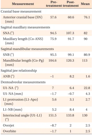
(7) Park et al • Mangement of CCD. logram confirmed the skeletal class III relationship due to maxillary underdevelopment (Figure 1F).. Treatment The treatment involved five steps and was expected to continue until skeletal maturity, when the under developed maxilla was to be surgically corrected. The first step was to treat multiple primary and newly erup ted permanent teeth for caries due to poor oral hygiene. At age 14, nine retained primary teeth (i.e., 53, 62, 63, 74, 73, 72, 82, 83, and 84) were extracted and seven supernumerary teeth were surgically removed under general anesthesia (Figure 2E). All the permanent canines were also removed, because they were determined to be nonretrievable due to severe impaction. The permanent teeth were observed for possible spontaneous eruption over 9 months. The mandibular lateral incisors and maxillary right lateral incisor failed to erupt spontaneously. These teeth were surgically exposed and bonded with brackets and gold chains tied to maxillary and mandibular lingual arches for guided eruption (Figure 2E). Simultaneously, a mesiodens and the maxillary right primary second molar were extracted. Shortly thereafter, full maxillary and mandibular fixed orthodontic appliances were placed for alignment and coordination of the dental arches (Figure 2C and 2D). The maxillary right lateral incisor was repositioned in the canine space (Figure 3E). To correct the transverse maxillary discrepancy, a tran spalatal arch was used. At skeletal maturity (age 19), the maxilla was advanced and downfractured to correct 6 mm of the sagittal maxillary discrepancy and vertical deficiency (Figure 3B and 3F). The postsurgical healing was good with excellent patient compliance and the orthodontic treatment was completed (Figure 3A, 3C, and 3D). The final step was implant placement and prosthodontic rehabilitation. Implants were placed for the maxillary right lateral incisor and left canine and mandibular canines (Figure 3E). The mandibular right caninepremolar space required two separate implants. Because the second molars were the last teeth to develop and the patient had been in treatment for a long time, these teeth were not retrieved. Treatment was completed with the delivery of the final crowns for the implants at age 24. Outcome The final extraoral and intraoral photographs are shown in Figure 4. Fifty-three hard-tissue and softtissue landmarks were digitized by an orthodontist using Dolphin software (version 11.5; Dolphin Imaging and Management Solutions, Chatsworth, CA, USA). The pre treatment and posttreatment lateral cephalograms were. 254. superimposed to illustrate treatment-induced changes (Figure 5). Comparison of the ANB, overbite, and overjet values between these time points (Table 2) showed a dramatic improvement in the sagittal jaw relationship after the maxillary advancement surgery and orthodontic treatment. Case 2. Features A 14-year-old Caucasian girl diagnosed with CCD pre sented for orthodontic treatment with a desire to have greater number of teeth to be visible. Her mother and younger sister also had CCD. Her medical and surgical histories revealed mitral valve prolapse, tethered spinal cord, arthritis, Crohn’s disease, thyroid reduction surgery, tonsillectomy, and Z-plasty of the lingual frenulum. The initial evaluation revealed jaw pain, bilateral temporo mandibular joint clicking, and facial muscle pain. Mouth. Table 2. Comparison of the pre-treatment and posttreatment cephalometric measurements in case 1 Measurement. PrePosttreatment treatment Mean. Cranial base measurement Anterior cranial base (SN) (mm). 57.6. 60.6. 76.1. SNA (o). 94.5. 107.3. 82. Maxillary length (Co-ANS) (mm). 75.9. 91.7. 90. Sagittal maxillary measurements. Sagittal mandibular measurements SNB (o) Mandibular length (Co-Pg) (mm). 95.5. 99.1. 104.6. 120.3. 80.9 113. Sagittal jaw relationship ANB (o). −1. 8.2. 1.6. Dentoalveolar measurements U1-NA (o). 7. 6.4. 22.8. −1.7. −0.7. 4.3. L1 protrusion (L1-Apo) (mm). 5.6. 3.1. 2.7. L1-NB (mm). 5.2. 8.4. 4. 151.5. 133.8. 130. Overjet. −8.7. 2. 2.5. Overbite. −1.7. 1. 2.5. U1-NA (mm). Interincisal angle (U1-L1) (o). Refer to Table 1 for details of the cephalometric points and measurements.. http://dx.doi.org/10.4041/kjod.2013.43.5.248. www.e-kjo.org.
(8) Park et al • Mangement of CCD. Figure 6. Initial record of patient 2 at age 14 years. A, Frontal facial view; B, lateral facial view; C, frontal intraoral view; D, lateral intraoral view; E, panoramic radiograph; and F, lateral cephalogram. breathing, tongue thrusting, and clenching habits were also noted. The patient had a concave facial profile and decreased facial height due to maxillary underdevelopment. Her lips were retrusive, resulting in an edentulous appearance (Figure 6A and 6B). The intraoral examination showed that she had five retained primary teeth (i.e., 55, 63, 75, 74, and 73) and 15 fully erupted permanent teeth (Figure 6C and 6D). She had a negative sagittal jaw relationship with class III malocclusion due to the hypoplastic maxilla (Figure 6D). On the panoramic radiograph, 14 impacted permanent teeth were identified and the left mandibular second molar was determined to be congenitally missing. www.e-kjo.org. http://dx.doi.org/10.4041/kjod.2013.43.5.248. (Figure 6E). The lateral cephalogram confirmed maxillary hypoplasia (Figure 6F). Multiple impacted supernumerary teeth had been extracted 2 years earlier along with the mandibular right first molar, due to extensive caries.. Treatment Orthodontic treatment was initiated with the following objectives: maxillary expansion, removal of the retained primary and supernumerary teeth, guided eruption or extrusion of the impacted permanent teeth, and cor rect ion of the class III jaw relationship by maxillary advancement surgery. A hyrax expander was placed for 2 months, which. 255.
(9) Park et al • Mangement of CCD. Figure 7. Progress record of patient 2 at age 17 years (A and B), 15 years (C and D), and 16 years (E and F). A, Frontal facial view; B, lateral facial view; C, frontal intraoral view; D, lateral intraoral view; E, panoramic radiograph; and F, lateral cephalogram. achieved 11 mm of expansion (Figure 7C). Heavy upper and lower wires were placed; the retained primary teeth were removed; and impacted permanent teeth 11, 15, 21, 22, 23, 35, and 47 were surgically exposed and bonded with gold chains placed close to the cusp tips for guided eruption (Figure 7C and 7D). All the bonded teeth except 21 erupted after approximately 11 months. The maxillary left central incisor was extracted because of exposure of the root, which was very short. Orthodontic alignment and coordination of the dental arches were complete in 14 months (Figure 7E). Le Fort I osteotomy was performed at age 18 (Figure 8D). With. 256. the removal of the remaining orthodontic appliances and placement of an implant and a fixed prosthesis for the maxillary left central incisor, the treatment was completed (Figure 8A-8D). However, the patient’s poor oral hygiene during the retention stage led to complete loss of the maxillary dentition.. Outcome Although no post treatment extraoral and intraoral photographs were obtainable, because of the loss of the maxillary teeth, the Dolphin software was used to digitize and superimpose the pretreatment and. http://dx.doi.org/10.4041/kjod.2013.43.5.248. www.e-kjo.org.
(10) Park et al • Mangement of CCD. Figure 8. Final record of patient 2 at age 18 years. A, Frontal facial view; B, lateral facial view; C, frontal intraoral view; D, lateral intraoral view. postsurgical lateral cephalograms (Figure 9). Comparison of the ANB and overjet values between these time points indicated improvement in the sagittal jaw relationship after the maxillary advancement surgery and orthodontic treatment (Table 3). The overbite also improved significantly, from −6.7 mm to 0.1 mm. Figure 9. Superimposition of the pretreatment (dotted line) and posttreatment (solid line) lateral cephalograms in case 2.. www.e-kjo.org. http://dx.doi.org/10.4041/kjod.2013.43.5.248. Clinical features of individuals with CCD We reviewed our database (FileMaker Pro 10.0 V3; FileMaker Inc., Santa Clara, CA, USA) of all the indi viduals with CCD who had been diagnosed at the UCSF. Table 4 summarizes their clinical features. Seventeen (nine female and eight male) affected indivi duals were identified. Four of 15 individuals (the height of two individuals was not recorded) had short stature. Further, 57% had a family history of CCD, with one-tothree generations of CCD passed down in the family. Eight of the 17 individuals had vertebral changes such as scoliosis. The characteristic features of this condition, such as missing or hypoplastic clavicles, persistent metopic sutures, frontal bossing, and hypertelorism, were observed in 93%, 94%, 88%, and 69% of these subjects, respectively. A high incidence of otitis media that required surgery to place pressure-equalization tubes was noted. Seven patients had speech problems and received speech therapy (Table 4).. 257.
(11) Park et al • Mangement of CCD. Table 3. Comparison of the pre-treatment and posttreatment cephalometric measurements in case 2 Measurement. PrePosttreatment treatment Mean. Cranial base measurement Anterior cranial base (SN) (mm). 58.4. 56.3. 75.1. SNA (o). 88.7. 86.4. 82. Maxillary length (Co-ANS) (mm). 71.9. 75.5. 90. 80.9. Sagittal maxillary measurements. Sagittal mandibular measurements SNB (o). 91. 85.2. Mandibular length (Co-Pg) (mm). 95. 98.4. 113. Regarding the dental features, congenital hypodontia was observed in 12% of this population. The number of retained primary teeth ranged from 0 to 20, with the mandibular canine being the most frequently retained primary tooth (Table 5). On average, the mandibular primary teeth were slightly more likely to be retained than the maxillary ones. The number of impacted permanent teeth ranged from 3 to 27, with the incisors and canines being the most frequently impacted in the maxillary and mandibular arches, respectively (Table 6). Zero to 18 supernumerary teeth were observed. Noteworthily, 14 of the 17 subjects had maxillary hypoplasia, 88% had class III malocclusion due to the hypoplastic maxilla, and 10 patients required Le Fort I maxillary advancement surgery.. DISCUSSION. Sagittal jaw relationship ANB (o). –2.3. 1.1. 1.6. 46.7. 10.9. 22.8. 1.5. 0.7. 4.3. –0.4. 3.1. 2.7. Dentoalveolar measurements U1-NA (o) U1-NA (mm) L1 protrusion (L1-Apo) (mm) L1-NB (mm). –0.2. 1.4. 4. 118.9. 153.4. 130. Overjet. 0.6. 0.7. 2.5. Overbite. –6.7. 0.1. 2.5. Interincisal angle (U1-L1) (o). Refer to Table 1 for details of the cephalometric points and measurements.. Management of the orofacial manifestations of CCD is lengthy and challenging, and requires careful planning and execution by an interdisciplinary team. The length of treatment ranges widely and is generally not completed until growth has seized. By reviewing these cases, we appreciate the importance of interdisciplinary teamwork for a successful outcome. We have also learned that it is critical to maintain the patient’s motivation and keep the treatment within the patient’s tolerance limit for a good outcome. Failure to meet these criteria may lead to deleterious results, as seen in case 2. The UCSF approach in case 1 is similar to the Bronx approach. Extraction of all the retained primary and supernumerary teeth was followed by an observation. Table 4. Clinical features of 17 individuals diagnosed with CCD at the UCSF Clinical features Gender. Summary (fractions or % from evaluating 17 individuals). Dental features. Summary (fractions or % from evaluating 17 individuals). 9F/8M. Maxillary hypoplasia. 82. Short stature. 27. Class III malocclusion. 88. Positive family history. 57. Individuals with congenitally missing teeth. 12. Vertebral alteration. 47. Number of over-retained primary teeth. 0 to 20. Missing or hypoplastic clavicles. 93. Number of impacted/delayed eruption of permanent teeth (excluding impacted 3rd molars). 3 to 27. persistant metopic suture. 94. Number of supernumerary teeth. 0 to 18. Frontal bossing. 88. Le Fort I advancement performed (+)/ anticipated. 59. Hypertelorism. 69. Speech pathology. 41. ENT pathology (otitis media)/ PE tubes. 88. CCD, Cleidocranial dysplasia; UCSF, the University of California, San Francisco; EENT, ear nose throat; PE, pressure equalizer; F, female; M, male.. 258. http://dx.doi.org/10.4041/kjod.2013.43.5.248. www.e-kjo.org.
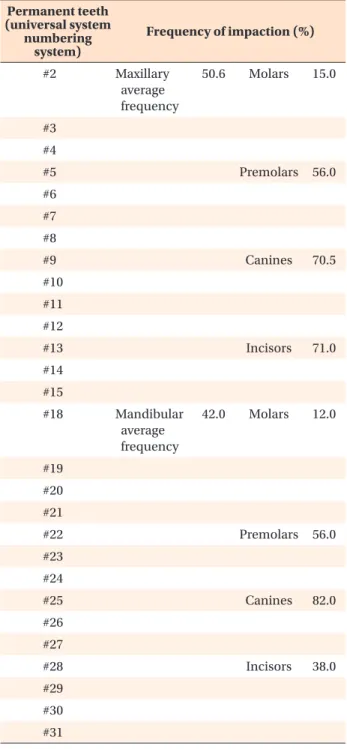
(12) Park et al • Mangement of CCD. Table 5. Frequency of retention of primary teeth in 16 individuals with CCD Primary teeth (universal system letter) A. Permanent teeth (universal system numbering system). Frequency of primary teeth retention (%, out of 16) Maxillary average frequency. 52.7. Table 6. Frequency of impaction or delayed eruption of permanent teeth in 17 individuals with CCD. Molars. 48.5. Canines. 63.0. #2. B C. #5. F. #6. G. #7. H. Incisors. 51.8. #9. J. #10 Mandibular 57.9 average frequency. Molars. 62.8. 15.0. Premolars. 56.0. Canines. 70.5. Incisors. 71.0. Molars. 12.0. Premolars. 56.0. Canines. 82.0. Incisors. 38.0. #11 #12. L. #13. M. #14. N. #15 Canines. 69.0. #18. Incisors. 55.0. #19. P Q S. #20. T. #21. Mandibular average frequency. #22. period and a second surgical phase of exposing and bonding the impacted permanent teeth. After ortho dontic alignment and coordination, patient 1 underwent a third surgical phase to correct the skeletal class III malocclusion. The treatment was completed with the restorative phase using implants and fixed prostheses. Case 2 was approached by using a method similar to the Belfast-Hamburg approach. In a single surgical operation, all the retained and supernumerary teeth were extracted and surgical exposure and bonding of the impacted permanent teeth were completed. However, as with 14 out of 17 patients described in our summary of clinical features of CCD, case 2 showed maxillary hypoplasia and required a second surgical phase to correct the skeletal class III malocclusion. We have summarized the clinical features of CCD observed in 17 individuals to aid in early diagnosis and, thereby, better management of this rare condition. A greater number of genetic and molecular studies are needed to explain the features, patterns, and variation in the severity of the syndrome.. www.e-kjo.org. Molars. #8. I. R. 50.6. #4. E. O. Maxillary average frequency. #3. D. K. Frequency of impaction (%). http://dx.doi.org/10.4041/kjod.2013.43.5.248. 42.0. #23 #24 #25 #26 #27 #28 #29 #30 #31 CCD, Cleidocranial dysplasia.. Recent advances in dentistry have yielded better thera peutic options and outcomes for individuals with CCD. For example, dental implants can be successfully used not only for orthodontic tooth movement with reduced side effects but also for replacing teeth that cannot be guided into the arch orthodontically, as presented in cases 1 and 2. Although the cases presented here were completed. 259.
(13) Park et al • Mangement of CCD. before the introduction of cone-beam computed tomo graphy (CBCT), this technology is now routinely used to provide a three-dimensional image of the dentition, reducing guesswork and ensuring better anatomical localiz ation of supernumerary and impacted teeth. CBCT can provide pertinent information such as the precise location of a supernumerary tooth in relation to important structures such as the cortex of the nasal floor, labial cortex of the nasal ridge, nasopalatine duct, and adjacent root apices.14 Such developments will con tinue to improve the management of complex dental cases.. CONCLUSION Individuals with CCD require extensive diagnostic work-up and a long-term treatment plan. The UCSF treatment protocol incorporates the Bronx and the Belfast-Hamburg approaches and can be organized into five steps. It involves the appropriate timing of removal of supernumerary and retained primary teeth, surgical exposure of impacted permanent teeth, ortho dontic extrusion and alignment, followed by Le fort I advancement and implant retained prosthesis. The two cases reported prove the efficacy of this protocol in resolving the clinical dentofacial features of CCD.. ACKNOWLEDGEMENTS The authors are grateful to UCSF Craniofacial Team and Dr. Gerald Nelson.. REFERENCES 1. Ishii K, Nielsen IL, Vargervik K. Characteristics of jaw growth in cleidocranial dysplasia. Cleft Palate Craniofac J 1998;35:161-6. 2. Lou Y, Javed A, Hussain S, Colby J, Frederick D, Pratap J, et al. A Runx2 threshold for the cleido cranial dysplasia phenotype. Hum Mol Genet 2009; 18:556-68. 3. Mundlos S, Otto F, Mundlos C, Mulliken JB, Ayls worth AS, Albright S, et al. Mutations involving the transcription factor CBFA1 cause cleidocranial dysplasia. Cell 1997;89:773-9. 4. Ducy P, Zhang R, Geoffroy V, Ridall AL, Karsenty G.. 260. Osf2/Cbfa1: a transcriptional activator of osteoblast differentiation. Cell 1997;89:747-54. 5. Baumert U, Golan I, Redlich M, Aknin JJ, Muessig D. Cleidocranial dysplasia: molecular genetic analysis and phenotypic-based description of a Middle European patient group. Am J Med Genet A 2005;139A:78-85. 6. Yamashiro T, Aberg T, Levanon D, Groner Y, Thesleff I. Expression of Runx1, -2 and -3 during tooth, palate and craniofacial bone development. Mech Dev 2002;119(Suppl 1):S107-10. 7. Lossdörfer S, Yildiz F, Götz W, Kheralla Y, Jäger A. Anabolic effect of intermittent PTH(1-34) on the local microenvironment during the late phase of periodontal repair in a rat model of tooth root resorption. Clin Oral Investig 2010;14:89-98. 8. Manjunath K, Kavitha B, Saraswathi TR, Sivapatha sundharam B, Manikandhan R. Cementum analysis in cleidocranial dysostosis. Indian J Dent Res 2008; 19:253-6. 9. Counts AL, Rohrer MD, Prasad H, Bolen P. An assessment of root cementum in cleidocranial dysplasia. Angle Orthod 2001;71:293-8. 10. Tanaka JL, Ono E, Filho EM, Castilho JC, Moraes LC, Moraes ME. Cleidocranial dysplasia: importance of radiographic images in diagnosis of the condition. J Oral Sci 2006;48:161-6. 11. Suda N, Hattori M, Kosaki K, Banshodani A, Kozai K, Tanimoto K, et al. Correlation between genotype and supernumerary tooth formation in cleidocranial dysplasia. Orthod Craniofac Res 2010;13:197-202. 12. Becker A, Lustmann J, Shteyer A. Cleidocranial dysplasia: Part 1-General principles of the ortho dontic and surgical treatment modality. Am J Or thod Dentofacial Orthop 1997;111:28-33. 13. Roberts T, Stephen L, Beighton P. Cleidocranial dysplasia: a review of the dental, historical, and practical implications with an overview of the South African experience. Oral Surg Oral Med Oral Pathol Oral Radiol 2013;115:46-55. 14. Liu DG, Zhang WL, Zhang ZY, Wu YT, Ma XC. Threedimensional evaluations of supernumerary teeth using cone-beam computed tomography for 487 cases. Oral Surg Oral Med Oral Pathol Oral Radiol Endod 2007;103:403-11.. http://dx.doi.org/10.4041/kjod.2013.43.5.248. www.e-kjo.org.
(14)
수치


+6


관련 문서
Conclusion : The treatment using facet joint block with lumbar surgical operation seems to have short term clinical effects, however can't prevent
The objective of this study is to analyze the shear bond strength of orthodontic buttons according to light tip distance and optic fiber diameter when an extended optic
In the analysis of soluble solids, the treatment group with material grains and the combined groups of glutinous millet and barley appeared high, and
The effects of recycling on the tensile bond strength of new and clinically used stainless steel orthodontic brackets: an in vitro study. Material testing of
The average tensile strength value before heat treatment of PLA output was 9.67 N/㎟, and the average tensile strength value after heat treatment was 24.17 N/㎟, which
The purpose of this study was to provide primary material in discussing methods to activate the treatment effects of diabetes and reduce the rate of
This study aims to suggest a basic data for the activation of aromatherapy pimple treatment program with the pimple skin care and treatment by testing the actual
Clinical evaluation of the use of decalcified freeze-dried bone allograft with guided tissue regen- eration in the treatment of molar furca- tion invasions.. Wallace SC,
