DOI:10.4047/jap.2011.3.2.76
ORIGINAL ARTICLE J Adv Prosthodont 2011;3:76-80
Corresponding author: Joo-Hee Lee
Department of Prosthodontics, Asan Medical Center, College of Medicine, University of Ulsan
86, Asanbyeongwon-gil, Songpa-gu, Seoul, 138-736, Korea Tel. 82 2 3010 5803: e-mail, [email protected]
Received April 5, 2011 / Last Revison April 26, 2011 / Accepted April 27, 2011
ⓒ 2011 The Korean Academy of Prosthodontics
This is an Open Access article distributed under the terms of the Creative Commons Attribution Non-Commercial License (http://creativecommons.org/licenses/by- nc/3.0) which permits unrestricted non-commercial use, distribution, and reproduction in any medium, provided the original work is properly cited.
INTRODUCTION
Under defined conditions, early and immediate loading protocols have been considered as attractive options compared with the classical 1- or 2-stage delayed loading approaches.1 Compared with delayed loading, early or immediate loadings mean not only an instant reduction in oral handicap, but also result in shorter treatment time and less service. The decision whether or not to continue with one of these treatment options at the time of implant placement is based on the assessment of implant stability. Several authors suggested that primary sta- bility may be a useful predictor for osseointegration2,3and that a high primary stability makes immediate loading more pre- dictable.4 A number of methods have been introduced to assess implant stability using both invasive and non-invasive ways. Though assessment of removal torque and histologic and histomorphometric evaluation provide reliable data on the strength
of the interface and the quality of implant anchorage in peri- implant bone,4these destructive measures are applicable in an experimental environment only. Clinical settings require non- destructive techniques. Non-destructive conventional methods, such as clinical evaluation through manipulation with forceps or judgment of percussion sound are highly subjective and lack the reliability. Peak insertion and removal torque values may be used,5but torque measurements can be performed only dur- ing implant insertion. Other objective methods like Periotest (Gulden, Bensheim, Germany) or the Dental Fine Tester (Kyocera, Kyoto, Japan) have been proposed, and they can mon- itor implant stability over the healing period. However, their lacks of resolution, poor sensitivity and susceptibility to operator variables have been criticized.2
In recent years, OstellTM device for resonance-frequency analysis (RFA) has been advocated to provide an objective mea- surement of implant primary stability and to monitor implant
The relationship between initial implant stability quotient values and bone-to-implant contact ratio
in the rabbit tibia
In-Phill Park1, DDS, MSD, Seong-Kyun Kim1, DDS, PhD, Shin-Jae Lee2, DDS, PhD, Joo-Hee Lee3*, DDS, PhD
1Department of Prosthodontics and Dental Research Institute, School of Dentistry, Seoul National University,
2Department of Orthodontics and Dental Research Institute, School of Dentistry, Seoul National University,
3Department of Prosthodontics, Asan Medical Center, College of Medicine, University of Ulsan, Seoul, Korea
PURPOSE. Implant stability quotient (ISQ) values have been supposed to predict implant stability. However, the relationship between ISQ values and bone-to-implant contact ratio (BIC%) which is one of the predictors of implant stability is still unclear. The aim of the present study was to evaluate initial ISQ values in relation to BIC% using rabbit model. MATERIALS AND METHODS. Four New Zealand white rabbits received a total of 16 implants in their tibia. Immediately after implant placement ISQ values were assessed. The measurements were repeat- ed at the time of sacrifice of the rabbits after 4 weeks. Peri-implant bone regeneration was assessed histomorphometrically by measuring BIC% and bone volume to total volume values (bone volume %). The relationships between ISQ values and the histomorphometric output were assessed, and then, the osseointegration prediction model via the initial ISQ values was processed. RESULTS. Initial ISQ values showed significant correlation with the BIC%. The bone volume % did not show any significant association with the ISQ values. CONCLUSION. In the limitation of this study, resonance frequency analysis is a useful clinical method to predict the BIC% values and examine the implant sta- bility. [J Adv Prosthodont 2011;3:76-80]
KEY WORDS. Implant stability, Resonance frequency analysis, Initial ISQ values, BIC%, Rabbit tibia
*This study was supported by a grant (2008-434) from the Asan Institute for Life Sciences, Seoul, Korea.
stability over the healing period3,6,7and in the long term in a non destructive manner.8This approach uses a transducer that is fixed to the implant and vibrated using a piezoceramic element with a frequency range from 5 to 15 KHz.9,10The resulting vibra- tion of the abutment-implant system produces a sharp increase in amplitude when the resonance frequency of the system is reached. This resonance frequency changes according to the stiffness of the excited abutment-implant system. Increasing bone anchorage of an implant would alter the resonance characteristics because of changes in stiffness of abutment-implant system in its peri-implant bone.10Thus, changes in resonance frequency of an implant could indicate changes in anchorage of the implant and allow for conclusions on implant stability.
A number of experimental and clinical studies showed increas- ing RFA values during healing after implant placement.
These increased Implant Stability Quotient (ISQ) values were attributed to increased bone anchorage.11-13Thus, changes in resonance frequency (RF) of an implant may possibly reflect changes in anchorage of the implant. Several factors influ- encing the RF of a dental implant have been proposed.14 Factors such as implant length and design, location of the first bone contact, degree of bone-to-implant contact (BIC), alve- olar bone trabecular pattern, thickness of cortical bone and bone density have been investigated in different model studies, animal experiments and human studies.14-18However, a correlation between RFA and other factors still remains conflicting and con- fusing.
This study attempted to verify the relationship between the ISQ values by RFA immediately after the implant place- ment for the future bone implant contact, BIC%.
MATERIALS AND METHODS
The animal study was approved by the Institutional Animal Care and Use Committee (ASANH 200802105) in Asan Institute of Life Science and followed the routine guidelines of the Laboratory of Animal Research of Asan Medical Center (Seoul, Korea).
Implant preparation
Threaded implants were manufactured via the machining of commercially pure titanium (Grade 4) (warentec Co., Seoul, Korea). The implants had lengths of 7.0 mm, outer diameters of 3.75 mm, and pitch-heights of 0.6 mm. The anodic oxida- tion treatment of the implant was performed at 300 V in an aque- ous electrolytic solution of 0.02 M calcium glycerophos- phate and 0.15 M calcium acetate. All procedures were per- formed at room temperature and a single implant anodization time was three minutes. A total of 16 implants were washed with distilled water, dried, and sterilized in ethylene oxide (EO) gas prior to animal surgery.
Surgical procedures
Four New Zealand white rabbits, each weighing 3 to 3.5 kg, were used in this study. For surgery, general anesthesia was induced through the intramuscular injection of 10 mg/kg of Zoletil (Vibac, Carros Cedex, France) and 0.15 ml/kg of Rompun (Bayer Korea, Ansan, Korea). Both rear legs of each rabbit were shaved and washed with iodine solution. Two percent lidocaine (1.0 ml) (Yu-han; 1:100,000) was administered at the tibial area. Using sterile surgical techniques, an incision was made into the skin to expose the proximal aspect of each tibia. The muscles were then dissected to allow for the elevation of the periosteum.
The flat surface on the lateral aspect of the proximal tibia was selected for implant placement. Holes were drilled into the tib- ia with a low-speed rotary instrument under constant irrigation with sterile saline. Tapping procedures were omitted. Each rab- bit had two implants inserted into each tibia, and a total of 16 implants were inserted. Muscle and fascial layers were closed in layers with Vicryl resorbable sutures (Woori Medical, Namyangju, Korea), while the skin was sutured with black silk for primary closure. Postoperatively, all animals received 50 mg/kg of Cefazolin sodium (Chong Kun Dang Pharm., Seoul, Korea) intramuscularly.19,20
Resonance frequency analysis
Instantly after the implant placement, RFA was performed using OstellTMMentor (Integration Diagnostics AB, Go¨teborg, Sweden). The Smartpeg (Integration Diagnostics AB, Go¨teborg, Sweden) was attached to the fixture with the aid of a mount with 4 - 5 NCm of torque. Subsequently, the probe was held close to the peg in vertical and horizontal direction during the pulsing time (Fig. 1). After the processing time, the ISQ val- ue was presented on the display. The measurement was repeated twice, once on the proximal side and the other on the inner side, respectively.10Then averages were calculated to reduce
Fig. 1. Photograph of the OstellTMMentor resonance frequency analy- sis transducer attached to an implant immediately after implant placement.
measurement errors. Four weeks after the implant insertion, the ISQ values were measured twice again before the rabbits were sacrificed.
Preparation of specimens and histomorphologic evaluation
The implants and the surrounding bone were harvested en bloc and fixed in neutral buffered formalin, dehydrated in 70%, 90%, 95%, and 100% alcohol, and embedded in a light-curing resin (Technovit 7200 VLC; Kulzer, Wehrheim, Germany). An Exakt sawing machine with grinding equipment (Exakt Apparatebau, Norderstedt, Germany) was used to cut and grind sections approximately 50 um thick that were then stained with 1% toluidine blue prior to evaluation under a light microscope.21All animals underwent histologic examination with the aid of an Olympus BX microscope (Olympus, Tokyo, Japan) connected to a computer. Image Tool Ver. 3.0 (San Antonio Dental School, University of Texas Health Science Center, USA) software was used to calculate the percent- age of BIC% in whole implant surface and the bone vol- ume % inside the thread. All the measurements were calculated under 100× magnification. A higher magnification objective and zoom were used to help determine whether or not the bone was in contact with the implant surface.
Statistics
To measure the agreement for the repeated measurements of ISQ values, the root mean squared values and the intra class classification coefficient (ICC) was calculated. It showed 2.8 and 2.2 for the initial and final ISQ values, respectively. In terms of ICC, the values were 0.954 and 0.966 for the initial and final ISQ values, respectively, which indicated an excel- lent agreement.
Paired t tests were used to compare the difference between
the initial and final ISQ values. In order to develop a BIC% pre- diction model, the normality assumption and equality of variance (homoscedasticity) were checked. In addition, indi- vidual rabbit’s random effect was tested through random group resampling method.22The rabbit random effect was not significant. There was no significant variation in the BIC% across the 4 rabbits in our sample. Thus, using a linear model looked sufficient. Multiple linear regression analyses were performed to determine associations between the ISQ values and the two histomorphometric values, BIC% and bone volume ratio.
The language R (R Development Core Team, 2009) was used to perform the data analysis. All values were considered sig- nificant when P<.05.
RESULTS
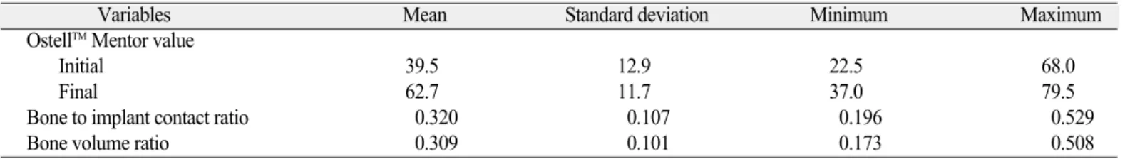
ISQ values, BIC ratio and bone volume ratio are shown in Table 1. Four weeks after the implant placement, the ISQ val- ues were increased (P<.001) showing the average of 23.2 of the difference. However, there was no significant correla- tion between the initial and final ISQ values. In addition, there were no significant correlation either between the BIC%
and bone volume%.
Development and formulation for the prediction model
Results of the multiple regression analyses for the resultant 2 histomorphometric variables are presented in Table 2. The adjusted R2 for the ISQ values included in the regression analy- sis for the BIC% was 0.473. The initial ISQ values showed sig- nificant correlation with the BIC ratio and showed a positive association with the BIC%. On the contrary the final ISQ val- ues did not show significant association on the BIC%. The bone volume% did not show any significant association with the ISQ values.
Table 2. Results of the multiple regression analyses for the resultant 2 bone-implant variables (P<.05)
Variables Significance Adjusted R2
Bone to implant contact ratio Initial OstellTMMentor value 0.014* 0.473*
Final OstellTMMentor value 0.066
Bone volume ratio Initial OstellTMMentor value 0.431 0.02
Final OstellTMMentor value 0.308 Table 1. ISQ values, BIC ratio and bone volume ratio
Variables Mean Standard deviation Minimum Maximum
OstellTMMentor value
Initial 39.5 12.9 22.5 68.0
Final 62.7 11.7 37.0 79.5
Bone to implant contact ratio 0.320 0.107 0.196 0.529
Bone volume ratio 0.309 0.101 0.173 0.508
DISCUSSION
Stability both at placement and during function is an impor- tant criterion for the success of dental implant.15Cadaver studies had shown that RFA values did correlate with the sur- face of BIC, and with the height of the crestal cortical bone pen- etrated by the implants in the oral aspects of the implant sites.16,23 On the contrary, many researchers have failed to show a correlation between the degree of implant-bone con- tacts and RFA measurement after healing period. Schliephake et al. could not find any correlation between BIC% and ISQ values of 80 implants in 10 foxhounds after healing periods of 1 or 3 months.17Ito et al. presented similar results from an exper- iment where 24 implants placed in the tibia of four mini-pigs were analyzed with RF and histology after 1, 2 and 4 weeks.24 The present study confirmed both positive and negative results that had been reported previously about RFA value in clinical and experimental settings. The RFA values which were measured just before the rabbits were sacrificed showed no cor- relation with the histomorphometric parameters. At first we had expected to find more or less closer relationship between the final RFA values and the BIC%. On the contrary, the initial ISQ values showed a significant effect on the BIC% unlike the final RFA values. After looking at the raw data again, the final ISQ values showed a relatively narrower range than the initial ISQ values did. Statistically, homogeneity of an inde- pendent variable increases the variation in the regression coefficient for the output variable, making it difficult to assess the effect of the independent variable on the output vari- able. This is partly why the final RFA values showed the larg- er probability values than P>.05.
In addition, the upper portion of the rabbit tibia for experi- ments is composed of 1 - 2 mm thickness of cortical layer sur- rounding the medullary canal. It could be speculated that the differences of primary RFA value came from differences of thickness of cortical bone under same fixtures and same sur- gical skills. Miyamoto et al. found a strong linear correlation between resonance frequency and the thickness of cortical bone measured by computed tomography.25However, in modern implant dentistry using moderately rough implants, the surface is often covered by a thin layer of bone, which is probably not important for the biomechanical support of implants. Hence, the final RFA values did not influence much on the BIC%.14 If some values immediately after an implant placement could predict a future retentive stability, it would provide important information to clinicians. Because of the considerable variation in the body reaction after a placement of a dental pros- thetic implant, it seemed doubtful whether any single mea- surement for the initial stability would be suitable for predicting future BIC characteristics. This study suggested the possibility of primary stability measured by RFA using OstellTMMentor as a predicting factor of future osseointegration, although it had
a limitation that the experiment were conducted with the rabbit tibia.
CONCLUSION
In this study, initial ISQ values measured by RFA showed sig- nificant correlation with the BIC% after 4 weeks of healing.
And BIC prediction model which used ISQ values to predict future osseointegration status showed reliable results in a rabbit tibia model. Conclusively the ISQ values might be a use- ful clinical reference to predict the degree of osseointegration when placing an implant.
REFERENCES
1. Esposito M, Grusovin MG, Willings M, Coulthard P, Worthington HV. Interventions for replacing missing teeth: different times for loading dental implants. Cochrane Database Syst Rev 2007;
2:CD003878.
2. Meredith N. Assessment of implant stability as a prognostic de- terminant. Int J Prosthodont 1998;11:491-501.
3. Friberg B, Sennerby L, Linden B, Gro¨ndahl K, Lekholm U.
Stability measurements of one-stage Bra�nemark implants dur- ing healing in mandibles. A clinical resonance frequency analy- sis study. Int J Oral Maxillofac Surg 1999;28:266-72.
4. Szmukler-Moncler S, Piattelli A, Favero GA, Dubruille JH.
Considerations preliminary to the application of early and im- mediate loading protocols in dental implantology. Clin Oral Implants Res 2000;11:12-25.
5. Kim SK, Heo SJ, Koak JY, Lee JH, Kwon JY. Development of predictable stability test for assessment of optimum loading time in dental implant. J Korean Acad Prosthodont 2008;46:628-33.
6. Rasmusson L, Kahnberg KE, Tan A. Effects of implant design and surface on bone regeneration and implant stability: an ex- perimental study in the dog mandible. Clin Implant Dent Relat Res 2001;3:2-8.
7. Bischof M, Nedir R, Szmukler-Moncler S, Bernard JP, Samson J. Implant stability measurement of delayed and immediately loaded implants during healing. Clin Oral Implants Res 2004;15:
529-39.
8. Heo SJ, Sennerby L, Odersjo¨M, Granstro¨m G, Tjellstro¨m A, Meredith N. Stability measurements of craniofacial implants by means of resonance frequency analysis. A clinical pilot study.
J Laryngol Otol 1998;112:537-42.
9. Meredith N. A review of nondestructive test methods and their application to measure the stability and osseointegration of bone anchored endosseous implants. Crit Rev Biomed Eng 1998;26:275-91.
10. Sim CP, Lang NP. Factors influencing resonance frequency analy- sis assessed by Osstell mentor during implant tissue integration:
I. Instrument positioning, bone structure, implant length. Clin Oral Implants Res 2010;21:598-604.
11. Meredith N, Book K, Friberg B, Jemt T, Sennerby L. Resonance frequency measurements of implant stability in vivo. A cross- sectional and longitudinal study of resonance frequency mea- surements on implants in the edentulous and partially dentate max- illa. Clin Oral Implants Res 1997;8:226-33.
12. Meredith N, Shagaldi F, Alleyne D, Sennerby L, Cawley P. The application of resonance frequency measurements to study the stability of titanium implants during healing in the rabbit tibia.
Clin Oral Implants Res 1997;8:234-43.
13. Sennerby L, Persson LG, Berglundh T, Wennerberg A, Lindhe J. Implant stability during initiation and resolution of experimental
periimplantitis: an experimental study in the dog. Clin Implant Dent Relat Res 2005;7:136-40.
14. Sennerby L, Meredith N. Implant stability measurements using resonance frequency analysis: biological and biomechanical aspects and clinical implications. Periodontol 2000 2008;47:
51-66.
15. Meredith N, Alleyne D, Cawley P. Quantitative determina- tion of the stability of the implant-tissue interface using resonance frequency analysis. Clin Oral Implants Res 1996;7:261-7.
16. Nkenke E, Hahn M, Weinzierl K, Radespiel-Tro¨ger M, Neukam FW, Engelke K. Implant stability and histomorphometry: a correlation study in human cadavers using stepped cylinder im- plants. Clin Oral Implants Res 2003;14:601-9.
17. Schliephake H, Sewing A, Aref A. Resonance frequency mea- surements of implant stability in the dog mandible: experi- mental comparison with histomorphometric data. Int J Oral Maxillofac Surg 2006;35:941-6.
18. Alsaadi G, Quirynen M, Michiels K, Jacobs R, van Steenberghe D. A biomechanical assessment of the relation between the oral implant stability at insertion and subjective bone quality assessment.
J Clin Periodontol 2007;34:359-66.
19. Lee SY, Koak JY, Heo SJ, Kim SK, Lee SJ, Nam SY.
Osseointegration of anodized titanium implants coated with poly(lactide-co-glycolide)/basic fibroblast growth factor by
electrospray. Int J Oral Maxillofac Implants 2010;25:315-20.
20. Yoon HI, Yeo IS, Yang JH. Effect of a macroscopic groove on bone response and implant stability. Clin Oral Implants Res 2010;21:1379-85.
21. Donath K, Breuner G. A method for the study of undecalcified bones and teeth with attached soft tissues. The Sa¨ge-Schliff (saw- ing and grinding) technique. J Oral Pathol 1982;11:318-26.
22. Bliese P. Multilevel Modeling in R (2.3). A Brief Introduction to R, the multilevel package and the nlme package. Retrieved February 13, 2009. from http://www.R-project.org.
23. Gedrange T, Hietschold V, Mai R, Wolf P, Nicklisch M, Harzer W. An evaluation of resonance frequency analysis for the determination of the primary stability of orthodontic palatal im- plants. A study in human cadavers. Clin Oral Implants Res 2005;
16:425-31.
24. Ito Y, Sato D, Yoneda S, Ito D, Kondo H, Kasugai S. Relevance of resonance frequency analysis to evaluate dental implant stability: simulation and histomorphometrical animal experiments.
Clin Oral Implants Res 2008;19:9-14.
25. Miyamoto I, Tsuboi Y, Wada E, Suwa H, Iizuka T. Influence of cortical bone thickness and implant length on implant stability at the time of surgery-clinical, prospective, biomechanical, and imaging study. Bone 2005;37:776-80.