다기관을 침범한 4세 Erdheim-Chester 환자에서 인터페론 알파와 스테로이드의 치료효과
황정원1ㆍ안창훈1ㆍ이민경1ㆍ황평한1,2
1전북대학교 의과대학 소아과학교실, 2전북대학교병원 임상의학연구소
Successful Treatment of Erdheim-Chester Disease with Multisystemic Involvement in a 4-year-old Child by Interferon- and Corticosteroid
Jung Won Hwang, M.D.1, Chang Hoon An, M.D.1, Min Kyung Lee, R.N.1 and Pyoung Han Hwang, M.D.1,2
1Department of Pediatrics, Chonbuk National University Medical School, 2Research Institute of Clinical Medicine of Chonbuk National University-Biomedical Research Institute of Chonbuk National University Hospital, Jeonju, Korea
Erdheim-Chester disease (ECD) is a rare form of proliferative non-Langerhans cell histio- cytosis that involves multiple organs and is associated with a high mortality. The prog- nosis of ECD is variable, and it mainly depends on the involved anatomic sites. The treatment modalities have not been standardized, but interferon- (IFN-) has been re- ported to be effective in the management of ECD. ECD usually affects middle aged in- dividuals with a slight male predominance but is extremely rare in children. We present an uncommon case of a 4-year-boy diagnosed with ECD who was treated with IFN-
and corticosteroid. He remained disease-free for 3 years after the completion of treat- ment.
pISSN 2233-5250 / eISSN 2233-4580 https://doi.org/10.15264/cpho.2017.24.1.69 Clin Pediatr Hematol Oncol 2017;24:69∼74
Received on December 18, 2016 Revised on February 10, 2017 Accepted on March 23, 2017
Corresponding Author: Pyoung Han Hwang Department of Pediatrics, Chonbuk National University Hospital, 20 Baekje-daero, Deokjin-gu, Jeonju 54907, Korea
Tel: +82-63-250-1472 Fax: +82-63-250-1464 E-mail: [email protected] ORCID ID: orcid.org/0000-0003-0108-0922
Key Words: Erdheim-Chester disease, Non-Langerhans cell histiocytosis, Interferon-, Corticosteroid
Introduction
Erdheim-Chester Disease (ECD) is a rare multisystemic non-Langerhans cell histiocytosis of unknown origin that occurs mostly in adults and rarely in children. Clinical fea- tures range from focal asymptomatic processes to fatal mul- tisystemic conditions, depending on the location and extent of disease. The primary site of histiocytic infiltration is the
bone; the bony lesion displays the characteristics of bi- lateral symmetric sclerosis of the metaphyseal regions of the long bones [1].
Other extra-skeletal lesions can be found in the pituitary, eye orbit, heart, lung, retroperitoneum, and central nervous system. Its diverse presentations depend on the involved organ: exophthalmos, diabetes insipidus, xanthelasma, in- terstitial lung disease, adrenal enlargement, and retro- peritoneal fibrosis [2].
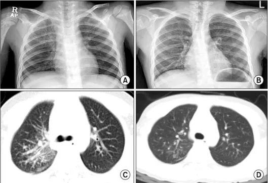
Fig. 1. Chest radiograph shows reticular opacities in right lungs before treatment (A). The reticular opacities disappeared after treat- ment with INF-alpha (B). Chest CT shows peribronchial thickening, nodular densities and ground-glass opacities in the right upper lobe before treatment (C), and improve- ment of lesion after treatment with IFN-alpha (D).
Treatment options for ECD include corticosteroids, che- motherapy, radiotherapy, surgery, and immunotherapy, but overall prognosis remains dull as these treatments do not improve the survival of ECD patients. The high mortality rate associated with ECD is thus a critical issue. Although alternative treatments have been shown to be effective re- cently, IFN- is still the first line therapy for ECD with vari- able efficacy and limited tolerance [3]. Herein, we report a case of ECD in a 4-year-old boy with the involvement of the central nervous system, lung, and bone. The boy was treated with IFN- and prednisolone, and he has re- mained disease-free 3 years after the completion of treat- ment.
Case Report
A 4-year-old boy was referred to our hospital for right hemifacial palsy of 1 week duration and intermittent right arm pain. After admission, he presented with productive cough, and according to the chest X-ray, there was haziness at the right upper lung field. Laboratory results, including white blood cell count and C-reactive protein, were within normal limits.
Diffuse reticular opacities and parahilar haziness were
observed in the right lung on chest X-ray (Fig. 1A). In addi- tion, chest computed tomography (CT) clearly demon- strated interlobular septal thickening and peribroncho- vascular thickening with centrilobular nodular opacities and some ground-glass opacities in the right lung (Fig. 1C), as well as multiple bone lesions of both humeri.
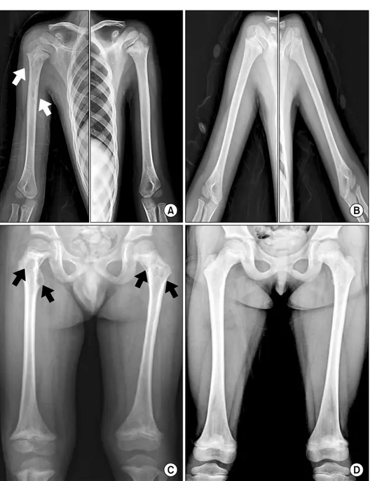
Radiographs revealed well-defined osteolytic lesions with a diameter of less than 1 cm and osteosclerosis of the prox- imal metadiaphyses of both humeri, with greater severity on the right (Fig. 2A). Osteosclerosis and cortical thicken- ing with osteolytic lesions were observed in the right clavi- cle and right ribs. Moreover, both proximal metadiaphyseal lesions and distal metaphyseal lesions were present in both femurs (Fig. 2C). However, these lesions did not show any uptakes on the bone scan.
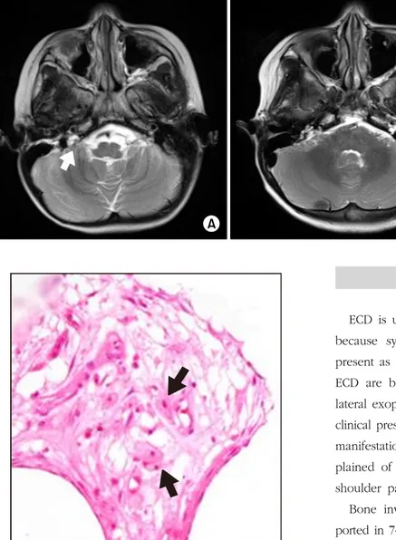
Temporal MR imaging revealed an extra-axial mass of 1.0×0.9 cm, in the right cerebellopontine angle that ex- tended to the right internal auditory canal which was hypo- intense on T1- and T2-weighted MR images and heteroge- neously enhanced (Fig. 3). The right facial nerve was supe- riorly displaced by the mass without significant thickening or enhancement.
Biopsy of the right humeral bone and subsequent im- munohistochemical staining showed positive CD68, neg-
Fig. 2. Well-defined osteolytic le- sions (arrows) and osteosclerosis are observed in the proximal metadiaphyses of the bilateral hu- meri before treatment (A), and osteosclerosis with osteolytic le- sions are improved after treatment with IFN-alpha (B). Symmetrical osteosclerosis with osteolytic lesions (arrows) are also seen in the pro- ximal and distal metaphyses of the bilateral femurs before treatment (C), and bony lesions are improved after treatment with IFN-alpha (D).
ative CD1a and nonspecific positive reaction to S-100 pro- tein; based on which he was presumptively diagnosed with Langerhans cell histiocytosis (LCH). Chemotherapy was ini- tiated with prednisolone, vinblastine, and etoposide based upon the LCH-II treatment protocol for 4 weeks, but he did not show any significant improvement; he developed blur- red vision and seizure and, based on non-convulsive status epilepticus pattern of electroencephalography (EEG), was treated with antiepileptic drugs. Brain magnetic resonance imaging (MRI) after a 2-month follow-up showed diffuse brain atrophy and bilateral optic nerve atrophy.
After a careful re-review of the radiologic images, it was understood that, despite the young age of the patient, the probability of LCH was low, but that of ECD high. A retro- spective pathological review was performed again, which revealed some portions of intertrabecular fibrosis with foamy histiocytes (Fig. 4), and positive CD68, negative CD1a and negative S-100 protein. As a result of the high prevalence of BRAF V600E mutation in ECD, we performed Sanger sequencing of BRAF exon 15 from bone marrow DNA sample of the patient; however, BRAF V600E mutation was not detected.
Fig. 3. T2 axial MR image revealed lobulated heterogeneous enhan- cing extra-axial mass (arrow) in the right cerebellopontine angle (A), and the right cerebellopontine mass showed no interval change after treatment (B).
Fig. 4. Bone biopsy showed foamy histiocytes (arrows) in the intertrabecular fibrosis of bone (H&E×400).
Finally, the patient was treated for ECD through chemo- therapy with IFN- (3×10⁶ units 2-3 times per week) and prednisolone (1 mg/kg/day). Chest radiographs showed a significant improvement of reticular opacities and parahilar haziness (Fig. 1B, D). However, bony lesions showed little change (Fig. 2B, D), and bone pain and other manifes- tations improved slowly throughout the treatment period.
After 24 months of treatment, IFN- was discontinued, and after the end of the treatment duration, no relapse was ob- served for 3 years.
Discussion
ECD is usually not suspected during the initial workup because systemic pediatric histiocytic diseases generally present as LCH in children. The classic triad symptoms of ECD are bone pain, diabetes insipidus, and painless bi- lateral exophthalmos with visual impairment [4]. The initial clinical presentation may show a broad spectrum of clinical manifestations in children. In our case, the child com- plained of right hemifacial palsy accompanied with right shoulder pain.
Bone involvement is the most common symptom, re- ported in 74% of the ECD populations [4], presenting as bi- lateral symmetrical osteosclerosis at the metaphyseal and di- aphyseal portions of the long bones, sparing the epiphyses [1,2]. Bone pain usually manifests around the knees and ankles. Although the classical hallmark of skeletal involve- ment for ECD is osteosclerosis, it has previously been re- ported to occasionally manifest as mixed sclerotic and lytic lesions, as in our case. Osteolytic lesions may be explained by recent infiltrations of histiocytes, giant cells, and unossi- fied fibrosis, while osteosclerosis is related to fibrosis [5].
Central nervous system (CNS) involvement was reported in 56% of the overall population and was the second most common site of ECD [1]. As multifocal involvement of CNS and orbits was a frequent manifestation, pituitary involve- ment, retro-orbital masses, and infiltrative axial lesions have been proposed as a neuroradiological triad strongly sugges- tive of ECD, when presented with a positive bone scan or
osterosclerosis of the facial sinus walls [4]. The meningeal mass is usually hypointense or isointense on T1- and T2-weighted images with homogeneous intense enhance- ment, and the cerebellopontine angle is a common site [6].
However, a gradual development of brain atrophy as in our case is unusual. We consider that it may be related to che- motherapy or valproic acid administration for seizure attack.
Mediastinum, heart, lungs, gastrointestinal tract, kidneys, retroperitoneum, CNS and orbits are the extra-skeletal sites that have been reported to date. The sheathing of the whole thoraco-abdominal aorta called the “coated aorta”
and the perirenal fascia infiltration taking the appearance of “hairy kidneys” are highly suggestive features of ECD [4].
Orbital involvement of ECD is also a common manifes- tation, and presents as intraorbital masses resulting in bi- lateral exophthalmos and visual impairment. Optic nerve atrophy secondary to the intraorbital masses has also been reported as in our case [7].
Pulmonary and renal impairments of ECD are un- common but often contribute to the death of patients [4].
The characteristic radiological finding is a pattern of inter- stitial lung disease with symmetrical smooth thickening of the interlobular septa, peribronchovascular thickening, cen- trilobular nodular opacities, and pleural effusion. These dis- tinctive findings, which are different from that of LCH rep- resented by cystic lesions and centrilobular nodules, caused us to consider ECD as an alternative diagnosis [8]. Radiolo- gical finding of pulmonary involvement often precedes clin- ical symptoms. Our case also showed interstitial infiltrations without specific symptoms. However, these findings can al- so be observed in other interstitial disorders, such as lym- phangitic disorders, pulmonary edema, and idiopathic fibrosis. Bilateral symmetrical osteosclerosis of the long bones, in addition to the interstitial pulmonary infiltration, can be the most important contrasting point, and a final diagnosis should be made by histopathological findings such as diffuse infiltrates of foamy histiocytes accompanied by lymphocytes, monocytes and multinucleated giant cells of the Touton type. Touton giant cells, immunochemically positive for CD68, are typical of ECD and Birbeck granules, seen on electron microscopy, and positive staining for S-100 protein, is absent in ECD [8].
To date, various treatments have been attempted to ach- ieve remission in ECD patients. However, there has not been a standard treatment method with good efficacy and improved survival, and more than half of the patients show relapse within 3 years. Since ECD is extremely rare in chil- dren, evidence regarding its treatment is limited to case re- ports [1].
Treatment for ECD has previously been carried out with steroids, various cytotoxic agents, and tandem autologous hematopoietic-stem-cell transplantation. Corticosteroids are the traditional first-line therapy; however, they are generally either ineffective or only transiently effective. Chemotherapy can induce transient partial responses but is often in- effective [2,9]. Recent studies suggest IFN- is as the most efficacious agent and recommended as the first-line therapy for ECD [4,9]. Our observations suggest that this well-tol- erated therapy with steroid and IFN- (starting dose: 3 to 6×10⁶ units, maintenance: 1×10⁶ units 2 to 3 times per week) has a significant effect on the outcome of ECD with- out BRAF V600E mutation.
Several mechanisms have been proposed to explain the biological effects of IFN in ECD, such as induction of func- tional maturation and activation of dendritic cells, immune- mediated destruction of histiocytes and direct antiprolifera- tive effects on histiocytes [10].
Anakinra [11] and infliximab [12] have also achieved good results and should be taken into consideration for treating ECD when IFN- treatment fails. More recently, the BRAF-inhibitor vemurafenib has been used in ECD patients with desirable efficacy [13]. Nevertheless, its adverse effects and limited data on its long-term efficacy discourages many clinicians to use this as a first-line therapy option. Further prospective studies about new therapy using monoclonal antibodies for BRAF inhibitor and TNF- inhibitor are needed.
References
1. Diamond EL, Dagna L, Hyman DM, et al. Consensus guide- lines for the diagnosis and clinical management of Erdheim- Chester disease. Blood 2014;124:483-92.
2. Mazor RD, Manevich-Mazor M, Shoenfeld Y. Erdheim-Chester disease: a comprehensive review of the literature. Orphanet
J Rare Dis 2013;8:137.
3. Mathis S, Godenèche G, Haroche J, et al. Long-term outcome of basilar stenosis in Erdheim-Chester disease: A case report.
Medicine (Baltimore) 2016;95:4813.
4. Cives M, Simone V, Rizzo FM, et al. Erdheim-Chester disease:
a systematic review. Crit Rev Oncol Hematol 2015;95:1-11.
5. Dion E, Graef C, Miquel A, et al. Bone involvement in Erdheim-Chester disease: imaging findings including periostitis and partial epiphyseal involvement. Radiology 2006;238:632-9.
6. Arnaud L, Hervier B, Néel A, et al. CNS involvement and treatment with interferon-a are independent prognostic factors in Erdheim-Chester disease: a multicenter survival analysis of 53 patients. Blood 2011;117:2778-82.
7. Hoffmann EM, Müller-Forell W, Pitz S, Radner H. Erdheim- Chester disease: a case report. Graefes Arch Clin Exp Ophthalmol 2004;242:803-7.
8. Arnaud L, Pierre I, Beigelman-Aubry C, et al. Pulmonary in- volvement in Erdheim-Chester disease: a single-center study of thirty-four patients and a review of the literature. Arthritis
Rheum 2010;62:3504-12.
9. Braiteh F, Boxrud C, Esmaeli B, Kurzrock R. Successful treat- ment of Erdheim-Chester disease, a non-Langerhans-cell his- tiocytosis, with interferon-alpha. Blood 2005;106:2992-4.
10. Haroche J, Amoura Z, Trad SG, et al. Variability in the efficacy of interferon-alpha in Erdheim-Chester disease by patient and site of involvement: results in eight patients. Arthritis Rheum 2006;54:3330-6.
11. Tran TA, Pariente D, Lecron JC, Delwail A, Taoufik Y, Meinzer U. Treatment of pediatric Erdheim-Chester disease with inter- leukin-1-targeting drugs. Arthritis Rheum 2011;63:4031-2.
12. Munoz J, Janku F, Cohen PR, Kurzrock R. Erdheim-Chester disease: characteristics and management. Mayo Clin Proc 2014;89:985-96.
13. Haroche J, Cohen-Aubart F, Emile JF, et al. Dramatic efficacy of vemurafenib in both multisystemic and refractory Erdheim- Chester disease and Langerhans cell histiocytosis harboring the BRAF V600E mutation. Blood 2013;121:1495-500.