Neurobiology of Disease
HMGB1, a Novel Cytokine-Like Mediator Linking Acute
Neuronal Death and Delayed Neuroinflammation in the
Postischemic Brain
Jung-Bin Kim,
1Joon Sig Choi,
2Young-Mi Yu,
1Kihoon Nam,
3Chun-Shu Piao,
1Seung-Woo Kim,
1Min-Hyung Lee,
4Pyung-Lim Han,
5Jong-sang Park,
3and Ja-Kyeong Lee
11Department of Anatomy and Center for Advanced Medical Education (BK21 project), Inha University School of Medicine, Inchon 400-712, Korea, 2Department of Biochemistry, Chungnam National University, Daejeon 305-764, Korea,3School of Chemistry and Molecular Engineering, Seoul National University, Seoul 151-742, Korea,4Department of Bioengineering, School of Engineering, Hanyang University, Seoul 133-791, Korea, and5Division of Nano Sciences and Department of Life Sciences, Ewha Womans University, Seoul 110-783, Korea
Cerebral ischemic injury proceeds with excitotoxicity-induced acute neuronal death in the ischemic core and with delayed damage
processes in the penumbra. However, knowledge concerning the direct mediators that connect these two processes is limited. Here, we
demonstrate that high-mobility group box 1 (HMGB1), a nonhistone DNA-binding protein, is massively released into the extracellular
space immediately after ischemic insult and that it subsequently induces neuroinflammation in the postischemic brain. Short hairpin
(sh)RNA-mediated HMGB1 downregulation in the postischemic brain suppressed infarct size, microglia activation, and
proinflamma-tory marker induction, indicating that HMGB1 plays a crucial role in the inflammaproinflamma-tory process. The proinflammaproinflamma-tory cytokine-like
function of extracellular HMGB1 was further verified in primary cortical cultures and microglial cultures. HMGB1 was found to
accumu-late in NMDA-treated primary cortical culture media, and supernatants collected from these cultures were found to trigger microglia
activation, the hallmark of brain inflammation. Moreover, treatment with recombinant HMGB1 also induced microglial activation, but
HMGB1-depleted supernatant produced by anti-HMGB1 antibody treatment or by HMGB1 shRNA expression did not, thus
demonstrat-ing the essential role of HMGB1 in microglial activation. Together, these results indicate that HMGB1 functions as a novel
proinflamma-tory cytokine-like factor that connects excitotoxicity-induced acute damage processes and delayed inflammaproinflamma-tory processes in the
post-ischemic brain.
Key words: HMGB1; MCAO; ischemia; inflammation; shRNA; PAMAM-Arg
Introduction
Cerebral ischemia leads to brain injury through a complex series
of pathophysiological events, which result in neuronal death and
subsequent neurological dysfunction. The major pathogenic
mechanisms may include glutamate excitotoxicity, Zn
⫹2toxic-ity, peri-infarct depolarization, inflammation, and apoptosis.
Ex-citotoxicity and Zn
⫹2toxicity result in acute and massive
neuro-nal death in the ischemic core (Lipton, 1999). A wealth of
evidence suggests that this acute neuronal damage is followed by
a second round of neuronal injury, called delayed neuronal death
(Kirino, 2000). Of the many pathophysiological events that may
contribute to this delayed injury, the cell-mediated processes
as-sociated with postischemic inflammation and apoptosis have
been studied extensively (Graham and Chen, 2001). In particular,
inflammatory reactions have been suggested to contribute to the
late stages of ischemic injury and to result in a worsening of
neurological outcome through multiple mechanisms, wherein
microglia plays a crucial role (Giulian, 1997; Dirnagl et al., 1999).
However, it remains to be determined whether mediators
con-nect the early acute neuronal death phase and delayed
neuroinflammatory-related events in the postischemic brain.
High-mobility group box 1 (HMGB1) is a nonhistone
DNA-binding protein that possesses two HMG boxes that are DNA
binding domains (Landsman and Bustin, 1993). HMGB1 is
widely expressed in various tissues including the brain. As a
chro-mosomal protein, HMGB1 has been implicated in diverse
intra-cellular functions, including the stabilization of nucleosomal
structure and the facilitation of gene transcription (Bustin, 1999).
Moreover, recent evidence identifies HMGB1 as a cytokine-like
mediator of delayed endotoxin lethality and acute lung injury
(Wang et al., 1999a; Abraham et al., 2000). HMGB1 is actively
secreted by macrophages and monocytes or released by necrotic
cells into the extracellular milieu, where it might be involved in
the triggering of inflammation (Wang et al., 1999a; Abraham et
al., 2000; Scaffidi et al., 2002; Bonaldi et al., 2003). Recombinant
HMGB1 has been found to induce acute inflammation in animal
models of lung injury and endotoxemia (Wang et al., 1999a;
Abraham et al., 2000), and anti-HMGB1 antibody attenuated
endotoxin-induced lethality even when administration of
anti-body was delayed until after early cytokine response (Wang et al.,
Received Sept. 8, 2005; revised April 11, 2006; accepted April 11, 2006.
This work was supported byKorea Science and Engineering Foundation Grant 2005-01096 to J.-K.L. Correspondence should be addressed to Dr. Ja-Kyeong Lee, Department of Anatomy, Inha University School of Medicine, 7-241 Shinheung-dong, Jung-Gu, Inchon 400-712, Republic of Korea. E-mail: jklee@inha.ac.kr.
DOI:10.1523/JNEUROSCI.3815-05.2006
1999a; Yang et al., 2004). In addition, high serum levels of
HMGB1 in patients with sepsis or hemorrhagic shock have been
reported to be associated with increased mortality and disease
severity (Ombrellino et al., 1999; Wang et al., 1999a). Moreover,
in the brain, HMGB1 has recently been reported to be released
after cytokine stimulation and to be involved in the inflammatory
process after it was administered intracerebroventricularly
(Wang et al., 1999b; Agnello et al., 2002).
The present study shows that HMGB1 is massively released
during the excitotoxicity-induced acute damaging process in the
postischemic brain and that extracellular HMGB1 triggers
in-flammatory processes, such as microglia activation. Thus, we
provide evidence that HMGB1 acts as a novel mediator that links
excitotoxicity-induced acute damage and subsequent
inflamma-tory processes in the postischemic brain.
Materials and Methods
Surgical procedures for middle cerebral artery occlusion. All experiments were performed in accordance with the animal research guidelines issued by the Inha University School of Medicine. Male Sprague Dawley rats (250 –300 g) were anesthetized with 5% isoflurane in a mixture of 30% oxygen and 70% nitrous oxide, and the middle cerebral artery (MCA) was occluded for 1 h using the suture occlusion method described pre-viously (Longa et al., 1989; Kim et al., 2004). The left femoral artery was cannulated for monitoring arterial blood pressure (Blood Pressure Ana-lyzer; Digi Med, Louisville, KY) and for blood sampling to analyze pH, PaO2, PaCO2, and glucose concentration (I-STAT; Sensor Devises, Waukesha, WI). Regional cerebral blood flow was monitored using a laser Doppler flowmeter (Periflux System 5000; Perimed, Jarfalla, Sweden). A thermoregulated heating pad and an overhead heating lamp were used to maintain a rectal temperature of 37⫾ 0.5°C. In sham-operated rats, an incision was made over the MCA, but the artery was not occluded.
Intracranial injection. Rats were anesthetized with an intramuscular injection (100 –150l/100 g body weight) of a mixture of ketamine and xylazine hydrochloride (3:1) and placed on a stereotaxic apparatus (Na-rishige Scientific Instrument Laboratory, Tokyo, Japan). The skull was leveled between bregma and lambda. The plasmid/arginine-grafted poly-amidoamine dendrimer (PAMAM-Arg) complex (10l) was injected stereotaxically into the striatum (coordinates in millimeters with refer-ence to bregma: anteroposterior, 0; lateral, 5.0; ventral, 4.0) using a 26 gauge Hamilton microsyringe (80330; Hamilton, Reno, NV). Two-millimeter-thick coronal brain slices, which localized between 8 and 10 mm from the frontal pole, were taken from the brain using a brain matrix (RBM-40000; ASI Instruments, Houston, TX), and brain regions af-fected by short hairpin (sh)RNA-expressing plasmid were dissected. The tissue samples were kept at⫺80°C until required.
Preparation of mixed neuron– glia cultures. Mixed cortical cells, includ-ing neurons and astrocytes, were prepared from embryonic day 15.5 mouse cortices and cultured as described by Kim et al. (2004). Dissoci-ated cortical cells were plDissoci-ated at a density of five hemispheres per 24-well plate (⬃4 ⫻ 105cells per well), which was poly-D-lysine (100g/ml) and laminin (100g/ml) coated. Cultures were maintained without antibi-otics in minimal essential medium (MEM) with 5% fetal bovine serum (FBS) and 5% horse serum. At 6 d in vitro (DIV), when astrocytes had reached confluence underneath neurons, cytosine arabinofuranoside was added to a final concentration of 10Mand the culture was main-tained for 2 d to halt microglial growth. FBS and glutamine were not supplemented from day 6, and the medium was changed twice per week after day 8. Cultures were used at 13–15 DIV.
Primary microglial culture. Primary microglial cultures were prepared as described previously. In brief, cells dissociated from the cerebral hemi-spheres of 1- to 3-d-old postnatal rat brains (Sprague Dawley strain) were seeded into 75 cm2flasks at a density of 1.2⫻ 106cells/ml in MEM (Sigma, St. Louis, MO) containing 10% FBS (Hyclone, Logan, UT). Cul-ture media were changed after 24 h and then twice per week. After 2 weeks, microglia were then detached from the flasks by mild shaking and filtered through a nylon mesh to remove astrocytes. After centrifugation
(1000⫻ g) for 5 min, the cells were resuspended in a fresh DMEM supplemented with 10% FBS and plated at a final density of 4⫻ 104 cells/well on a 24-multiwell culture dish. The following day, cells were subjected to different treatments. The microglial cultures used were ⬎96% pure.
NMDA and staurosporin treatment and cell supernatant preparation. Primary cortical cells were treated with serum-free MEM containing 30 MNMDA (Sigma) and 10Mstaurosporin (Sigma) for 1 h. The me-dium was then removed and replaced with fresh MEM, and cells were cultured for 24 h. Four hundred microliters of the culture medium were then collected and concentrated to 40l by using Centricon 10 (Milli-pore, Billerica, MA) and either immunoblotted or used as a culture me-dium for activating primary microglial cultures.
Sampling of CSF. Rats were anesthetized, and the head was placed in a stereotaxic apparatus. The skin was incised, and the occipital bone was cleared of muscle to expose the atlanto-occipital membrane. A 27 gauge needle was inserted into the cisterna magna. When the tip of the needle was inserted 1–1.5 mm, reflux of the CSF was observed. Approximately 100l of CSF was withdrawn.
Generation of the HMGB1 shRNA transgene. To generate shHMGB1-pU6, two 64-mer sense and antisense oligonucleotides containing a 19 nucleotide inverted repeat corresponding to the HMGB1 coding region were synthesized (see Fig. 3A). The inverted motif, which also contained the seven-nucleotide spacer and five Ts (TTTTT), was subcloned into the BamHI and HindIII sites, immediately downstream of the U6 promoter of the pU6 plasmid (Ambion, Austin, TX). A mutant plasmid, MshHMGB1-U6, was constructed in the same way, except that six nucleotides within the target region were substituted. The sequences inserted were as follows (only the sense sequences are shown): shHMGB1, 5 ⬘-GATCCC- GAAGCACCCGGATGCTTCTTTCAAGAGAAGAAGCATCCGGGTGC-TTCTTTTTTGGAAA-3⬘; MshHMGB1, 5⬘-GATCCCGAAGCACTACTGC-GCTTCTTTCAAGAGAAGAAGCGCAGTAGTGCTTCTTTTTTGGAAA-3⬘. Transient transfections. All plasmids, including shHMGB1-pU6 and MshHMGB1-pU6, were mixed with PAMAM-Arg at a 1:3.5 charge ratio and administered to rat brain or transfected into primary cortical cul-tures. After 48 h, cell or tissues were subjected to various biochemical assays.
RNA preparation and reverse transcription-PCR. Total RNA was pre-pared using TRIzol reagent (Invitrogen, Gaithersburg, MD) according to the manufacturer’s instructions, and 1g RNA samples were used for cDNA synthesis. First-strand cDNA synthesis was primed with random hexamers and conducted according to the manufacturer’s specifications (RT-PCR kit; Roche, Mannheim, Germany). The cDNA equivalent to 200 ng of total RNA was subjected to PCR using the manufacturer’s protocol (PCR core kit; Roche). The primer sequences of rat HMGB1, tumor necrosis factor-␣ (TNF␣), interleukin-1 (IL-1), inducible ni-tric oxide (iNOS), and cytochrome oxidase (COX)-2 have been de-scribed previously (Yu et al., 2003; Kim et al., 2004).
Northern analysis. Northern analysis was performed as described pre-viously (Kim et al., 2004). Each lane was loaded with 50g of total RNA. After being transferred to a nylon membrane (Roche), RNA blots were hybridized with a 64 bp DNA probe corresponding to HMGB1 shRNA. Probes were labeled with P32-dCTP using a random prime labeling sys-tem (Amersham Biosciences, Piscataway, NJ) and purified using a robe-Quant G-50 micro column (Amersham Biosciences). A total of 1⫻ 107 cpm of labeled probe was applied to each membrane in hybridization buffer (ULTRAhyb; Ambion) and incubated at 42°C for 18 h. Mem-branes were washed and exposed to film.
2,3,5-Triphenyl tetrazolium chloride staining. Rats were decapitated af-ter 2 d of reperfusion, and entire brains were dissected coronally into 2 mm brain slices using a metallic brain matrix (RBM-40000; ASI Instru-ments). Slices were immediately stained by immersion in 1% 2,3,5-triphenyl tetrazolium chloride (TTC) at 37°C for 15 min and then in 4% paraformaldehyde for preservation.
Lectin histochemistry. Vibratome sections were incubated with horse-radish peroxidase (HRP)-conjugated Griffonia simplicifolia isolectin-B4 (GSA I-B4) (Sigma) at 10g/ml in PBS at room temperature for 3 h. After three washes in PBS, isolectin binding sites were visualized using HRP/3,3⬘-diaminobenzidine.
5-Bromo-4-chloro-3-indolyl--D-galactopyranoside histochemistry. Ani-mals were perfused transcardially with 4% paraformaldehyde, and brain slices (50m thick) were prepared. After three washes in 0.01% sodium deoxycholate, 0.02% Nonidet P-40, 2 mMMgCl2, and 0.1MNaHPO4, pH 7.3, 5-bromo-4-chloro-3-indolyl--D-galactopyranoside (X-gal) stain-ing was performed over 12–16 h at 37°C in X-gal buffer [2 mMMgCl2, 5 mMK3Fe(CN)6, 5 mMK4Fe(CN)6, 0.1 mMNaHPO4, pH 7.3, 0.01% so-dium deoxycholate, and 0.02% Nonidet P-40] containing 1 mg/ml X-gal. Two-dimensional gel electrophoresis. Seven-centimeter linear pH 3–10 non-linear immobilized pH gradient strips were loaded with proteins by passive rehydration overnight. Isoelectric focusing was performed on an IPGphor device (Amersham Biosciences) and was performed up to a total of 66 kVh. The IPG strips were rinsed thoroughly with distilled water and quickly dried on filter paper, and focused proteins were re-duced (50 mMTris/HCl, pH 6.8, 6 m urea, 2% w/v SDS, 30% v/v glycerol, and 2% w/v DTT) and alkylated (50 mMTris/HCl, pH 6.8, 6 m urea, 2% w/v SDS, 30% v/v glycerol, and 4.5% w/v iododacetamide) for 20 min each. Strips were then placed on top of 12% SDS- polyacrylamide gels and ran at 25 mA constant current per gel.
Immunoblotting. Brain homogenates were immunoblotted as de-scribed previously (Kim et al., 2004). Rats were deeply anesthetized with an overdose of sodium pentobarbital (100 mg/kg, i.p.), and brain slices were prepared as described above. Blood samples (1 ml) were drawn by cardiac puncture after 15 min of centrifugation at 1500⫻ g, and serum was collected and stored at⫺80°C in sterile polypropylene tubes until required for assay. Tissue and serum samples were incubated in 50 mM HEPES, pH 7.4, 0.1% Nonidet P-40, and 150 mm NaCl supplemented with a protease inhibitor mixture (BD Biosciences, Franklin Lakes, NJ) for 15 min on ice and centrifuged at 6000⫻ g at 4°C. Protein concentra-tions were determined using Bradford’s method (Bio-Rad, Hercules, CA). Lysates were incubated for 10 min in the presence of SDS sample buffer containing 2-mercaptoethanol and centrifuged at 16,000⫻ g for 5 min at 4°C, and supernatants were loaded onto SDS-PAGE. Primary antibodies were diluted as follows, 1:1000 for anti-HMGB1 (BD Bio-science) and 1: 500 for p38 mitogen-activated protein kinase (MAPK) and phospho-p38 MAPK, and detected by using a chemiluminescence
kit (BD Bioscience) using donkey anti-rabbit HRP-conjugated second-ary antibody (Pierce, Rockford, IL).
NO measurement. Primary microglia (4⫻ 104) plated on 24-well plates were treated with lipopolysaccharide (LPS) (100 ng/ml) for 48 h. To measure the amount of NO produced by microglia, 100l of condi-tioned medium was mixed with an equal volume of Griess reagent (0.5% sulfanilamide and 0.05% N-1-naphthylethylenediamine) and incubated for 10 min at room temperature. Absorbance of the mixture at 550 nm was measured using a microplate reader.
HMGB1 measurements. Serum samples or cell-conditioned media were fractionated by SDS-PAGE, and HMGB1 levels were determined by immunoblotting using a standard curve for recombinant HMGB1 as a reference (Sigma). Briefly, serum samples or cell-conditioned media (100 –200l) were ultrafiltered using a Centricon 10 (Millipore), frac-tionated by SDS-PAGE, and transferred to polyvinylidene difluoride membranes (Bio-Rad). The primary antibodies for anti-HMGB1 (BD Bioscience) and anti-histone (Chemicon, Temecula, CA) were diluted 1:1000 and 1:500, respectively. Detection was performed using a chemi-luminescence kit (BD Bioscience).
Treatment of recombinant HMGB1. Primary microglia (4 ⫻ 104) plated on 24-well plates were treated with various amounts of recombi-nant human HMGB1 (rhHMGB1) (Sigma). At different time points, cells and cell supernatants were harvested and used in reverse transcrip-tion (RT)-PCR and Griess assay, respectively. The endotoxin content of rhHMGB1 was determined with a standard endotoxin-specific Limulus amebocyte lysate reagent (Endosafe, Wilmington, MA). Endotoxic con-tent was always⬍0.05 EU/ml.
Results
Plasma HMGB1 rapidly increases after transient focal
cerebral ischemia
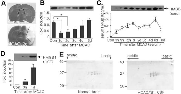
We observed that the HMGB1 level in the infarction area in
ipsi-lateral sides (Fig. 1 A, bottom) significantly declined at 1 d after
MCA occlusion (MCAO)/reperfusion to below the basal level
(Fig. 1 B). HMGB1 levels gradually increased over 4 d to above
Figure 1. Plasma and CSF HMGB1 immediately increased after MCAO. A, TTC staining was performed with coronal brain sections, which were obtained 2 d after sham operation or after 1 h of MCAO. B, HMGB1 levels in ischemic hemispheres (indicated region in bottom panel in A) were determined by immunoblotting at various times after 1 h of MCAO. C, D, HMGB1 levels in serum (C) and in CSF (D) were examined by immunoblotting. E, Total protein extracts obtained from normal brain tissue or from CSF after MCAO were loaded onto two-dimensional gels, blotted, and immuno-detected with anti-HMGB1 antibody. Data are presented as means⫾ SEM (n ⫽ 3). *p ⬍ 0.05.
basal levels (Fig. 1 B). In contrast, plasma
HMGB1 levels rapidly increased from 3 h
after 1 h of MCAO, continued to increase
for 6 d (Fig. 1C), and then declined slowly.
Rapid HMGB1 accumulation was also
de-tected in CSF 3 h after MCAO, and further
accumulation was observed at 1 d after
MCAO (Fig. 1 D). To examine whether
HMGB1 in CSF is hyper-acetylated, as is
the case for HMGB1, which is actively
se-creted by other cell types (Bonaldi et al.,
2003), two-dimensional electrophoresis
was performed on proteins purified from
CSF obtained 3 h after MCAO. The
pat-terns of HMGB1 spots detected on blots
were similar to those obtained for normal
brain tissue, and no hyper-acetylated spot
was present (Fig. 1 E), indicating that the
rapid accumulation of HMGB1 in the CSF
was not achieved by active secretion but
probably by passive release.
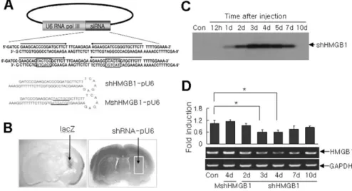
shRNA-mediated suppression of
HMGB1 expression in the brain
To investigate the functional significance
of the rapid accumulation of HMGB1 in
the serum and CSF of the postischemic
brain, we knocked down HMGB1 mRNA
using a plasmid expressing the shRNA of
the HMGB1 gene. The hairpin-forming 64 bp insert in this
plas-mid (shHMGB1-pU6) harbors an inverted repeat of 19 bp
cor-responding to the HMGB1 coding region, into which a 7 bp
spacer was inserted between the inverted sequences (Fig. 2 A). A
mutant shHMGB1-expressing plasmid, containing a
six-nucleotide substitution within the 19 bp region, was also
con-structed (MshHMGB1-pU6). A novel cationic polymer,
PAMAM-Arg, which was generated by conjugating primary
amines located on the surface of PAMAM dendrimer with
L
-arginine (Choi et al., 2004), was used as a gene carrier. We
reported previously that PAMAM-Arg enables high transfection
efficiencies in a neuronal cell line, Neuro 2A (Choi et al., 2004),
and in primary cortical cells (Kim et al., 2006).
The extent of the diffusion of the injected plasmid from the
needle path was approximated by injecting a mixture of a
lacZ-expressing plasmid and PAMAM-Arg and then
-gal staining
(Fig. 2 A, left). Northern blot analysis of a sample from the
plasmid-affecting area (Fig. 2 B, white box in the right panel)
revealed the presence of the shRNA transcript at 24 h after
injec-tion and its persistence for
⬎7 d (Fig. 2C). Moreover, this shRNA
was found to suppress endogenous HMGB1 expression to 56.3
⫾
8.9% (n
⫽ 3; p ⬍ 0.05) of the control level on day 4, but then
HMGB1 expression recovered gradually to its baseline level at
10 d after injection (Fig. 2 D). In contrast, HMGB1 expression
was
unaffected
in
mutant
shRNA-expressing
plasmid
(MshHMGB1-pU6)-administered animals (Fig. 2 D), thus
dem-onstrating the specificity of shHMGB1-pU6 plasmid and
allevi-ating concerns about the possible toxic effects of PAMAM-Arg
and injected plasmid.
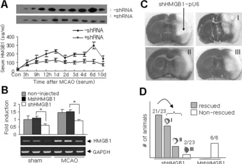
shRNA-mediated HMGB1 suppression had a neuroprotective
effect in the postischemic brain
The shHMGB1/pU6/PAMAM-Arg complex was administered
24 h before the 1 h of MCAO, into the striatum, which
encom-passes infarction core later (Fig. 3C, top left). Serum HMGB1
levels were found to be significantly reduced (Fig. 3A), and the
suppressive effect of shRNA was sustained for 10 d by a single
administration of shHMGB1-pU6 plasmid (Fig. 3A), which
agrees well with the observed duration of shRNA expression (Fig.
2C). The administration of shHMGB1-pU6 plasmid also
re-pressed delayed HMGB1 induction in the infarct core (Fig. 1 B),
but this was not detected in animals administered
MshHMGB1-pU6 (Fig. 3B). Moreover, TTC(TTC) staining revealed that
shHMGB1-pU6 administration at 24 h before MCAO
signifi-cantly suppressed infarct volumes in 21 of 23 animals (Fig. 3D).
Seventy-six percent of shHMGB1-pU6-administered animals
ex-hibited infarct suppression along the syringe path (⬎70%
sup-pression of infarct; type I) (Fig. 2 B, white box region), and the
remainder showed infarct suppression in the entire striatum
(type II) (Fig. 3C). In contrast, no infarct suppression (type III)
was observed in animal administered MshHMGB1-pU6 (Fig.
3D).
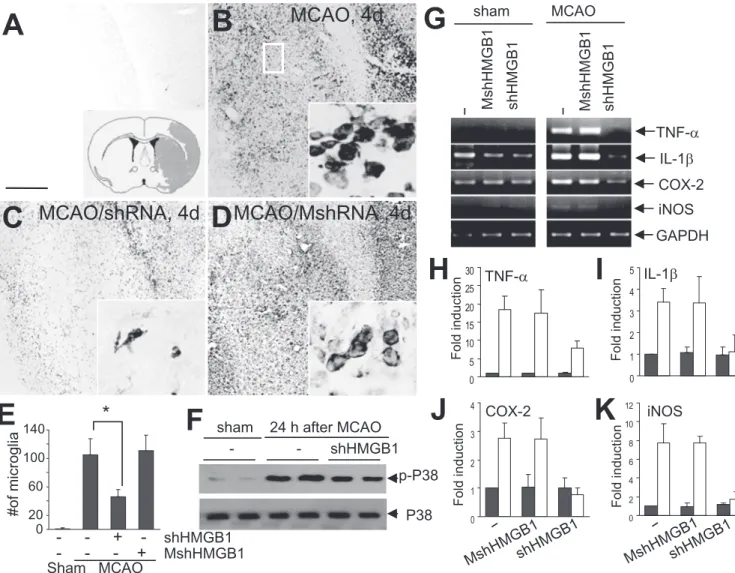
Anti-inflammatory effect of HMGB1 suppression in the
postischemic brain
Microglia activation is a hallmark of brain inflammation. The
numbers of activated or phagocytic microglia were notably
in-creased 4 d after 1 h of MCAO (Fig. 4 B, E, black box in the inset in
Fig. 4 A), but not in animals pre-administered shHMGB-pU6,
which showed relatively few ramified microglia in the infarction
core (Fig. 4C,E). In contrast, numerous activated or phagocytic
microglia were detected in animals administered
MshHMGB-pU6 (Fig. 4 D, E), thus indicating an anti-inflammatory effect of
HMGB1 suppression in the postischemic brain. In addition, p38
MAPK activation (detected as the phosphorylated form) was
sig-nificantly suppressed in shHMGB-pU6-administered animals
(Fig. 4 F, small box in the inset in Fig. 4 A). Moreover, the
induc-tions of the proinflammatory factors TNF-␣, IL-1, COX-2, and
iNOS were repressed in the injected area (Fig. 4 A, inset, small
Figure 2. shRNA-mediated silencing of HMGB1 gene expression in the normal brain. A, Schematic diagram of the HMGB1 shRNA transgene showing its sense and antisense regions and the predicted transcript, a hairpin structure with a 7 bp loop. shHMGB1-pU6 and MshHMGB1-pU6 represent wild-type and mutant shRNA (six-nucleotide substitution in red)-expressing plas-mid, respectively. siRNA, Small interfering RNA. B,-Galactosidase staining was performed 2 d after lacZ-expressing plasmid transfection using PAMAM-Arg as a gene carrier (left), and the boundary of the region expressing the exogenous gene was indicated (right, white box). shHMGB1-pU6 or MshHMGB1-pU6 was injected into the indicated region, and biochemical assays were done on samples prepared from the indicated area in the right panel. C, Northern blotting for shHMGB1 transcripts in shHMGB1-pU6-transfected brains was performed at the indicated times. Each lane contained total RNA obtained from five ani-mals, and blots were probed with the32P-labeled 64 nucleotide sense oligonucleotide. Con, Control. D, The levels of HMGB1 expression in shHMGB1-pU6- or MshHMGB1-pU6-administered animals were determined by RT-PCR at the indicated times after transfection. GAPDH, Glyceraldehyde-3-phosphate dehydrogenase. Data are presented as means⫾ SEM (n ⫽ 4). *p ⬍ 0.05.
box) 24 h after MCAO/reperfusion (Fig. 4G–K ), indicating that
HMGB1 plays a role in proinflammatory molecule release during
this stage. Together, these results indicate that HMGB1
suppres-sion has an anti-inflammatory effect in the postischemic brain.
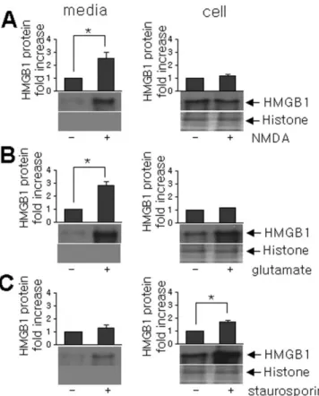
Massive HMGB1 release after excitotoxicity-induced
neuronal cell death in primary cortical cultures
We next investigated the function of extracellular HMGB1,
which accumulates during the acute damaging stage in the
post-ischemic brain, using primary cortical cultures. Levels of HMGB1
in the cell homogenates and supernatants of primary cortical
cultures were measured after NMDA, glutamate, or staurosporin
treatment. Under normal conditions, HMGB1 was exclusively
localized to the nuclei of neurons and astrocytes (data not
shown). Interestingly, HMGB1 was found to accumulate in
cul-ture medium after incubating primary cortical cells with NMDA
for 24 h (Fig. 3A), and a similar enrichment of HMGB1 was
observed for glutamate-treated culture medium (Fig. 5B). These
results indicate that HMGB1 is rapidly released into the
extracel-lular space after excitotoxicity-induced neuronal death.
How-ever, in marked contrast, HMGB1 levels in staurosporine-treated
cells were increased in cell homogenates but not in medium (Fig.
5C). In all cases, no histone was detected in culture medium, and
its levels in cell homogenates were unaffected by these treatments
(Fig. 5).
Activation of microglia by extracellular HMGB1
To examine whether HMGB1 released into excitotoxin-treated
cell supernatants is capable of triggering microglial activation,
primary microglia cultures were incubated with media collected
from NMDA-treated primary cortical cells. To remove residual
NMDA in supernatant, cells were cultured in fresh medium after
being exposed to NMDA (30
M) for 1 h.
Supernatants obtained from
NMDA-treated cortical cultures were found to
in-duce microglial activation, as
demon-strated by NO secretion (Fig. 6 A).
However, NMDA-treated supernatants
complemented with HMGB1
anti-body showed no microglial activation
(Fig. 6 A), indicating that HMGB1 in
me-dium plays an important role in microglial
activation. Regardless of the presence of
anti-HMGB1
antibody,
staurosporin-treated cell supernatant was incapable of
activating microglia (Fig. 6 A). Moreover,
NMDA-induced HMGB1 release into
cul-ture medium was notably reduced in
shHMGB1-pU6-transfected cells but not
in
MshHMGB1-pU6-transfected
cells
(Fig. 6 B), and as was expected,
superna-tants from shHMGB1-pU6-expressing
primary cortical cells were incapable of
ac-tivating microglia (Fig. 6C), further
sup-porting the notion that HMGB1 in culture
medium plays a crucial role in microglial
activation. In addition, microglial
activa-tion by excitotoxin-treated cell
superna-tant was further confirmed by the marked
inductions of proinflammatory markers
[e.g., iNOS, interferon
␥ (IFN␥), COX-2,
and TNF␣) (Fig. 6D). As was expected,
supernatants
from
shHMGB1-pU6-expressing primary cortical cells were incapable of inducing
proinflammatory markers (Fig. 6 D). Together, these results
demonstrate that extracellular HMGB1 released by
excitotoxin-treated neurons plays a crucial role in microglial activation in
vitro (Fig. 6) and in vivo (Fig. 4).
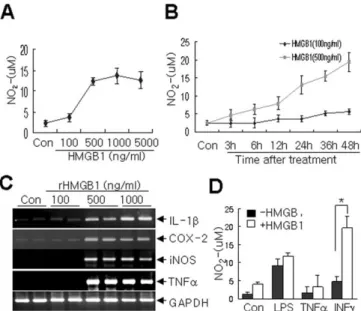
Purchased rhHMGB1 dose-dependently induced microglial
NO release, and this effect was maximal at 500 ng/ml (Fig. 7 A, B).
Marked proinflammatory factor induction was also observed in
rhHMGB1-treated cells, and the levels of proinflammatory factor
induction at different rhHMGB1concentrations paralleled those
of NO release (Fig. 7C). Moreover, the amount of NO released
from rhHMGB1-treated cells was lower than those released from
LPS- or IFN␥-treated cells (Fig. 7D), which were treated with low
doses of LPS (50 ng/ml), IFN␥ (25 ng/ml) or ⌻NF␣ (50 ng/ml) to
examine synergistic interactions. Interestingly, cotreatment with
a battery of proinflammatory cytokines revealed that rhHMGB1
appeared to act synergistically with IFN␥ to drive NO release (Fig.
7D).
Discussion
The present study demonstrates that HMGB1 is massively
re-leased into the extracellular milieu during the acute damaging
phase and that HMGB1 connects acute excitotoxicity-induced
neuronal death to delayed damaging processes, such as
inflam-mation, in the postischemic brain. Extracellular HMGB1 might
function as a proinflammatory cytokine, activate microglia, and
hence stimulate the release of other cytokines and aggravate brain
injury. Therefore, the shHMGB1-mediated protective effect
shown in Figure 3 might be derived from a reduction in the
amount of HMGB1 released during the acute phase. This
sugges-tion is in accord with previous studies that reported Hmgb1
⫺/⫺necrotic cells have a greatly reduced ability to promote
inflam-Figure 3. Neuroprotection by the shRNA-mediated silencing of HMGB1 induction in the postischemic brain. shHMGB1-pU6/ PAMAM-Arg or MshHMGB1-pU6/PAMAM-Arg complexes were administered into the indicated region (C) 24 h before 1 h of MCAO.
A, The serum levels of HMGB1 in shHMGB1-pU6-administered animals were determined by immunoblotting at the indicated times
after 1 h of MCAO. B, The levels of HMGB1 in the brain tissues (Fig. 2 B, right, white box) of shHMGB1-pU6- or MshHMGB1-pU6-administered animals were determined by RT-PCR 24 h after 1 h of MCAO. C, A TTC-stained infarction area in a coronal brain section 2 d after1 h of MCAO. The arrow indicates the administration point of the shHMGB1-pU6/PAMAM-Arg complex 24 h before 1 h of MCAO. Representative pictures showing suppressed infarct formation (types I and II) and no suppression (type III) are presented.
D, Numbers of rescued or nonrescued animals are presented in the bar graph. Data are presented as means⫾SEM(n⫽3).*p⬍
mation (Scaffidi et al., 2002). Recently, Tsung et al. (2005)
re-ported that HMGB1 appears to mediate hepatic injury after
mu-rine liver ischemia/reperfusion, wherein HMGB1 modulates
inflammatory signaling pathways.
Excitotoxicity is a leading cause of neuronal death after focal
cerebral ischemia. The activation of glutamate receptors, by the
attendant failure of ion homeostasis and increase in intracellular
Ca
2⫹concentrations, is a major factor in the initiation of
isch-emic cell death (Dirnagl et al., 1999). Because early increases in
plasma HMGB1 levels were suppressed by HMGB1 shRNA (Fig.
3A) and HMGB1 was released in glutamate- or
NMDA-challenged primary cortical cultures (Fig. 5), HMGB1 release
during acute excitotoxic insults in the postischemic brain might
be derived from neurons. Furthermore, we observed the rapid
disappearance of positive neurons but not of
HMGB1-positive glia in infarct core at 3 h after MCAO (J. B. Kim and J. K.
Lee, unpublished data). A massive release of HMGB1 from
dam-aged neurons would serve as a danger signal and evoke
inflam-matory reactions, such as the activations of glial cells, various
blood immune cells, and endothelial cells. The mediator-like
function of HMGB1 with respect to linking early and secondary
damaging processes in the postischemic brain might also be
rel-evant to other neuropathological conditions, such as epilepsy and
spinal cord injury, which also involve similar delayed progresses
after acute and massive neuronal death (Rosenzweig and
Mc-Donald, 2004; Weise et al., 2005). We found that the
preadmin-istration of the HMGB1 shRNA-expressing plasmid
shHMGB1-pU6 (the same plasmid as used in the present study) into the
hippocampal CA3 region of mouse brain prominently
sup-pressed kainic acid-induced neuronal death and after gliosis (S.
W. Kim, J. B. Kim, J. K. Lee, unpublished observation). The above
results also support the notion that HMGB1 functions as a
proin-flammatory cytokine in the CNS.
Although the mechanism by which HMGB1 exerts its
proin-flammatory cytokine-like effects in the CNS is unknown, the
re-sults of the present study indicate that HMGB1 directly triggers
microglial activation probably by directly activating signaling
cascades and that it also indirectly regulates the expressions of
Figure 4. Suppression of inflammatory markers by HMGB1 shRNA in the postischemic brain. A–D, Microglia activation 4 d after 1 h of MCAO was visualized by anti-GSA I-B4immunostaining (large rectangle area in the inset in A). shHMGB1-pU6 (C) or MshHMGB1-pU6 (D) was administered 24 h before MCAO. Activated microglia were detected in the ischemic core (B). Activated microglia numbers were notably reduced in wild-type shRNA-expressing brains (C) but not in mutant shRNA-expressing brains (D). The insets in A–D are high-magnification micrographs. E, The numbers of GSA I-B4-positive cells in the indicated areas (0.2⫻ 0.3 mm) in B were obtained by scoring in a blind manner the GSA I-B4-positive cells in 12 photographs taken from three independent experiments. F, Activation of p38 MAPK was examined 24 h after 1 h of MCAO by immunoblotting using anti-phosphorylated p38 MAPK antibody. G–K, The expressions of proinflammatory markers in the presence of shHMGB1-pU6 or MshHMGB1-pU6 were examined 1 d after 1 h of MCAO. Solid bars, Sham; open bars, MCAO (H–K). Data are presented as means⫾SEM(n⫽3).*p⬍0.05.Scale bar: A–D, 500m. GAPDH, Glyceraldehyde-3-phosphate dehydrogenase.
classic proinflammatory cytokines. The present and other reports
show that the activations of several MAPKs and nuclear factor
B
(NF-B) are involved in the proinflammatory effect of HMGB1
(Huttunen et al., 1999; Taguchi et al., 2000; Park et al., 2003) (Fig.
4 F). These are downstream molecules of RAGE (receptor for
advanced glycation end product) or toll-like receptor family
members, which are important receptors in the HMGB1
signal-ing process (Schmidt et al., 2000; Huttunen and Rauvala, 2004;
Park et al., 2004; Tsung et al., 2005). Although we found that
RAGE, TRL2, and TRL4 are present in microglia (data not
shown), interactions between them and the relative
contribu-tions they make to different signaling pathways require
addi-tional investigation. In addition, the inductions of
proinflamma-tory factors by HMGB1 were notable in vivo (Fig. 4G–K ) and in
vitro (Fig. 6 D). We believe that HMGB1 functions synergistically
with other cytokines, particularly with IFN␥ during NO
produc-tion in activated microglia. Synergistic effects could be achieved
by HMGB1 secretion upregulation by IFN␥ or by IFN␥ secretion
upregulation by HMGB1, as has been described in natural killer
cells (DeMarco et al., 2005).
Despite the fact that the level of HMGB1-induced NO release
in microglia was relatively low compared with those induced by
LPS or IFN␥, reductions in infarct sizes and inflammatory
pro-cess suppressions were remarkable in the HMGB1
shRNA-expressing brain (Fig. 3). This may be because extracellular
HMGB1 in vivo acts on target cells other than microglia. HMGB1
potently stimulates the release of various proinflammatory
cyto-kines in cultured macrophages, monocytes, or neutrophils
(Andersson et al., 2000; Park et al., 2003). In addition, the
activa-tion of human microvascular endothelial cells by HMGB1 has
been reported to result in the inductions of various adhesion
molecules, proinflammatory cytokines, and RAGE and in the
activations of MAPKs and NF-B (Fiuza et al., 2003; Treutiger et
al., 2003). Therefore, HMGB1 might function pleiotropically,
and its effects may be interconnected and affect various target
cells in vivo. For that reason, inhibition of HMGB1 by shRNA at
early time points might influence neighboring cells, although
de-tailed a mechanism needs to be explored, so that protected brain
tissue could be extended beyond the shRNA-injected area (Fig.
3C,D).
In the present study, we found that rapidly accumulating
HMGB1 in CSF is not hyper-acetylated (Fig. 1 E), indicating that
it is not actively secreted but rather released from damaged cells.
However, in view of the findings that serum HMGB1 levels were
sustained for 10 d after reperfusion and that serum HMGB1 levels
showed a delayed surge at around 6 d after 1 h of MCAO (Fig.
1C), HMGB1 might also be actively secreted by inflammatory
cells subject to delayed activation in the postischemic brain. In
this regard, we observed HMGB1 induction in phagocytic
micro-glia in the infarction core 3– 4 d after MCAO/reperfusion (data
not shown), which might be responsible for the observed delayed
surge in HMGB1 levels in ischemic hemispheres (Fig. 1 B).
HMGB1 is highly acetylated by nuclear acetyltransferase before
secretion, whereas passively released HMGB1 is not acetylated
Figure 5. Accumulation of HMGB1 in bath medium containing primary cortical cell cultures undergoing excitotoxicity-induced cell death. Primary cortical cultures were incubated in serum-free MEM containing 30MNMDA (A), 50Mglutamate (B), or 10Mstaurosporin (C) for 1 h. After 24 h, both culture media and cell homogenates were analyzed by immunoblotting with anti-HMGB1 or anti-histone antibody (A–C). Data are presented as means⫾SEM(n⫽3). *p⬍ 0.05.
Figure 6. Microglial activation by extracellular HMGB1. A, Primary microglial cultures (1⫻ 104cells) were incubated with media collected from NMDA- or staurosporine (stauros)-treated primary cortical cultures for 24 h in the presence or absence of anti-HMGB1 antibody, and NO production was determined using the Griess method. shHMGB1-pU6 or MshHMGB1-pU6 was transiently transfected into primary cortical cells 24 h before NMDA treatment. Con, Control. B, The amounts of HMGB1 in NMDA-treated cells and in medium bathing were determined by RT-PCR after 24 h of NMDA treatment. C, Primary microglia cultures were treated with media collected from NMDA-treated primary cortical cultures, transfected or not transfected with shRNA-expressing plasmid or mutant shRNA-expressing plasmid, and NO production was de-termined using the Griess method. D, Changes in the RNA levels of the proinflammatory mark-ers TNF-␣,COX-2,andIL-1inthepresenceofHMGB1shRNA-expressingplasmidorofmutant shRNA-expressing plasmid were followed by RT-PCR. LPS (100 ng/ml) treatment was used as a positive control. GAPDH, Glyceraldehyde-3-phosphate dehydrogenase. The results of four inde-pendent experiments are presented as means⫾ SEM.
(Bonaldi et al., 2003). Because it is highly possible that acetylated
HMGB1 has biological properties and functions that differ from
released or recombinant HMGB1, it may be that HMGB1 derived
from microglia exerts different effects via various signaling
mol-ecules and pathways.
The therapeutic applications of shRNA require the sustained
intracellular production of shRNA in targeted tissues. Here, we
demonstrate that a single administration of shRNA-expressing
plasmids provided the stable expression of a silencing trigger for
⬎7 d (Fig. 2B). This is the first report to show persistent shRNA
activity in the brain, although a previous report addressed the
duration of interfering RNA activity in undifferentiated and
dif-ferentiated P19 cells (Omi et al., 2004). Accumulating
informa-tion regarding the impact of delayed neuronal death after
tran-sient global (Kirino, 1982; Abe et al., 1995) or focal (Wang et al.,
2004) ischemia raises the possibility that delayed damage may be
a therapeutic target. The sustained suppression of a targeted gene
for
⬎6 d in the postischemic brain by a single treatment suggests
that shHMGB1-pU6 might provide an effective means of
con-trolling transient cerebral ischemia. Moreover, we have reported
on efficient gene delivery using a cationic dendrimer in the brain.
In this study, we found that the PAMAM-Arg dendrimer
achieved transfection levels
⬎35% in primary cortical cultures,
and these were remarkably higher than the levels obtained using
commercially available reagents, such as Lipofectamine or
poly-ethyleneimine (Kim et al., 2006). Furthermore, this effectiveness
of PAMAM-Arg was observed in glial cells and in neurons (Kim
et al., 2006). These results demonstrate the wide-ranging
appli-cation potentials of shRNA-mediated gene modulation in the
brain.
References
Abe K, Aoki M, Kawagoe J, Yoshida T, Hattori A, Kogure K, Itoyama Y (1995) Ischemic delayed neuronal death. A mitochondrial hypothesis. Stroke 26:1478 –1489.
Abraham E, Arcaroli J, Carmody A, Wang H, Tracey KJ (2000) HMG-1 as a mediator of acute lung inflammation. J Immunol 165:2950 –2954. Agnello D, Wang H, Yang H, Tracey KJ, Ghezzi P (2002) HMGB-1, a
DNA-binding protein with cytokine activity, induces brain TNF and IL-6 pro-duction, and mediates anorexia and taste aversion. Cytokine 18:231–236. Andersson U, Wang H, Palmblad K, Aveberger AC, Bloom O, Erlandsson-Harris H, Janson A, Kokkola R, Zhang M, Yang H, Tracey KJ (2000) High mobility group 1 protein (HMG-1) stimulates proinflammatory cytokine synthesis in human monocytes. J Exp Med 192:565–570. Bonaldi T, Talamo F, Scaffidi P, Ferrera D, Porto A, Bachi A, Rubartelli A,
Agresti A, Bianchi ME (2003) Monocytic cells hyperacetylate chromatin protein HMGB1 to redirect it towards secretion. EMBO J 22:5551–5560. Bustin M (1999) Regulation of DNA-dependent activities by the functional motifs of the high-mobility-group chromosomal proteins. Mol Cell Biol 19:5237–5246.
Choi JS, Nam K, Kim J-B, Lee J-K, Park J (2004) Enhanced transfection efficiency of PAMAM dendrimer by surface modification withL-arginine. J Control Release 99:445– 456.
DeMarco RA, Fink MP, Lotze MT (2005) Monocytes promote natural killer cell interferon gamma production in response to the endogenous danger signal HMGB1. Mol Immunol 42:433– 444.
Dirnagl U, Iadecola C, Moskowitz MA (1999) Pathobiology of ischemic stroke: an integrated view. Trends Neurosci 22:391–397.
Fiuza C, Bustin M, Talwar S, Tropea M, Gerstenberger E, Shelhamer JH, Suffredini AF (2003) Inflammation-promoting activity of HMGB1 on human microvascular endothelial cells. Blood 101:2652–2660. Giulian D (1997) Microglia and inflammatory responses. In: Primer on
ce-rebrovascular disease (Welch KMA, Caplan LR, Reis DJ, Siesjo BK, Weir B, eds), pp117–124. San Diego: Academic.
Graham SH, Chen J (2001) Programmed cell death in cerebral ischemia. J Cereb Blood Flow Metab 21:99 –109.
Huttunen HJ, Rauvala H (2004) Amphoterin as an extracellular regulator of cell motility: from discovery to disease. J Int Med 255:351–366. Huttunen HJ, Fages C, Rauvala H (1999) Receptor for advanced glycation
end products (RAGE)-mediated neurite outgrowth and activation of NF-kB require the cytoplasmic domain of the receptor but different downstream signaling pathways. J Biol Chem 274:19919 –19924. Kim JB, Piao CS, Lee KW, Han PL, Ahn JI, Lee YS, Lee JK (2004) Delayed
genomic responses to transient middle cerebral artery occlusion in the rat. J Neurochem 89:1271–1282.
Kim JB, Choi JS, Nam K, Lee M, Park JS, Lee JK (2006) Enhanced transfec-tion of primary cortical cultures using arginine-grafted PAMAM den-drimer, PAMAM-Arg. J Control Release, in press.
Kirino T (1982) Delayed neuronal death in the gerbil hippocampus follow-ing ischemia. Brain Res 239:57– 69.
Kirino T (2000) Delayed neuronal death. Neuropathology 20:S95–S97. Landsman D, Bustin MA (1993) Signature for the HMG-1 box
DNA-binding proteins. BioEssays 15:539 –546.
Lipton P (1999) Ischemic cell death in brain neurons. Physiol Rev 79:1431–1568.
Longa EZ, Weinstein PR, Carlson S, Cummins R (1989) Reversible middle cerebral artery occlusion without craniotomy in rats. Stroke 20:84 –91. Ombrellino M, Wang H, Ajemian MS, Talhouk A, Scher LA, Friedman SG,
Tracey KJ (1999) Increased serum concentrations of high-mobility-group protein 1 in haemorrhagic shock. Lancet 354:1446 –1447. Omi K, Tokunaga K, Hohjoh H (2004) Long-lasting RNAi activity in
mam-malian neurons. FEBS Lett 558:89 –95.
Park JS, Arcaroli J, Yum HK, Yang H, Wang H, Yang KY, Choe KH, Strassheim D, Pitts TM, Tracey KJ, Abraham E (2003) Activation of gene expression in human neutrophils by high mobility group box 1 protein. Am J Physiol Cell Physiol 284:C870 –C879.
Park JS, Svetkauskaite D, He Q, Kim J-Y, Strassheim D, Ishizaka A, Abraham E (2004) Involvement of Toll-like receptors 2 and 4 in cellular activation by high mobility group box 1 protein. J Biol Chem 279:7370 –7377. Rosenzweig ES, McDonald JW (2004) Rodent models for treatment of
spi-nal cord injury: research trends and progress toward useful repair. Curr Opin Neurol 17:121–131.
Figure 7. Induction of microglia activation by recombinant HMGB1. A, B, Primary microglia cultures were treated with the indicated concentrations of rHMGB1 for 48 h (A) or treated with 100 or 500 ng of rHMGB1 for the indicated times (B), and NO production was determined by measuring nitrite in medium using the Griess method. C, Changes in the RNA levels of the pro-inflammatory markers iNOS, TNF-␣, COX-2, and IL-1 in the presence of increasing amounts of rHMGB1 were followed by RT-PCR. D, Primary microglia cultures were treated with rHMGB1 (100 ng/ml), LPS (100 ng/ml), TNF␣ (50 ng/ml), or IFN␥ (25 ng/ml) individually or in combination for 48 h, and NO production was determined. The data of four independent exper-iments are presented as means⫾ SEM. *p ⬍ 0.05. Con, Control; GAPDH, glyceraldehyde-3-phosphate dehydrogenase.
Scaffidi P, Misteli T, Bianchi ME (2002) Release of chromatin protein HMGB1 by necrotic cells triggers inflammation. Nature 418:191–195. Schmidt AM, Yan SD, Yan SF, Stern DM (2000) The biology of the receptor
for advanced glycation end products and its ligands. Biochim Biophys Acta 1498:99 –111.
Taguchi A, Blood DC, del Toro G, Canet A, Lee DC, Qu W, Tanji N, Lu Y, Lalla E, Fu C, Hofmann MA, Kislinger T, Ingram M, Lu A, Tanaka H, Hori O, Ogawa S, Stern DM, Schmidt AM (2000) Blockade of RAGE-amphoterin signalling suppresses tumour growth and metastases. Nature 405:354 –360.
Treutiger CJ, Mullins GE, Johansson A-SM, Rouhiainen A, Rauvala HME, Erlandsson-Harris H, Andersson U, Yang H, Tracey KJ, Andersson J, Palmblad JEW (2003) High mobility group 1 B-box mediates activation of human endothelium. J Int Med 254:375–385.
Tsung A, Sahai R, Tanaka H, Nakao A, Fink MP, Lotze MT, Yang H, Li J, Tracey KJ, Geller DA, Billiar T (2005) The nuclear factor HMGB1 me-diates hepatic injury after murine liver ischemia-reperfusion. J Exp Med 201:1135–1143.
Wang H, Bloom O, Zhang M, Vishnubhakat JM, Ombrellino M, Che J, Fra-zier A, Yang H, Ivanova S, Borovikova L, Manogue KR, Faist E, Abraham E, Andersson J, Andersson U, Molina PE, Abumrad NN, Sama A, Tracey
KJ (1999a) HMG-1 as a late mediator of endotoxin lethality in mice. Science 285:248 –251.
Wang H, Vishnubhakat JM, Bloom O, Zhang M, Ombrellino M, Sama A, Tracey KJ (1999b) Proinflammatory cytokines (tumor necrosis factor and interleukin 1) stimulate release of high mobility group protein-1 by pituicytes. Surgery 126:389 –392.
Wang W, Redecker C, Bidmon HJ, Witte OW (2004) Delayed neuronal death and damage of GDNF family receptors in CA1 following focal ce-rebral ischemia. Brain Res 1023:92–101.
Weise J, Engelhorn T, Dorfler A, Aker S, Bahr M, Hufnagel A (2005) Expres-sion time course and spatial distribution of activated caspase-3 after ex-perimental status epilepticus: contribution of delayed neuronal cell death to seizure-induced neuronal injury. Neurobiol Dis 18:582–590. Yang H, Ochani M, Li J, Qiang X, Tanovic M, Harris HE, Susarla SM, Ulloa L,
Wang H, DiRaimo R, Czura CJ, Wang H, Roth J, Warren HS, Fink MP, Fenton MJ, Andersson U, Tracey KJ (2004) Reversing established sepsis with antagonists of endogenous high-mobility group box 1. Proc Natl Acad Sci USA 101:296 –301.
Yu YM, Han P-L, Lee J-K (2003) JNK pathway is required for retinoic acid-induced neurite outgrowth of human neuroblastoma, SH-SY5Y. Neuro-Report 14:941–945.