J. Exp. Biomed. Sci. 11 (2005) 421–427
Growth Inhibitory Patterns by Adenoviral p16 Transduction in HCC Cell Lines with Different pRB Status
Keun-Cheol Kim†
Division of Life Sciences, College of Natural Sciences, Kangwon National University, Chuncheon, Kangwon-do, 200-701, Korea
To evaluate the diagnostic significance of p16 overexpression in human hepatocellular carcinoma (HCC), we analyzed p16 status and growth inhibitory patterns by p16 overexpression in HCC cell lines having different pRB status. SKHep1 and SNU449 cells show homozygous deletion of p16. The p16 gene in SNU398 cell is inactivated at posttranscription level. Adenoviral-p16 (Ad-p16) infection inhibits the cell growth in Hep3B, SNU398, and SNU449. Failure of growth inhibition in SKHep1 results from the low transduction efficiency of adenovirus. The p16-mediated growth inhibition shows G1 phase arrest in pRB-positive SNU449 but not in pRB-negative Hep3B. These results suggest that therapeutic efficacy of p16 gene might be considered on the transduction efficiency and the toxicity of adenoviral vector. Beside, growth inhibitory effect of p16 could be exerted through either pRB-dependent or -independent pathway.
Key Words: p16, pRB, Hepatocellular carcinoma; Growth inhibition; Adenovirus
INTRODUCTION
Hepatocellular carcinomas (HCCs) are one of the most fatal malignancies among Korean, Chinese, and sub-African (Di Bisceglie et al., 1988; Chin et al., 1999). These malign- ancies are generally associated with the exposure of aflato- xin B1, chronic liver inflammation, cirrhosis, or viral in- fection (Bressac et al., 1991). Surgical resection is curative treatment in case of localized HCC but has many difficul- ties in case of the metastatic stage. Thus, new therapeutic approaches including gene therapy are trying to increase curability (Kwon et al., 2001; Park et al., 2003). Efficient gene delivery in HCC has been achieved constructing re- combinant adenovirus or retrovirus (Gerolami et al., 2000;
Harada et al., 2004).
The remarkable characteristic of the human malignancy is the aberration of p16/cdk4/cyclin D1/pRB control path- way. The p16 protein has been identified as a cell-cycle
regulatory protein, which is a potent inhibitor of cdk4/cdk6 (Serrano et al., 1993; Sherr, 1996). The p16 regulates target gene expression by modulating the phosphorylation status of pRB in G1-S transition of cell cycle (Madema et al., 1995; Vernell et al., 2003). The p16 is inactivated by dele- tion, mutation, or promoter methylation in many tumor types with broad frequencies, which has been identified in primary tumor tissues of diverse origin, including melanoma, glioma, leukemia, and pancreatic adenocarcinoma (Okamoto et al., 1994; Foulkes et al., 1997; Schutte et al., 1997). In case of HCC, promoter methylation and posttranscriptional alteration are important mechanism of p16 inactivation, in- dicating that functional mutation of p16 is associated with hepatocarcinogenesis (Hui et al., 1996; Qin et al., 2004). It was also reported that p16 gene is mutated at codon- 94Ala→Glu, codon 104Arg→Gly or frameshift in HCC (Kita et al., 1996). These results have suggested that p16 has an important role for HCC.
In these regards, p16 may play as a good anticancer agent and provide a rational basis on the treatment in the HCC.
Thus, we were to examine the diagnostic significance of p16 as a therapeutic agent against HCC. Adenoviral vector could yield a high infectivity in human cancer cell lines (Lee et al., 2000). A recombinant Ad-p16 is capable of pro- ducing a high expression level of p16, which will evaluate
*Received: October 31, 2005
Accepted after revision: November 26, 2005
†Corresponding author: Keun-Cheol Kim, Division of Life Sciences, College of Natural Sciences, Kangwon National University, Chuncheon, Kangwon-do, 200-701, Korea.
Tel: 82-33-250-8532, Fax: 82-33-251-3990 e-mail: [email protected]
the therapeutic efficacy of Ad-p16 as a candidate for gene therapy against HCC.
MATERIALS AND METHODS
1. Cell lines and adenovirus
Chang's liver, SKHep1, HepG2, and Hep3B cell lines were obtained from ATCC. Cells were cultured in MEM (GIBCO-BRL, Gaithersburg, MD) supplemented with 10%
fetal bovine serum (Hyclone, Logan, UT). SNU182, SNU- 398, and SNU449 cell lines were purchased from Korean Cell Line Bank. These cell lines were maintained in RPMI- 1640 supplemented with 7% fetal bovine serum. Ad-LacZ, Ad-Luc, and Ad-p16 viruses were obtained from Dr. H Kwon (KCCH, Korea).
2. Southern blot and northern blot
Genomic DNA was prepared with DNA extraction buffer (100 mM NaCl, 10 mM Tris 8.0, 25 mM EDTA 8.0, 0.5%
SDS, 0.2 mg/ml proteinase K). DNA was digested with EcoRI, and was separated on 0.8% agarose gels. Total RNA was isolated with extraction buffer containing NP-40 deter- gents. RNA samples were separated on 1.2% formaldehyde gels. It was blotted to Hybond membranes in 20X SSC (0.15 M NaCl, plus 0.015 M sodium citrate). After UV cross-linking, the blot was hybridized for 32P-labeled p16 cDNA in 50% deionized formamide, 5X SSC, 0.5% SDS, 5X Denhart's solution at 42℃.
3. Western blot
Whole cell lysates were prepared in TNN buffer (40 mM Tris 8.0, 120 mM NaCl, 0.1% NP-40). Protein concentra- tions were determined by the Bio-Rad DC protein assay (Bio-Rad, Hercules, CA). Protein samples were separated by SDS-PAGE. The gel were blotted to nitrocellulose filter, and hybridized with Anti-p16 (Santa Cruz, San Diego, CA) or anti-pRB (Pharmingen, San Diego, CA).
4. X-gal staining
Cells of 1×10⁵ were cultured in 6 well plate containing medium of 2 ml. After cells were infected with Ad-LacZ for 1 hour, cells were changed with normal medium. The cells were fixed with 0.5% glutaraldehyde and incubated in reaction buffer containing X-gal (1 mg/ml), potassium ferricyanide/ferrocyanide (5 mM), and 2 mM MgCl2.
5. Sulforhodamine B (SRB) assay
Cells of 1×10⁵ were cultured in 6 well plate containing medium of 2 ml. After cells were infected with recombinant adenoviruses for 1hour, cells were changed with normal medium. Thereafter, infected cells were replated into 96 well at triplicated manners. The plate was fixed with 10%
TCA at the indicated time and then stained with 0.1% SRB dissloved in 1% acetic acid. After rinsing the plate with 1%
acetic acid, it was solubilized with 10 mM Tris buffer (pH 10.5). Cell growth was analyzed with ELISA reader (Bio- Rad, Hercules, CA) at OD540.
6. FACS analysis
Cells of 5×10⁵ were cultured in a 100 mm culture dish containing medium of 10 ml. After infection with adeno- virus of 20 M.O.I. (virus number/cell number) for 1 hour in serum free condition, cells were changed with normal me- dium. After 48 hours infection, cells were harvested and stained with propidium iodide. Cell cycle distribution was analyzed by flow cytometric analysis (Becton Dickinson, Mountain View, CA) according to Becton Dickinson pro- tocols.
RESULTS AND DISCUSSION
1. Functional inactivation of p16 in high frequencies in HCC cell lines
We analyzed the status of p16 gene in seven HCC cell lines. Two cell lines (SKHep1 and SNU449) showed homo- zygous deletion of p16 locus from southern blot analysis.
Northern blot analysis revealed that Chang's liver, Hep3B,
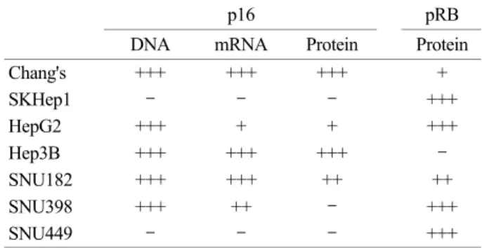
Table 1. Status of p16 or pRB in HCC cell lines. Table was obta- ined from southern blot, northern blot, and western blot analysis for p16 and pRB
p16 pRB
DNA mRNA Protein Protein
Chang's +++ +++ +++ +
SKHep1 - - - +++
HepG2 +++ + + +++
Hep3B +++ +++ +++ -
SNU182 +++ +++ ++ ++
SNU398 +++ ++ - +++
SNU449 - - - +++
+++: high, ++: moderate, +: low
SNU182, and SNU398 cells expressed remarkable p16 gene. The p16 band in HepG2 was detected in RT-PCR but not in northern blot analysis, implying the presence of low expression of p16 gene in this cell line (Table 1). We detec- ted faint band from HepG2 cells by long exposure of the film (data not shown). In case of SNU398 cells, p16 pro- tein was not detected. From our results, we confirmed that p16 is inactivated by homozygous deletion or posttranscri- ptional alteration in HCC cells. Transcriptional failure by promoter methylation was not detected in our data. Ho- wever, we could not exclude inactivation mechanism by the promoter methylation shown in other systems of HCC (Qin et al., 2004). These results suggest that the functional inactivation of p16 was shown in high frequencies in HCCs.
To compare the expression of p16 and pRB in the protein
level, western blot analysis was adjusted to pRB expression.
The pRB was expressed in all HCC cell lines except Hep3B
Fig. 1. Western blot analysis of p16 and pRB. The p16 protein was detected in Chang's liver, Hep3B and SNU182. pRB was shown in all HCC cell lines except Hep3B.
Fig. 2. Transduction efficiency of recombinant adenovirus. (A) LacZ staining in Ad-LacZ infected HCC cell lines. After the infection at the M.O.I. of 1, 10, or 50 for 48 hours, the cells were stained and counted. (B) Western blot in Ad-p16 infected SK-Hep-1, HepG2, or Hep3B. Dose and time dependent expressions of p16 were successfully detected in Ad-p16 infected HepG2 and Hep3B. Mock means the control for 24 hours under the normal medium. Expression of p16 by recombinant adenovirus infection results in slower migration than that of the endogenous p16 on SDS-PAGE.
B A
(Fig. 1). However, it has been reported that HepG2 had deletion in some exons in pRB gene but expressed pRB involving the leucine zipper region, presumably indicating functional mutation of pRB in this cell line (Kaino, 1997).
2. High transduction efficiency in the most HCC cells but exception in SKHep1
Adenoviral vector has many advantages in using the gene therapy (Lee et al., 2000; Kwon et al., 2001). The expression system was produced by high titer in virus-producing cell Fig. 3. Growth inhibition of Ad-p16 infected HCC cell lines. Chang's liver, HepG2, Hep3B, SNU182, SNU398, and SNU449 were in- fected with either control (filled squares), Ad-Luc (open squares), or Ad-p16 (open circles). Cell growths of Hep3B, SNU398, and SNU449 were significantly inhibited by Ad-p16 infection. This experiment was performed over at least 5 times.
lines, in addition to high transduction efficiency in dividing or resting cells (Lee et al., 2004). To explore transduction efficiency by adenovirus infection, we introduced Ad-LacZ virus into HCC cell lines. All cell lines except SKHep1 showed high transduction efficiency at M.O.I of 10≤ (Fig.
2A). Low transduction efficiency in SKHep-1 showed poor X-gal staining at even high concentration of Ad-LacZ. These results might be indicated as the lack of recognition for adenoviral protein (Cristiano et al., 1998). Consistently, in- troduction of Ad-p16 was not expressed in SKHep1 cells (Fig. 2B). However, introduction of Ad-p16 successfully
expressed in dose and time dependent manners in HepG2 and Hep3B cells. These results suggest that transduction efficiency of p16 might be limited on gene therapy subjec- ted to HCC.
3. Distinct growth inhibitory patterns in cell context- dependent manners
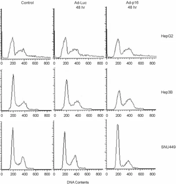
SRB assay was performed to determine therapeutic effi- cacy of Ad-p16 in HCC cells. Although we used the appro- priate concentration (20 M.O.I.) for effective killing esta- blished from previous study (Kim et al., 2001). As shown in
Fig. 4. Changes of cell population after Ad-p16 infection in HCC cell lines. Cells were grown in the infection of Ad-p16 for 48 hours.
Cell population of HepG2 was unaffected by Ad-p16 infection. Ad-p16 infected SNU449 resulted in a significant G1 arrest but Ad-p16 infected Hep3B did not.
Fig. 3, HepG2, and SNU182 cells did not show any growth inhibition by Ad-Luc or Ad-p16 infection. Cytotoxicity by Ad-Luc transduction was seen in SNU398 cells, even though in 10 M.O.I. ≤ (data not shown). Cell growths of Hep3B, SNU398, and SNU449 were significantly inhibited. Consi- dering the endogenous pRB status, SNU449 and SNU 398 cells are pRB-positive cells and Hep3B cells a pRB-negative cell. So, we have focused on the change of cell population in pRB-negative Hep3B cells. FACS analysis revealed the change of cell population. Dead cells were excluded in FACS analysis during harvesting the cells. G1 population in HepG2 cells, which did not show growth inhibition, remai- ned unchanged (Fig. 4). It was assumed that pRB resulted in functional inactivation although it is expressed in HepG2 (Kaino, 1997). In the pRB-expressed SNU449, G1 popula- tion was increased from 54% to 75%, suggesting that most of cells were arrested at G1 phase by Ad-p16 infection, However, G1 population in Hep3B cells was reduced from 59% to 39%. Instead, it seemed to be arrested in G2/M phase. Some investigators have suggested that p16 has a functional role in G2 phase (Gabrielli et al., 1999; Tamm et al., 2002). UVC-induced p16 correlated with CTD phosp- horylation at a late S-G2 delay in tumor cells with non- functional pRB (Gabrielli et al., 1999). In addition, Ad-p16 infection induced deformed polyploid cells in colon cancer cells, indicating that p16 act in G2/M phase (Tamm et al., 2002). These experiments provide the functional role of p16 in pRB-independent pathway.
As a result, these present data suggest that modality of p16 gene therapy might be considered on transduction effi- ciency and toxicity of adenoviral vector itself. Beside, growth inhibitory effect of p16 could be exerted through either pRB-dependent or -independent pathway.
Acknowledgements
Author appreciates Dr. Chang-Min Kim at National Cancer Center for his providing of technical comments.
This work was supported by a grant from Kangwon National University (3005044-1-1).
REFERENCES
Bressac B, Kew M, Wands J, Ozturk M. Selective G to T muta- tions of p53 gene in hepatocellular carcinoma from southern Africa. Nature 1991. 350: 429-431.
Chin PL, Chu DZJ, Clarke KG, Odom-Maryon T, Yen Y, Wagman LD. Ethnic differences in the behavior of hepatocellular carcinoma. Cancer 1999. 85: 1931-1936.
Cristiano RJ, Xu B, Nguyen D, Schmacher G, Kataoka M, Spitz FR, Roth JA. Viral and nonviral gene delivery vectors for cancer gene therapy. Cancer Det Prev. 1998. 22: 445-454.
Di Bisceglie AM, Rustgi VK, Hoofnagle JH, Dusheiko GM, Lotze MT. NIH conference. Hepatocellular carcinoma. Ann. Intern.
Med. 1988. 108: 390-401.
Foulkes WD, Flanders TY, Pollock PM, Hayward NK. The CDKN2A (p16) gene and human cancer. Mol Med. 1997. 3:
5-20.
Gabrielli BG, Sarcevic B, Sinnamon J, Walker G, Castellano M, Wang XQ, Ellem KA. A cyclin D-Cdk4 activity required for G2 phase cell cycle progression is inhibited in ultraviolet radiation-induced G2 phase delay. J Biol Chem. 1999. 274:
13961-13969.
Gerolami R, Uch R, Jordier F, Chapel S, Bagnis C, Brechot, Mannoni P. Gene transfer to hepatocellular carcinoma: trans- duction efficacy and transgene expression kinetics by using retroviral and lentiviral vectors. Cancer Gene Ther. 2000. 7:
1286-1292.
Harada N, Shimada M, Okano S, Suehiro T, Soejima Y, Tomita Y, Maehara Y. IL-12 gene therapy is an effective therapeutic strategy for hepatocellular carcinoma in immunosuppressed mice. J Immunol. 2004. 173: 6635-6644.
Hui A-M, Sakamoto M, Kanai Y, Ino Y, Gotoh M, Yokota J, Hirohashi S. Inactivation of p16INK4 in hepatocellular carcinoma. Hepatology 1996. 24: 575-579.
Kaino M. Alterations in the tumor suppressor genes p53, RB, p16/MTS1, and p15/MTS2 in human pancreatic cancer and hepatoma cell lines. J Gastroenterol. 1997. 32: 40-46.
Kim IA, Yang YJ, Yoon SC, Choi IB, Kay CS, Kwon HC, Kim CM, Joe YA, Kang JK, Hong YK. Potential of adenoviral p53 gene therapy and irradiation for the treatment of malig- nant gliomas. Int J Oncol. 2001. 19: 1041-1047.
Kita R, Nishida N, Fukuda Y, Azechi H, Matsuoka Y, Kodema T, Sando T, Nakao K, Ishizaki K. Infrequent alterations of the p16INK4A gene in liver cancer. Int J Cancer. 1996. 67: 176 -180.
Kwon HC, Kim JH, Kim KC, Lee KH, Lee JH, Lee BH, Lee KH, Jang JJ, Lee CT, Lee H, Kim CM. In vivo antitumor effect of herpes simplex virus thymidine kinase gene therapy in rat hepatocellular carcinoma: feasibility of adenovirus-mediated intra-arterial gene delivery. Mol Cells 2001. 11: 170-178.
Lee CT, Park KH, Yanagisawa K, Adachi Y, Ohm JE, Nadaf S, Dikov MM, Curiel DT, Carbon DP. Combination therapy with conditionally replicating adenovirus and replication de- fective adenovirus. Cancer Res. 2004. 64: 6660-6665.
Lee SH, Kim MS, Kwon HC, Park IC, Park MJ, Lee CT, Kim YW, Kim CM, Hong SI. Growth inhibitory effect on glioma cells of adenovirus-mediated p16/INK4a gene transfer in vitro and in vivo. Int J Mol Med. 2000. 6: 559-563.
Medema RH, Herrera RE, Lam F, Weiberg RA. Growth suppre- ssionby p16ink4 requires functional retinoblastoma protein.
Proc Natl Acad Sci USA. 1995. 92: 6289-6293.
Okamoto A, Demetrick DJ, Spillare EA, Hagiwara K, Hussain SP, Bennett WP, Forrester K, Gerwin B, Serrano M, Beach DH, Harris CC. Mutations and altered expression of p16INK4 in human cancer. Proc Natl Acad Sci USA. 1994. 91: 11045 -11049.
Park KH, Kim G, Jang SH, Kim CH, Kwon SY, Yoo CG, Kim YW, Kwon HC, Kim CM, Han SK, Shim YS, Lee CT. Gene therapy with GM-CSF, interleukin-4 and herpes simplex virus thymidine kinase shows strong antitumor effect on lung cancer. Anticancer Res. 2003. 23: 1559-1564.
Qin Y, Liu J-Y, Li B, Sun Z-L, Sun Z-F. Association of low p16INK4a and p15INK4b mRNAs expression with their CpG islands methylation with human hepatocellular carcino- genesis. World J Gastroenterol. 2004. 10: 1276-1280.
Schutte M, Hruban RH, Geradts J, Maynard R, Hilgers W, Rabindran SK, Moskaluk CA, Hahn SA, Scwarte-Waldhoff I, Schmiegel W, Baylin SB, Kern SE, Herman JG. Abrogation of the Rb/p16 tumor-suppressive pathway in virtually all pancreatic carcinomas. Cancer Res. 1997. 57: 3126-3130.
Serrano M, Hannon GJ, Beach D. A new regulatory motif in cell- cycle control causing specific inhibition of cyclin D/CDK4.
Nature 1993. 366: 704-707.
Sherr CJ. Cancer cell cycles. Science 1996. 274: 1672-1677.
Tamm I, Schumacher A, Karawajew L, Ruppert V, Arnold W, Nussler AK, Neuhaus P, Dorken B, Wolff G. Adenovirus- mediated gene transfer of P16INK4/CDKN2 into bax-negative colon cancer cells induces apoptosis and tumor regression in vivo. Cancer Gene Ther. 2002. 9: 641-650.
Vernell R, Helin K, Müller H. Identification of target genes of the p16INK4A-pRB-E2F pathway. J Biol Chem. 2003. 278:
46124-46137.