86
A schwannoma arising from median nerve at proximal forearm is rare.1 Most cases of schwannomas are asymptomatic.2 Neurologic symptoms resulted by compressing the nerve of its origin are rare presentations in schwannoma cases. Here, we report an unusual case of schwannoma originated from median nerve at proximal forearm that caused ulnar nerve symptom.
CASE REPORT
A 77-year-old male visited the orthopaedic clinic at our institution with hard palpable mass at volar aspect of proximal forearm occurred 10 years ago. The patient complained about mild pain and discomfort on proximal forearm and described the size of mass seemed to be increasing recently. On physical examination, the patient presented tingling sensation at ring and little finger by percussion on the mass. However, Tinel’s sign of the ulnar nerve at the cubital tunnel and the elbow flexion test were both negative. There was no evidence of suspicion for anterior interosseous nerve or other median
nerve disturbance. However, Froment’s test implying chronic ulnar nerve damage was positive. To evaluate the neuropathy, nerve conduction velocity (NCV) study was carried out. On NCV study, mild elongated latency of ulnar nerve below elbow level was revealed. Magnetic reasonance imaging (MRI) was performed to determine the exact location and the origin of the mass and to make sure there were no other lesions that might have caused similar symptoms. On preoperative enhanced MRI study, lesion was well circumscribed along the median nerve at proximal forearm. The mass showed similar intensity with muscle on T1-wighted images.
Old hemorrhages or calcifications-like heterogenic signals were found on T2-weighted images (Fig. 1A, B). Contrast- enhanced T1-weighted images revealed irregularly moderate enhancement of the lesion (Fig. 1C). On T2-weighted axial image, it seemed to be clear that the mass was originated from the median nerve. High signal change around the ulnar nerve was found. The mass was occupying a great amount of space on proximal forearm. The structures around the mass were
A Schwannoma Originating from Median Nerve at Proximal Forearm Caused Ulnar Nerve Symptom by Compression
Jeong Hyun Yoo, Joon Yub Kim*, Hyoung Soo Kim, Joo Hak Kim, Ki Hyuk Sung, Sang Hun Song, Ho Il Kwak
Department of Orthopaedic Surgery, Myongji Hospital, Goyang, Korea
CC This is an open-access article distributed under the terms of the Creative Commons Attribution Non-Commercial License (http://creativecommons.org/licenses/by-nc/3.0) which permits unrestricted noncommercial use, distribution, and reproduction in any medium, provided the original work is properly cited.
Copyright © 2014 by the Korean Society for Microsurgery. All Rights Reserved.
Received October 3, 2014 Revised November 5, 2014 Accepted November 6, 2014
*Correspondence to: Joon Yub Kim Department of Orthopaedic Surgery, Myongji Hospital, 55 Hwasu-ro, 14beon- gil, Deogyang-gu, Goyang 412-826, Korea Tel: +82-31-810-5114
Fax: +82-31-969-0500 E-mail: [email protected]
Financial support: None.
Conflict of interest: None.
A schwannoma is a benign soft tissue tumor arising from the nerve sheath of a Schwann cell. Clinically, a schwannoma is an asymptomatic mass rarely causing neurologic deficits. However, it can cause discomfort as well as motor and sensory disturbances by compressing the nerve of its origin. The authors encountered a huge schwannoma arising from the median nerve at the proximal forearm, which caused symptoms mainly in the ulnar nerve. The tingling sensation along the ulnar nerve disappeared completely after enucleation of the schwannoma originating from the median nerve.
Key Words: Schwannoma, Median nerve, Ulnar nerve, Forearm, Compression, Neurilemmoma
ARMS
Archives of Reconstructive MicrosurgeryCase Report
pISSN 2383-5257 eISSN 2288-6184 Arch Reconstr Microsurg 2014;23(2):86-88 http://dx.doi.org/10.15596/ARMS.2014.23.2.86
Jeong Hyun Yoo, et al. A Schwannoma Causing Neuropathy of Adjacent Nerve
www.e-arms.org 87
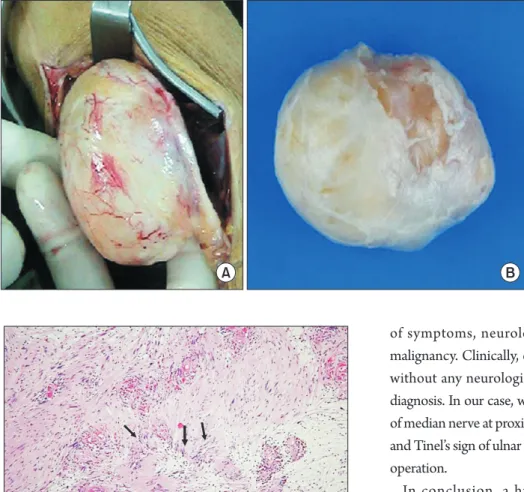
squeezed out from their original location (Fig. 2). During the operation, we observed finely encapsulated and marginated 5×4×8 cm sized mass (Fig. 3). Histological finding revealed an encapsulated schwannoma composed of densely packed spindle cells of Antoni type A tissue with nuclear palisading or Verocay bodies and loosely packed Antoni type B tissue (Fig.
4). A complete enucleation was performed and Tinel’s sign was disappeared immediately after the operation. However, Froment’s sign was still positive.
DISCUSSION
Schwannomas are benign soft tissue tumors that arising from schwann cells of nerve sheath encapsulated with epineurium.1 Schwannomas are slow-growing masses preserving nerve fibers with various sizes from 1 mm to 250 mm.3 Most cases are observed in the head and neck area as bilateral vestibular schwannomas. They can also occur in spinal roots, peripheral nerves on the flexor area of the upper extremities and the posterior aspect of the lower extremities.2 Malignant transformations are rare. Clinically, schwannomas occur incidentally as painless mass. But occasionally, schwannomas can exhibit neurological symptoms by compressing surrounding structures.3 They are usually located along the course of nerve which is not mobile longitudinally. Tinel’s sign might be positive on involved nerve.4 In the case of huge schwannoma, pain, numbness, paresthesia, and muscle-wasting can occur.
In our case, a schwannoma originated from proximal median nerve revealed Tinel’s sign of ulnar nerve without median nerve symptom. Intrinsic muscle wasting of the hand was also observed (Froment’s sign positive). It is unusual that the huge schwannoma cause neurologic symptom to adjacent nerve, not the nerve of its origin. For the diagnosis of schwannoma, MRI offers information about the origin and surrounding structures.5 Usually, schwannoma show intermediate signal intensity on T1- wighted images and intermediate heterogenous signal intensity in high signal intensity on T2-weighted images.4
Fig. 1. Magnetic reasonance imaging (MRI) from a schwannoma at proximal forearm arising from median nerve abutting flexor carpi ulnaris. A round mass with signal density similar to skeletal muscle was observed in coronal T1-weighted MRI of the forearm (A), coronal T2-weighted image revealed old hemorrhage and calcifications-like heterogenicity (B) and the mass showed irregular intermediate enhancement on contrast enhanced sagittal T1-weighted image (C).
A B C
Fig. 2. Axial T2-weighted image clearly showing the schwannoma was originated from the median nerve (white dotted arrow). High signal change was found around the ulnar nerve (white arrow). Great amount of space was occupied by the mass on proximal forearm. The structures around the mass were crowded out from their original location.
Arch Reconstr Microsurg Vol. 23. No. 2. November 2014
88
In our case, the well encapsulated huge round mass from the median nerve abutting flexor digitorum superficialis and flexor carpi ulnaris was revealed in MRI finding. The mass was deviated to ulnar side and flexor carpi ulnaris was compressed by the huge schwannoma. The ulnar nerve between the two heads of flexor carpi ulnaris seemed to be compressed.
Histologically, densely packed spindle cells of Antoni type A tissue, loosely packed Antoni type B tissue, Verocay bodies and thick walled capillaries are characteristics of schwannoma.
Either Antoni type A or B tissue may be appeared almost every histologic findings of cases.6 We found every histologic findings of the schwannoma in our case which confirmed the diagnosis.
Operative indications for schwannomas are aggravation
of symptoms, neurologic impairment, and suspicion of malignancy. Clinically, complete enucleation of schwannoma without any neurologic damage itself can help confirm the diagnosis. In our case, well encapsulated 5×4×8 cm sized mass of median nerve at proximal forearm was completely enucleated and Tinel’s sign of ulnar nerve was completely resolved after the operation.
In conclusion, a huge sized schwannoma can cause neurologic symptom by compressing adjacent nerve which may be very confusing for surgeons to clarify the origin of neurologic impairment. Careful evaluation of the lesion must be performed by the physicians.
REFERENCES
1. Hubert J, Landes G, Tardif M. Schwannoma of the median nerve. J Plast Surg Hand Surg 2013;47:75-7.
2. Phalen GS. Neurilemmomas of the forearm and hand. Clin Orthop Relat Res 1976;(114):219-22.
3. Mariottini A, Carangelo B, Peri G, Tacchini D, Mormouras V, Muya M, et al. Schwannoma of median nerve at the elbow. Case report and short review of the literature. G Chir 2011;32:55-8.
4. DiTrapani R, Rubin DI. An unusual presentation of a proximal median nerve schwannoma. Muscle Nerve 2012;46:983-4.
5. Rinaldi I. The value of preoperative angiography in the surgical management of cervical hourglass neurofibroma. Case report. J Neurosurg 1972;36:97-101.
6. Ikeda K, Osamura N, Kasashima S. Ossified median nerve schwannoma: a case report. Hand Surg 2012;17:371-3.
Fig. 3. A 5×4×8 cm sized well encapsul- ated mass originated from median nerve was observed during the surgery (A) and enucleated (B).
A B
Fig. 4. Histological finding revealing the encapsulated schwannoma composed of densely packed spindle cells of Antoni type A tissue (left upper portion of the picture) with nuclear palisading or Verocay bodies (black arrows) and loosely packed Antoni type B tissue (right lower portion of the picture) (H&E, ×100).