Efficacy and feasibility of laparoscopic subtotal cholecystectomy for acute cholecystitis
In Oh Jeong, Jang Yong Kim, Yun-Mee Choe, Sun Keun Choi, Yoon Seok Heo, Keon-Young Lee, Sei Joong Kim, Young Up Cho, Seung-Ik Ahn, Kee Chun Hong,
Kyung Rae Kim, and Seok-Hwan Shin
Department of Surgery, Inha University College of Medicine, Incheon, Korea
Backgrounds/Aims: For patients with acute cholecystitis, conversion from laparoscopic cholecystectomy to open surgery is not uncommon due to possibilities of serious hemorrhage at the liver bed and bile duct injury. Recent studies reported successful laparoscopic subtotal cholecystectomy for acute cholecystitis. The purpose of this study was to determine the efficacy and feasibility of such an operation based on the experience of surgeons at our facility. Methods: In this study, we enrolled 144 patients who had received either laparoscopic subtotal cholecystectomy (LSC), laparoscopic cholecystectomy (LC), or open cholecystectomy (OC) for acute cholecystitis from January 2004 to December 2009 at the Department of Surgery of our hospital. Their symptoms, signs, operative findings, pathologic results and post- operative results were compared and analyzed. Results: There were 26 patients in the LSC group 80 in the LC group and 38 in the OC group. There were no differences in mean age, sex, and symptoms of acute cholecystitis. The LSC group showed higher CRP levels (p<0.001) and a higher grade according to the Tokyo criteria (p=0.001). The mean operative time was 115.6 minutes and mean blood loss was 158.9 ml without intra-operative or postoperative transfusion. There weren't any bile duct injuries during the operation. No group suffered bile leakage. Drains were removed 3.3 days after the operation in the LC group, the shortest time compared to the other groups (p<0.001).
LC and LSC groups demonstrated shorter postoperative hospital days and time to diet resumption than the OC group (p<0.001). Conclusions: LSC appears to be a safe and effective treatment in cases of severe acute cholecystitis that require consideration of conversion to open surgery. (Korean J Hepatobiliary Pancreat Surg 2011;15:225-230) Key Words: Acute cholecystitis; Laparoscopic subtotal cholecystectomy
Received: August 4, 2011; Revised: September 20, 2011; Accepted: October 20, 2011 Corresponding author: Seung-Ik Ahn
Department of Surgery, Inha University College of Medicine, Sinheung-dong 3-ga, Jung-gu, Incheon 400-712, Korea Tel: +82-32-890-2114, Fax: +82-32-890-3999, E-mail: siahn@inha.ac.kr
Copyright Ⓒ 2011 by The Korean Association of Hepato-Biliary-Pancreatic Surgery Korean Journal of Hepato-Biliary-Pancreatic Surgery ∙ pISSN: 1738-6349
INTRODUCTION
Laparoscopic cholecystectomy (LC) was introduced in 1989 as a standard treatment for benign gallbladder dis- eases, but was not well enforced initially for acute cholecystitis. However, ever since the first treatment of acute cholecystitis with LC,1 experience with LC has ac- cumulated and techniques have improved, and LC has be- come the treatment of choice for acute cholecystitis.
Nevertheless, in cases with severe acute cholecystitis demonstrating severe swelling and edema of the gall- bladder, fibrosis of surrounding organ, fibrosis of Calot's triangle, and anatomic deformity, it is difficult to perform LC successfully. Furthermore, when massive bleeding from the liver bed and bile duct injury are suspected, sur-
geons are forced to switching to open conversion.2,3 As an alternative treatment, laparoscopic subtotal chol- ecystectomy (LSC) was introduced, but in the beginning there was controversy regarding its safety. Still, LSC has continuously been applied to acute cholecystitis, and stud- ies have shown this treatment to be feasible and safe.4-6 Hence, the purpose of this study was to analyze experi- ences with LSC and determine its safety and efficacy for acute cholecystitis.
METHODS
Patients
Among 1,069 patients who received LC at one medical center from January, 2004 to December, 2009 by a single
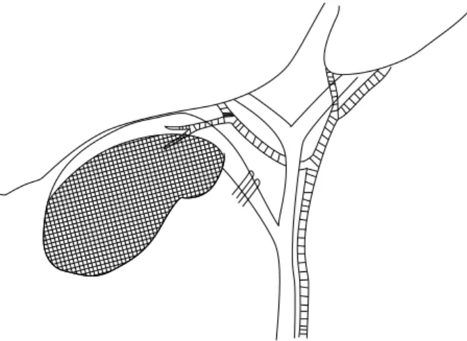
Fig. 1. Illustration of the surgical technique. The gallbladder infundibular serosa dissected and the cystic duct was clipped twice. The anterior wall of the gallbladder was dissected and mucosa of the remnant posterior wall was destroyed.
surgeon, 144 patients were diagnosed with acute chol- ecystitis and their clinical data were selected for this retro- spective study. Body temperature, blood tests, and more than one imaging study among abdomen ultrasonography, abdomen computed tomography (CT), and hepatobiliary scintigraphy were done for every patient suspected of hav- ing acute cholecystitis. Patients suspected, from imaging studies, of having a common bile duct stone were further investigated with preoperative endoscopic retrograde chol- angiopancreatography (ERCP) or endoscopic ultrasono- graphy (EUS). When a common bile duct stone was iden- tified, endoscopic stone extraction and endoscopic sphinc- terotomy were performed. Intraoperative cholangiography was not performed. When the patients demonstrated bile duct obstruction signs such as increased total bilirubin and alkaline phosphatase (ALP), postoperatively, examinations for remnant bile duct stones were carried out.
The diagnosis of acute cholecystitis was made accord- ing to the Tokyo guidelines7,8 based on local symptoms of inflammation, systemic signs, and imaging studies, and the severity of acute cholecystitis was divided into Grade I (mild), II (moderate), and III (severe). According to the methods of surgery, the 144 patients were divided into an LSC group, an LC group, and an open group, and their symptoms, signs, operative findings, pathologic results, and postoperative results were compared and analyzed.
The thickness of the gallbladder wall was assessed inside the operating theatre with a ruler, measuring the thickest part of the resected gallbladder's wall. For statistical anal- ysis, the chi-square test and a one-way ANOVA test were used. To distinguish the differences between the groups, Tukey's honestly significant difference (HSD) test was carried out, and a p-value of less than 0.05 was considered statistically significant.
Operative procedure
The operation was initially laparoscopic for all patients:
26 patients received LSC because LC was thought to be difficult to dissect the Calot's triangle due to severe in- flammation and to identify the anatomy of the cystic ar- tery and the cystic duct. Bile juice was aspirated from the gallbladder with a needle. When pus was evaluated, it was sent for microbial culture. Upon exploring Calot's triangle, either the cystic duct or the structure assumed to be the cystic duct after opening its serosa was ligated with an
endo-clip but not divided. The cystic artery was not sepa- rately dissected. Then, the infundibulum of the gallbladder was transsected near Hartmann's pouch, and the anterior wall of the gallbladder was removed with an electro- cautery device. As a result, there remained 25 to 35 per- cent of the gallbladder. Small gallbladder stones were suc- tioned out while big stones were removed using an endobag. The posterior wall attached to the liver bed was not removed. The mucosa of the posterior wall, however, was destroyed by electrocautery (Fig. 1). Finally, the op- erative field was washed with saline and after placing a drain, the operation was completed.
RESULTS
There were 26 patients who received LSC, 80 who re- ceived LC, and 38 who received open cholecystectomy.
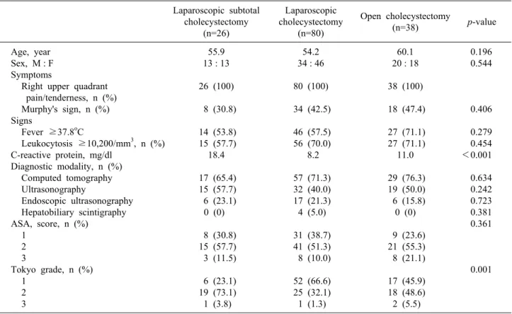
Their mean ages were 55.9 years, 54.2 years, and 60.1 year, respectively, and were not different (p=0.196). There were no differences in gender (p=0.544). As for symptoms of acute cholecystitis, all 144 patients suffered from right upper quadrant pain and tenderness, while Murphy's sign was demonstrated by 30.8% of the LSC group, 42.5% of the LC group, and 47.4% of the open group, again with- out a significant difference (p=0.406). Fever higher than 37.8oC was observed in 53.8% of the LSC group, 57.5%
of the LC group, and 71.1% of the open group, but these differences were not being significant (p=0.279). Patients with leukocytosis (a white cell count >10,200/mm3) was
Table 1. Preoperative characteristics of 144 patients who underwent cholecystectomy Laparoscopic subtotal
cholecystectomy (n=26)
Laparoscopic cholecystectomy
(n=80)
Open cholecystectomy
(n=38) p-value
Age, year Sex, M : F Symptoms
Right upper quadrant pain/tenderness, n (%) Murphy's sign, n (%) Signs
Fever ≥37.8oC
Leukocytosis ≥10,200/mm3, n (%) C-reactive protein, mg/dl
Diagnostic modality, n (%) Computed tomography Ultrasonography
Endoscopic ultrasonography Hepatobiliary scintigraphy ASA, score, n (%)
1 2 3
Tokyo grade, n (%) 1
2 3
55.9 13 : 13 26 (100) 8 (30.8) 14 (53.8) 15 (57.7)
18.4 17 (65.4) 15 (57.7) 6 (23.1)
0 (0) 8 (30.8)
15 (57.7) 3 (11.5) 6 (23.1) 19 (73.1) 1 (3.8)
54.2 34 : 46 80 (100) 34 (42.5) 46 (57.5) 56 (70.0)
8.2 57 (71.3) 32 (40.0) 17 (21.3) 4 (5.0) 31 (38.7) 41 (51.3) 8 (10.0) 52 (66.6) 25 (32.1) 1 (1.3)
60.1 20 : 18 38 (100) 18 (47.4) 27 (71.1) 27 (71.1)
11.0 29 (76.3) 19 (50.0) 6 (15.8)
0 (0) 9 (23.6)
21 (55.3) 8 (21.1) 17 (45.9) 18 (48.6) 2 (5.5)
0.196 0.544
0.406 0.279 0.454
<0.001 0.634 0.242 0.723 0.381 0.361
0.001
ASA, american society of anesthesiology.
Table 2. Operative findings of 26 patients who underwent lap- aroscopic subtotal cholecystectomy
Operative finding Number of patients (%) Adhesion
Thickening
Degree of thickening, mm2 (range) Necrosis
Pus
18 (69.2) 26 (100) 8.08 (4-15)
9 (34.6) 14 (53.8)
seen in 57.7%, 70.0%, and 71.1%, respectively (p=0.454).
Mean CRP levels were 18.4 mg/dl in the LSC group, 8.2 mg/dl in the LC group, and 11.0 mg/dl in the open group.
The LSC group showed the highest level and it was sig- nificantly different (p<0.001). Imaging studies included abdominal computed tomography, ultrasonography, and hepatobiliary scintigraphy, and showed no differences be- tween groups. There were also no significant differences in American Society of Anesthesiology (ASA) scores (p=0.361). Severity according to the Tokyo guidelines was highest in the LSC group (p=0.001) (Table 1), but there was not a linear correlation of severity among the groups (p=0.310).
Among 26 patients of the LSC group, there was an ad- hesion between the gallbladder and its surrounding tissues in 69.2%. All patients had gallbladder wall thickening, 8.1 mm being average. Necrosis of the gallbladder was evi- dent in 34.6%, and pus drainage in 53.8% (Table 2).
The mean operative time for the LSC group was 115.6 minutes, and the mean blood loss was 158.9 ml. There was no need for intra-operative or postoperative trans- fusion, and no intraoperative bile duct injury. The surgical diagnosis made from operative findings included 9 cases of acute cholecystitis, 14 cases of gallbladder congestion, 1 case of gallbladder perforation, and 2 cases of acalcu- lous cholecystitis (Table 3).
No group suffered from postoperative bile leakage.
There was 1 case of wound infection in the LC group who recovered with conservative management without reopera- tion. One patient who underwent open cholecystectomy received a transfusion due to bleeding from liver cirrhosis, and died of acute lung injury. The drain was removed 3.3 days on average after LC, which was the shortest time among the groups. The mean overall hospital stay, the postoperative hospital stay, and the mean time to restart
Table 4. Postoperative outcomes of 144 patients who underwent cholecystectomy Laparoscopic subtotal
cholecystectomy (n=26)
Laparoscopic cholecystectomy
(n=80)
Open cholecystectomy
(n=38) p-value
Bile leakage
Time to remove drain, days (range) Hospital stays, days (range)
Postoperative hospital stays, days (range) Diet resumption, days (range)
Operative complications, n (%) Reoperation, n (%)
Mortality, n (%)
0 8.7 (3-17) 11.1 (5-23) 6.0 (3-12) 1.7 (1-3)
0 (0) 0 (0) 0 (0)
0 3.3 (0-15) 10.3 (5-28) 4.6 (2-20) 1.7 (1-6) 1 (1.3) 0 (0) 0 (0)
0 7.7 (4-30) 17.7 (10-36) 10.8 (6-28)
4.3 (2-31) 1 (2.6) 0 (0) 1 (2.6)
<0.001
<0.001
<0.001
<0.001 0.693 0.444 Table 3. Operative results of 26 patients who underwent lapa-
roscopic subtotal cholecystectomy
Parameter Values
Operative time, mean (range) Intraoperative blood loss, ml (range) Intraoperative transfusion, ml Postoperative transfusion, ml Intraoperative bile duct injury, n Bacteriologic positive of bile, n (%) Operative diagnosis, n (%)
Acute cholecystitis Gallbladder empyema Gallbladder perforation Acalculous cholecystitis Pathologic diagnosis, n (%)
Acute cholecystitis Chronic cholecystitis
Acute and chronic cholecystitis Acute gangrenous cholecystitis Gallbladder empyema
115.6 (80-160) 158.9 (0-800)
0 0 0 2 (7.7) 9 (34.6)
14 (53.8) 1 (3.8) 2 (7.7) 5 (19.2) 5 (19.2) 8 (30.8) 4 (15.4) 4 (15.4) a food diet were lessed in the LC and LSC groups than in the open group (p<0.001) (Table 4).
DISCUSSION
LC has become the treatment of choice for acute cholecystitis. Yet, open conversion is common in many severe cases, where severe inflammation and edema of the gallbladder and adhesion between adjacent structures don’t indicate clear anatomical identification of cystic ar- tery and duct under laparoscopic vision, thus resulting in increased risk of bile duct injury and bleeding. Open con- version for these severe cases has been regarded as ad- equate to date.
In 1985, Bornman and Terblanche introduced open parti- al cholecystectomy as an alternative treatment for acute
cholecystitis,9 but there were concerns about accumulation of secretion from the remnant gallbladder's posterior wall mucosa and abscess formation. Also, LSC was noted for the possibility of missed gallstones during retraction and unconfirmed cystic duct resulting in bile leakage, bile ab- scess, and fistula.15 Soon thereafter, safety and efficacy of open partial cholecystectomy were verified by Johansson et al.,16 and recent papers have reported the usefulness of LC as a treatment for severe cholecystitis.5,10-14 Bile leak- age from LSCs range from 3.0%12 to 17.9%;14 intra-ab- dominal abscess and remnant gallstones range from 3.8%13 to 16.2%.11 These complications did not occur in our study. Remnant gallstones can be minimized by re- moving small stones with suction devices and big stones with an endobag. Also, even if the cystic duct cannot be clearly isolated, it can be ligated to minimize bile leakage.
We initially had concerns with abscess formation and ac- cumulation of secretions from remnant mucosa, which we electrocauterized to prevent such complications. These complications, if they occurred, were scheduled to be treated with conservative treatment or ERCP treatment such as bile duct stent insertion.
We ligated the infundibulum with an endoclip. Recent studies reported usage of endo-loops and a GIA stap- ler.2,4,6,11,12,18
Sinha et al. performed LSC without ligating the infundibulum, resulting in bile leakage in 17.9% of case, and was treated non-operatively with conservative treatment or stent insertion by ERCP.14 This suggests that LSC is feasible even without complete identification of anatomic structures.
Our study did not implicate that severer cholecystitis according to the Tokyo guideline requires certain types of surgery. However, the LSC group's grade was higher than
that of the LC group (p=0.001), while it was similar with that of the open group (p=0.260), which implies that con- sideration of LSC or open surgery is required for severer cholecystitis according to Tokyo guidelines. As open con- version is considered during LC due to severe chol- ecystitis, LSC may be tried first owing to the better post- operative results of laparoscopic surgery than open surgery. Furthermore, LSC is safe when performed by sur- geons experienced with laparoscopy surgery. LSC should be limited to severe cholecystitis in which identification of the cystic duct and artery is impossible due to severe edema and swelling of gallbladder, fibrosis and adhesion of Calot's triangle, and necrosis of gallbladder wall, and thus increased risk of injury of bile duct and bleeding is anticipated.
Patients who received open surgery, compared to lapa- roscopic surgery, tended to have a higher incidence of wound complications, more pain, and longer hospital stays, postoperative hospital stays, and time to diet resumption. There was one case of wound infection in the LC group, who was treated with conservative treatment.
There was no severe wound complication requiring reop- eration in any of the groups. Drains were removed earlier from the LC group than from other groups (p<0.001).
There was no difference between LSC and OC groups (p=0.488). It may have been due to concerns that the LSC group suffers from bile duct leakage and was observed for a longer period without removing the drain. There was no mortality in the laparoscopic group, but one patient in the open group died of a transfusion-related acute lung injury.
The transfusion was prescribed because of bleeding from liver cirrhosis. The operation for this patient was con- verted to an open technique due to massive bleeding from the liver bed. Since LSC does not involve dissection of the liver bed, bleeding is less. Thus, in the case of a pa- tient such as the one mentioned above who is predicted to develop massive bleeding due to liver cirrhosis or thrombocytopenia, LSC rather than open conversion is advised.4-6,12 Furthermore, since open conversion done on patients of old age or poor general condition may induce a prolonged operative time, aggravation of cardiopulmo- nary function from increased pain, and delayed recovery of bowel movement, LSC should be considered. Frazee et al. also reported fewer postoperative cardiopulmonary complications with laparoscopic surgery compared to
open surgery for patients with poor pulmonary function.19 When performing LSC for severe acute cholecystitis, suspected malignancy prior to operation with imaging studies such as CT and MRI (magnetic resonance imag- ing) should never be made light of. Open conversion is mandatory when malignancy is demonstrated during LSC.17 Furthermore, serial ultrasonography, CT, MRI, ERCP, and serum tumor markers should be carried out to follow-up on signs of malignancy. The LSC group in this study had a mean follow-up period of 2.3 months af- ter operation, during which no remnant gallbladder pro- gressed to carcinoma. There was one case in the LC group who suffered from postoperative cholangitis due to bile duct obstruction, for which examination was carried out to reveal a distal common bile duct cancer. The patient underwent pylorus preserving pancreatoduodenectomy. So far, there have not been any reports of remnant GB after LSC progressing to carcinoma. Yet, due to the short fol- low-up period, this cannot be confirmed and thus, further studies with longer follow-up periods are necessary.
The standard treatment of benign gallbladder disease is LC. Nonetheless, LC may not be so successful for severe acute cholecystitis. In most cases, open conversion has been inevitable. When LSC is applied, however, reduced pain, shorter hospital stays, and earlier diet resumption can be anticipated. Hence, when considering open con- version for severe acute cholecystitis, LSC is a safe and effective alternative.
ACKNOWLEDGEMENTS
This work was supported by INHA University Research Grant.
REFERENCES
1. Jacobs M, Verdeja JC, Goldstein HS. Laparoscopic cholecystec- tomy in acute cholecystitis. J Laparoendosc Surg 1991;1:175-177.
2. Cox MR, Wilson TG, Luck AJ, Jeans PL, Padbury RT, Toouli J. Laparoscopic cholecystectomy for acute inflammation of the gallbladder. Ann Surg 1993;218:630-634.
3. Palanivelu C, Rajan PS, Jani K, et al. Laparoscopic cholecystec- tomy in cirrhotic patients: the role of subtotal cholecystectomy and its variants. J Am Coll Surg 2006;203:145-151.
4. Chowbey PK, Sharma A, Khullar R, Mann V, Baijal M, Vashistha A. Laparoscopic subtotal cholecystectomy: a review of 56 procedures. J Laparoendosc Adv Surg Tech A 2000;10:31-34.
5. Michalowski K, Bornman PC, Krige JE, Gallagher PJ, Terblanche J. Laparoscopic subtotal cholecystectomy in patients
with complicated acute cholecystitis or fibrosis. Br J Surg 1998;
85:904-906.
6. Ransom KJ. Laparoscopic management of acute cholecystitis with subtotal cholecystectomy. Am Surg 1998;64:955-957.
7. Sekimoto M, Takada T, Kawarada Y, et al. Need for criteria for the diagnosis and severity assessment of acute cholangitis and cholecystitis: Tokyo Guidelines. J Hepatobiliary Pancreat Surg 2007;14:11-14.
8. Hirota M, Takada T, Kawarada Y, et al. Diagnostic criteria and severity assessment of acute cholecystitis: Tokyo Guidelines. J HBP Surg 2007;14:78-82.
9. Bornman PC, Terblanche J. Subtotal cholecystectomy: for the dif- ficult gallbladder in portal hypertension and cholecystitis. Surgery 1985;98:1-6.
10. Bickel A, Shtamler B. Laparoscopic subtotal cholecystectomy. J Laparoendosc Surg 1993;3:365-367.
11. Beldi G, Glättli A. Laparoscopic subtotal cholecystectomy for se- vere cholecystitis. Surg Endosc 2003;17:1437-1439.
12. Ji W, Li LT, Li JS. Role of laparoscopic subtotal cholecystec- tomy in the treatment of complicated cholecystitis. Hepatobiliary Pancreat Dis Int 2006;5:584-589.
13. Philips JA, Lawes DA, Cook AJ, et al. The use of laparoscopic
subtotal cholecystectomy for complicated cholelithiasis. Surg Endosc 2008;22:1697-1700.
14. Sinha I, Smith ML, Safranek P, Dehn T, Booth M. Laparoscopic subtotal cholecystectomy without cystic duct ligation. Br J Surg 2007;94:1527-1529.
15. Hashizume M, Sugimachi K, MacFadyen BV. The clinical man- agement and results of surgery for acute cholecystitis. Semin Laparosc Surg 1998;5:69-80.
16. Johansson M, Thune A, Nelvin L, Stiernstam M, Westman B, Lundell L. Randomized clinical trial of open versus laparoscopic cholecystectomy in the treatment of acute cholecystitis. Br J Surg 2005;92:44-49.
17. Nakajima J, Sasaki A, Obuchi T, Baba S, Nitta H, Wakabayashi G. Laparoscopic subtotal cholecystectomy for severe cholecy- stitis. Surg Today 2009;39:870-875.
18. Rosenberg J, Bisgaard T. The difficult gallbladder: technical tips for laparoscopic cholecystectomy. Surg Laparosc Endosc Percu- tan Tech 2000;10:249-252.
19. Frazee RC, Roberts JW, Okeson GC, et al. Open versus laparo- scopic cholecystectomy. A comparison of postoperative pulmo- nary function. Ann Surg 1991;213:651-653.