Role of common bile duct resection in T2 and T3 gallbladder cancer patients
Jin Hong Lim1, Jae Uk Chong2, Sung Hoon Kim3, Seung Woo Park4, Jin Sub Choi2, Woo Jung Lee2, and Kyung Sik Kim2
1Department of Surgery, Gangnam Severance Hospital, Yonsei University College of Medicine, Seoul,
2Department of Surgery, Severance Hospital, Yonsei University College of Medicine, Seoul,
3Department of Surgery, Wonju Severance Christian Hospital, Yonsei University College of Medicine, Wonju,
4Department of Internal Medicine, Severance Hospital, Yonsei University College of Medicine, Seoul, Korea
Backgrounds/Aims: Routine bile duct resection as part of the typical oncological resection for patients with advanced gallbladder cancer remains controversial with regard to, ultimately, curative value. The aim of this study was to compare oncological outcomes for patients undergoing surgery for gallbladder cancer with or without bile duct resection.
Methods: We recruited, for the purpose of this study, all patients who underwent surgical resection for T2 and T3 gallbladder cancer at Severance hospital, Seoul, Korea, during the period January 2000 and December of 2011. The patient data was reviewed retrospectively. Results: The patients (n=149) recruited to participate in the study were div- ided into two groups according to their bile duct resection status: The bile duct resection group (BDR group, n=54);
and, the bile duct non-resection group (BDNR group, n=95). Significant difference was found in lymph node retrieval between BDR and BDNR groups (15 vs. 5, respectively with p<0.001). There was no significant difference between the two groups with regard to the five year survival rate (43% in BDR group vs. 57% in BDNR group, p=0.339).
Following multivariate analysis, lymph node metastasis, advanced T-stage, and total retrieved lymph nodes <6 were independent prognostic factors for poor survival in patients with T2 and T3 gallbladder cancer. Conclusions: The find- ings revealed by the current study suggest that the role of bile duct resection might be limited to improved staging, and offers no advantage in long-term survival. However, in view of the foregoing and given the minimal increase in morbidity associated with BDR, it should be actively considered as a treatment option for patients who present with findings suspicious for invasion around hepatoduodenal ligament. (Ann Hepatobiliary Pancreat Surg 2018;22:42-51) Key Words: Gallbladder neoplasms; Common bile duct; Survival rate; Prognosis
Received: March 4, 2017; Revised: September 7, 2017; Accepted: September 17, 2017 Corresponding author: Kyung Sik Kim
Department of Surgery, Yonsei University College of Medicine, 50 Yonsei-ro, Seodaemun‐gu, Seoul 03722, Korea Tel: +82‐2‐2228‐2125, Fax: +82‐2‐313‐8289, E-mail: [email protected]
Copyright Ⓒ 2018 by The Korean Association of Hepato-Biliary-Pancreatic Surgery
This is an Open Access article distributed under the terms of the Creative Commons Attribution Non-Commercial License (http://creativecommons.org/
licenses/by-nc/4.0) which permits unrestricted non-commercial use, distribution, and reproduction in any medium, provided the original work is properly cited.
Annals of Hepato-Biliary-Pancreatic Surgery ∙ pISSN: 2508-5778ㆍeISSN: 2508-5859
INTRODUCTION
Gallbladder adenocarcinoma, historically, has been con- sidered an incurable malignancy with a dismal prognosis.1,2 Complete surgical resection seems to represent the only potentially effective curative treatment for resectable gall- bladder cancer. Regional lymph node dissection is indis- pensable and invaluable for the accurate staging of gall- bladder cancer.3,4 The extent of resection in gallbladder cancer is usually determined by the pre- and intraoperative assessment of the nature and extent of the tumor invasion.
According to the National Comprehensive Cancer Network (NCCN) guidelines,5 simple cholecystectomy is considered
appropriate to treat T1a gallbladder cancer. In patients with more advanced localized disease, en bloc hepatic re- section and lymphadenectomy are recommended.5 Whether routine bile duct resection is medically indicated and
“should be performed”, in many cases, remains a judgment call on the part of the surgeon because the surgical in- dication may remain unclear, even to direct inspection.
There is a lack of objective evidence to support an in- creased long-term survival rate after routine resection, while increased morbidity has been reported.6-8 Several re- cent studies have focused on identifying prognostic factors conductive to long-term survival in those patients afflicted with advanced gallbladder cancer. Although several varia-
bles such as lymphovascular invasion, the operative meth- od and total lymph node count of <6 have been proposed no definitive consensus has ever been achieved.3,9,10
Current guidelines5 suggest that a selective approach to bile duct resection be implemented and utilized, with the aim of ensuring a negative margin. Despite the relative paucity of rarified, substantial medical evidence, others recommend routine bile duct resection in an effort to in- crease chances of disease-free survival.9 The aim of this study was to more precisely assess and identify the role of bile duct resection for T2 and T3 gallbladder cancer, vis-à-vis the specific issues of oncological and long-term survival benefit.
MATERIALS AND METHODS
Patients
For the purpose of this study, all patients who under- went surgical resection for documented diagnoses of gall- bladder cancer at the Severance hospital, Seoul, Korea during the period January 2000 through December of 2011 were identified. Patients were included for further analysis if the pathological and surgical report confirmed T2 or T3 gallbladder cancer, and an R0 margin. The pa- tients recruited for the study were divided into two groups: The bile duct resected (BDR group); and, the bile duct non-resected group (BDNR group). Formal compar- ison was made between the clinicopathologic character- istics and the follow-up data referable to the patients in each of the two groups.
Preoperative studies
All patients underwent preoperative abdominal ultra- sonography and computed tomography. Endoscopic ultra- sonography was used to identify and assess the degree of penetration of the focal malignancy into the gallbladder wall in patients with suspected invasive lesions. Positron emission tomography was performed to evaluate distant metastases after laparoscopic or open cholecystectomy in patients with incidentally-diagnosed gallbladder cancer.
Blood levels of carbohydrate antigen 19-9 (CA 19-9) were evaluated as a tumor marker. The medical history of each participant patient was scrutinized, and all patients under- went physical examinations, baseline laboratory testing, electrocardiography and chest imaging.
Surgical strategy
The standard surgical procedure undertaken was radical cholecystectomy (involving cholecystectomy and segment 4b/5 liver resection around the gallbladder bed), with a margin of approximately 2 cm. However, some surgeons did not perform liver resections (in those cases where in- traoperative inspection and the findings did not reveal gross liver invasion). Lymph node (LN) dissection was classified as either “D1” or “D2” dissection. The D1 was defined as “dissection around the hepatoduodenal liga- ment (including LN removal around the cystic duct, bile duct, portal vein, and hepatic artery) and dissection of LNs around the gastrohepatic ligament”. The D2 dis- section was defined as “D1 dissection plus dissection of the celiac LNs, pancreaticoduodenal LNs, and para-aortic LNs”. Although the extent of lymph node dissection often depended on a judgement call by the individual surgeon, D1 dissection was routinely performed by all surgeons.
Dissection of para-aortic lymph nodes and other nodes be- longing to the N2 group were usually conducted when lymph node enlargement was observed intraoperatively.
Para-aortic lymph nodes were excluded from the total lymph node count. The nature and extent of additional or combined resection was determined by tumor extent.
Decision regarding bile duct resection depended on the surgeons’ visual and tactile assessment in the operating room. During the study period within the institution, no consensus was achieved among the surgeons regarding the issue of “routine” bile duct resection. Due to the lack of consensus, some surgeons performed routine bile duct re- section, while others opted to perform the procedures which they considered medically reasonable and necessary based upon dynamic assessment of the intraoperative find- ings during the actual surgery. Regardless, we also noted that complete clearance of the hepatoduodenal ligament including bile duct resection, was always performed in the patients with positive cystic duct resection margins, as well as those patients suspected to have direct invasion around the hepatoduodenal ligament as identified in pre- operative imaging studies.
Perioperative and follow-up data
During surgery, operative procedures, operative time, intraoperative blood loss and transfusion requirement were carefully documented. Complications were registered ac-
Fig. 1. Distribution of T-stage according to residual tumor.
cording to the Clavien-Dindo Classification of Surgical Complications.11 Patients with advanced T-stage (T≥3) or lymph node metastasis (N≥1) were recommended for ad- juvant chemotherapy, radiotherapy, or chemoradiation therapy. Patients were referred to medical oncologists, and each oncologist chose a different treatment regimen ac- cording to experience and preference. The adjuvant ther- apy was not provided if the patient declined the treatment, or if the patient’s performance status was greater than two based on the Eastern Cooperative Oncology Group guidelines. CA 19-9 levels were checked as a tumor mark- er, and an abdominal-pelvic computed tomography scan was performed at three months postoperative. If and when tumors recurred, chemotherapy and/or radiation therapy were initiated dependent upon patient status. Local re- currence of tumors, such as the trocar site recurrence, was controlled by surgical excision. The tumor recurrences were categorized as “local recurrences” or “distant meta- stases”. The documented date of the last follow-up was set at January 2015.
Statistical analysis
For each quantitative variable, the Shapiro-Wilk test was used as a test of normality. Continual data were com- pared using Student’s t-test or Mann-Whitney U test, as appropriate. Categorical variables were compared using the Chi-square test or Fisher’s exact test, as appropriate.
Survival curves were calculated by the Kaplan-Meier method; differences in the survival curves were compared by the log-rank test. Multivariate analysis identified prog- nostic factors of survival using the Cox proportional haz- ard model. Statistical analyses were performed with Statistical Package for the Social Sciences v.20 software (IBM-SPSS Inc., Chicago, IL USA). Statistical sig- nificance was defined by p-values <0.05.
RESULTS
Of the 271 patients who underwent surgery for gall- bladder cancer within the study period, 211 (78%) patients underwent R0 resection. Of these 211 patients, 149 (55%) had T2 or T3 disease confirmed pathologically and were included for further analysis (Fig. 1).
Clinicopathologic characteristics of patients divided in- to BDR group and BDNR group are summarized in Table
1. Eighty-nine patients (60%) received liver resection; of these subjects, 73 (49%) received partial or complete re- section of segment 4b/5. Trisectionectomy was performed in eight patients (5%), central bisectionectomy in three pa- tients (2%) and hemihepatectomy in five patients (3%).
Combined resection of adjacent organs was performed in six patients (4%) of BDR group and included the pancreas (n=3), duodenum (n=1), and colon (n=2) (Fig. 2). Of the 50 patients with T3 tumors, invasion into the liver (n=30), serosa (n=17) and bile duct (n=3) were diagnoseded and documented.
The median follow-up period for BDR group was 50 months (range: 0-145 months) and for BDNR group was 80 months (range: 0-152 months). During the follow-up period, recurrence occurred in 64 patients (43%). Local recurrences (n=8) occurred at the liver resection margin (n=4), hilar area (n=3) and trocar site (n=1).
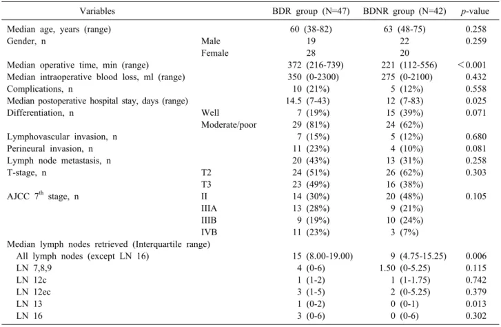
Given the clinicopathologic heterogeneity (Table 1) among the two groups, a subgroup analysis was per- formed for those who underwent liver resection (n=89) with or without BDR (Table 2). In this subset of patients, only eleven of 47 patients underwent BDR for suspected invasion.
While median operative time and postoperative hospital stay was significantly increased in the BDR group, com- plication rates were seemingly unaffected. Complications (according to Clavien-Dindo Classification) were com- parable between BDR and BDNR group (p=0.558).
Among 10 (21%) complications in BDR group, three were grade 1 and included ascites and seroma. One patient had
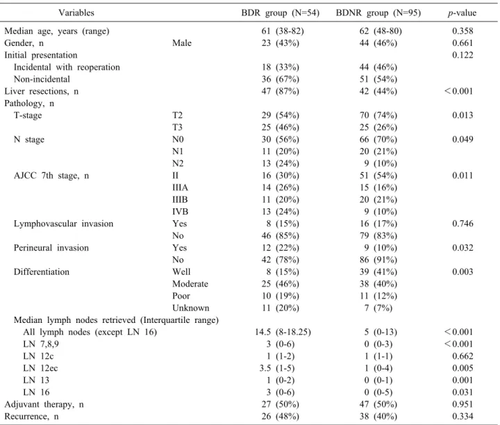
Table 1. Clinicopathologic characteristics
Variables BDR group (N=54) BDNR group (N=95) p-value
Median age, years (range) 61 (38-82) 62 (48-80) 0.358
Gender, n Male 23 (43%) 44 (46%) 0.661
Initial presentation 0.122
Incidental with reoperation 18 (33%) 44 (46%)
Non-incidental 36 (67%) 51 (54%)
Liver resections, n 47 (87%) 42 (44%) <0.001
Pathology, n
T-stage T2 29 (54%) 70 (74%) 0.013
T3 25 (46%) 25 (26%)
N stage N0 30 (56%) 66 (70%) 0.049
N1 11 (20%) 20 (21%)
N2 13 (24%) 9 (10%)
AJCC 7th stage, n II 16 (30%) 51 (54%) 0.011
IIIA 14 (26%) 15 (16%)
IIIB 11 (20%) 20 (21%)
IVB 13 (24%) 9 (10%)
Lymphovascular invasion Yes 8 (15%) 16 (17%) 0.746
No 46 (85%) 79 (83%)
Perineural invasion Yes 12 (22%) 9 (10%) 0.032
No 42 (78%) 86 (91%)
Differentiation Well 8 (15%) 39 (41%) 0.003
Moderate 25 (46%) 38 (40%)
Poor 10 (19%) 11 (12%)
Unknown 11 (20%) 7 (7%)
Median lymph nodes retrieved (Interquartile range)
All lymph nodes (except LN 16) 14.5 (8-18.25) 5 (0-13) <0.001
LN 7,8,9 3 (0-6) 0 (0-3) <0.001
LN 12c 1 (1-2) 1 (1-1) 0.662
LN 12ec 3.5 (1-5) 1 (0-4) 0.005
LN 13 1 (0-2) 0 (0-1) 0.001
LN 16 3 (0-6) 0 (0-5) 0.031
Adjuvant therapy, n 27 (50%) 47 (50%) 0.951
Recurrence, n 26 (48%) 38 (40%) 0.334
BDR group, bile duct resection group; BDNR group, bile duct non-resection group; LN 16, para-aortic lymph nodes; LN 7,8,9, lymph nodes around the gastrohepatic ligament; LN12c, pericholecystic lymph nodes; LN 12ec, lymph nodes around the hep- atoduodenal ligament, excluding pericholecystic nodes; LN 13, retropancreatic lymph nodes
Fig. 2. Type of operative pro- cedures. BDNR group, bile duct non-resection group; BDR group, bile duct resection group; Chole, cholecystectomy; LN, lymph node dissection; CBD, common bile duct resection; Radical chole+etc., Radical cholecystectomy and com- bined resection; PPPD, Pylorus- preserving pancreaticoduodenec- tomy.
Table 2. Comparison between bile duct resection and bile duct non-resection groups for combined liver resected patients
Variables BDR group (N=47) BDNR group (N=42) p-value
Median age, years (range) 60 (38-82) 63 (48-75) 0.258
Gender, n Male 19 22 0.259
Female 28 20
Median operative time, min (range) 372 (216-739) 221 (112-556) <0.001
Median intraoperative blood loss, ml (range) 350 (0-2300) 275 (0-2100) 0.432
Complications, n 10 (21%) 5 (12%) 0.558
Median postoperative hospital stay, days (range) 14.5 (7-43) 12 (7-83) 0.025
Differentiation, n Well 7 (19%) 15 (39%) 0.071
Moderate/poor 29 (81%) 24 (62%)
Lymphovascular invasion, n 7 (15%) 5 (12%) 0.680
Perineural invasion, n 11 (23%) 4 (10%) 0.081
Lymph node metastasis, n 20 (43%) 13 (31%) 0.258
T-stage, n T2 24 (51%) 26 (62%) 0.303
T3 23 (49%) 16 (38%)
AJCC 7th stage, n II 14 (30%) 20 (48%) 0.105
IIIA 13 (28%) 9 (21%)
IIIB 9 (19%) 10 (24%)
IVB 11 (23%) 3 (7%)
Median lymph nodes retrieved (Interquartile range)
All lymph nodes (except LN 16) 15 (8.00-19.00) 9 (4.75-15.25) 0.006
LN 7,8,9 4 (0-6) 1.50 (0-5.25) 0.115
LN 12c 1 (1-2) 1 (1-1.75) 0.742
LN 12ec 3 (1-5) 2 (0-5.25) 0.379
LN 13 1 (0-2) 0 (0-1) 0.013
LN 16 3 (0-6) 0 (0-6) 0.302
BDR group, bile duct resection group; BDNR group, bile duct non-resection group; LN 16, para-aortic lymph nodes; LN 7,8,9, lymph nodes around the gastrohepatic ligament; LN12c, pericholecystic lymph nodes; LN 12ec, lymph nodes around the hep- atoduodenal ligament, excluding pericholecystic nodes; LN 13, retropancreatic lymph nodes
a grade 2 complication with chyle drainage. Four patients had grade 3a complications and included pigtail insertions for fluid collection. Remaining two patients had grade 3b complications due to wound repair under general anesthesia.
Para-aortic lymph node (LN 16) dissection was per- formed more frequently in the BDR group (29 patients (62%) vs. 18 patients (43%), p=0.032).
Twenty-two patients (50%) in the BDR group and 22 patients (52%) in the BDNR group received adjuvant ther- apy, p=0.600.
In terms of recurrence, there was no significant differ- ence between the two groups (21 patients (45%) in the BDR group vs. sixteen patients (38%) in the BDNR group, p=0.529. Most of the recurrences were distant metastasis.
At the end of the follow-up period, 83 patients (56%) were still alive. There was a follow-up loss of three pa- tients (2%) after the discharge from the hospital. The
5-year disease-free survival and overall survival rates were 55% and 54%, respectively. The 5-year overall sur- vival rates of Stages II, IIIA, IIIB and IVB (according to cancer stage using the American Joint Committee on Cancer (AJCC) 7th Edition Staging) were 69%, 43%, 51%, and 16%, respectively. There were significant differences of overall survival rate according to the TNM stage, ex- cept between stage IIIA and IIIB (p=0.460). Comparisons of survival rates (according to TNM) stage are shown in Fig. 3.
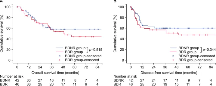
According to the survival analysis for bile duct re- section, there was no evidence of survival benefit of bile duct resection in each of cancer stage (Fig. 4). Further survival analysis for liver resected patients has also shown no significant role of bile duct resection in both overall survival and disease-free survival (Fig. 5).
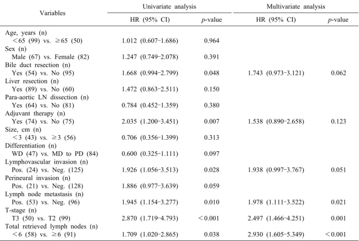
In univariate analysis for disease-free survival, bile duct resection (p=0.048) was one indicator of poor survival outcome. However, in multivariate analyses, lymph node
Fig. 3. Survival curves according to American Joint Committee on Cancer Staging Manuam 7th edition TNM stage. (A) Disease-free survival. (B) Overall survival.
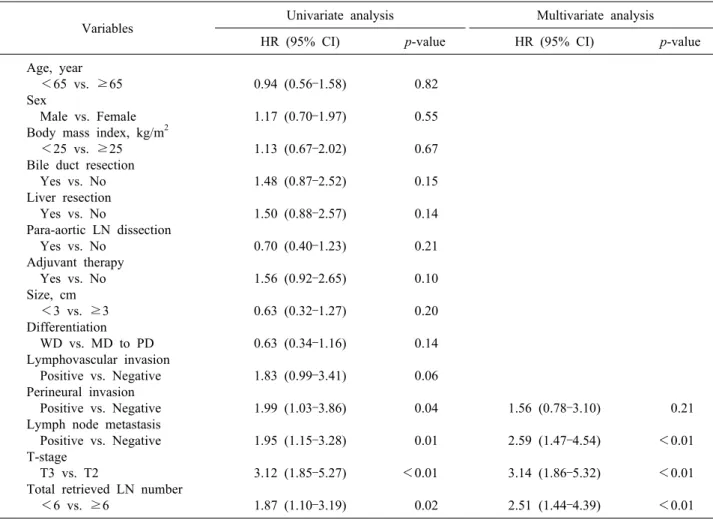
metastasis (p=0.021), T3 stage vs. T2 stage (p=0.001), and total retrieved lymph nodes <6 vs. ≥6 (p<0.001) were the independent prognostic factors for poor survival out- come (Table 3). In multivariate analysis for overall sur- vival, lymph node metastasis, T3 stage, and the total num- ber of retrieved lymph nodes <6 were the independent prognostic factors of poor survival outcome (Table 4).
DISCUSSION
The optimal extent of resection in advanced gallbladder cancer patients remains a debated issue. Chiefly, the clin- ical significance of concomitant bile duct resection re- mains controversial. Kokudo et al.7 reported no appreci- able survival benefit secondary to bile duct resection in patients with LN metastasis. Sakamoto et al.8 also re- ported that there was no perceptible survival benefit asso- ciated with bile duct resection in patients with ≥T2 gall- bladder cancer. Araida et al.6 analyzed 838 gallbladder cancer patients and reported that no survival difference re- sulted from bile duct resection in patients without direct infiltration of the hepatoduodenal ligament.
In the current study, there were no significant, docu- mented differences in the overall survival between the BDR group and BDNR group, even though the BDR group patients had more advanced tumors than the BDNR group patients. Advanced T-stage, presence of LN meta-
stasis and total LN count of <6 were the independent prognostic indicators of poor survival outcome in the mul- tivariate analysis. In other study groups, lymphovascular invasion, perineural invasion, LN metastasis, total LN count of <6 and poor differentiation have been reported to be significant prognostic factors in patients with gall- bladder cancer.3,9,10
Adequate LN assessment is important in patients with gastrointestinal cancers,12-14 because insufficient lymph node dissection during surgery impedes accurate staging.
Ito et al.3 reported that the minimum LN requirement for adequate gallbladder cancer staging was six nodes. In our study, the LN count of <6 was also the prognostic factor for poor survival outcome in gallbladder cancer patients (T2 and T3 stage) who received R0 resection. The BDR group had more LNs removed than the BDNR group.
Specifically, the LNs around the retropancreatic area were more frequently retrieved in the BDR group. Although bile duct resection did not affect the overall survival of gallbladder cancer patients, bile duct resection could be helpful for more accurately staging the disease.
Anatomically, the lymphatics around the hepatoduodenal ligament are the first lymphatic channels draining the gallbladder.15 Tumor cell-spread into the hepatoduodenal ligament is very frequently observed in advanced gall- bladder cancer. In 2014, pathologists in Japan reported 70.6% lymphatic/venous/perineural invasion of gallbladder
Fig. 4. Survival curves comparing the BDR and BDNR groups. (A) Overall survival for Stage II. (B) Overall survival for Stage IIIA. (C) Overall survival for Stage IIIB. (D) Overall survival for Stage IVB. BDNR group, Bile duct non-resection group;
BDR group, Bile duct resection group.
Fig. 5. Survival curves comparing the BDR and BDNR groups for liver resection patients. (A) Overall survival. (B) Disease-free survival. BDNR group, Bile duct non-resection group; BDR group, Bile duct resection group.
Table 3. Univariate and multivariate analyses of prognostic factors for disease-free survival
Variables Univariate analysis Multivariate analysis
HR (95% CI) p-value HR (95% CI) p-value
Age, years (n)
<65 (99) vs. ≥65 (50) Sex (n)
Male (67) vs. Female (82) Bile duct resection (n) Yes (54) vs. No (95) Liver resection (n) Yes (89) vs. No (60) Para-aortic LN dissection (n) Yes (64) vs. No (81) Adjuvant therapy (n) Yes (74) vs. No (75) Size, cm (n)
<3 (43) vs. ≥3 (56) Differentiation (n)
WD (47) vs. MD to PD (84) Lymphovascular invasion (n) Pos. (24) vs. Neg. (125) Perineural invasion (n) Pos. (21) vs. Neg. (128) Lymph node metastasis (n) Pos. (53) vs. Neg. (96) T-stage (n)
T3 (50) vs. T2 (99)
Total retrieved lymph nodes (n) <6 (58) vs. ≥6 (91)
1.012 (0.607–1.686) 1.247 (0.749–2.078) 1.668 (0.994–2.799)
1.472 (0.863–2.511) 0.784 (0.452–1.359)
2.035 (1.200–3.451) 0.706 (0.356–1.399) 0.600 (0.325–1.111)
1.926 (1.056–3.513) 1.886 (0.977–3.639)
1.945 (1.154–3.277) 2.870 (1.719–4.793) 1.709 (1.020–2.865)
0.964 0.391 0.048
0.150 0.380
0.007 0.313 0.097
0.028 0.059
0.010
<0.001 0.038
1.743 (0.973–3.121)
1.538 (0.890–2.658)
1.938 (0.997–3.767)
1.978 (1.111–3.522) 2.497 (1.466–4.251) 2.930 (1.605–5.349)
0.062
0.123
0.051
0.021 0.001
<0.001 HR, hazard ratio; 95% CI, 95% confidence interval; WD, well differentiated; MD, moderately differentiated; PD, poorly differ- entiated; Pos., positive; Neg., negative
cancer (T2 and T3).16 In our study, the BDR group had more lymph nodes retrieved around the retropancreatic area vs. the BDNR group. While other lymph node sta- tions did not yield more retrieved lymph nodes for the BDR group, total number of lymph nodes retrieved was significantly higher in the BDR group. These results in- dicate that lymph node dissection with bile duct resection allow more thorough removal of lymph nodes without leaving remnant lymph nodes and connective tissue around the hepatoduodenal ligament.
Gallbladder cancer can spread in a skipped manner.
Shimizu et al.17 in pathologic analysis of 50 patients with advanced gallbladder cancer, reported that 30 (60%) pa- tients with the hepatoduodenal ligament invasion includ- ing five patients with skipped lesion from the primary tumor. Among patients with hepatoduodenal ligament in- vasion, 25 (83%) patients showed cancer cells in extra- hepatic bile duct and 21 (70%) patients showed occult mi- croscopic extension. Ogura et al.18 also reported dis-
continuation of tumor invasion into the hepatic parenchyma. In terms of these findings, BDR has a poten- tial benefit of eradicating occult tumor spread at the com- mon bile duct from gallbladder cancer because of the pos- sibility of skipped lesions not involving the cystic duct margin. We could not, however, affirm any positive effect on the long-term survival rate attributable to, or associated with, BDR. As such, it may be that the role of BDR may be limited to improved staging of gallbladder cancer as discussed in our study.
Too, the patients in the BDR group were afflicted with more aggressive cancer than the BDNR group and accord- ingly, surgical resection in the BDR group was also more extensive than in the BDNR group. To minimize the bias from extensive surgical resection and aggressive cancer in BDR group, only the patients with liver resection were further analyzed. Bile duct resection involved longer oper- ative time and long postoperative hospital stays. These findings somewhat echo the findings reported by another
Table 4. Univariate and multivariate analyses of prognostic factors for overall survival
Variables Univariate analysis Multivariate analysis
HR (95% CI) p-value HR (95% CI) p-value
Age, year <65 vs. ≥65 Sex
Male vs. Female Body mass index, kg/m2 <25 vs. ≥25 Bile duct resection Yes vs. No Liver resection Yes vs. No
Para-aortic LN dissection Yes vs. No
Adjuvant therapy Yes vs. No Size, cm <3 vs. ≥3 Differentiation WD vs. MD to PD Lymphovascular invasion Positive vs. Negative Perineural invasion Positive vs. Negative Lymph node metastasis Positive vs. Negative T-stage
T3 vs. T2
Total retrieved LN number <6 vs. ≥6
0.94 (0.56–1.58) 1.17 (0.70–1.97) 1.13 (0.67–2.02)
1.48 (0.87–2.52) 1.50 (0.88–2.57)
0.70 (0.40–1.23) 1.56 (0.92–2.65) 0.63 (0.32–1.27)
0.63 (0.34–1.16) 1.83 (0.99–3.41)
1.99 (1.03–3.86) 1.95 (1.15–3.28) 3.12 (1.85–5.27)
1.87 (1.10–3.19)
0.82 0.55 0.67
0.15 0.14
0.21 0.10 0.20
0.14 0.06
0.04 0.01
<0.01
0.02
1.56 (0.78–3.10) 2.59 (1.47–4.54) 3.14 (1.86–5.32)
2.51 (1.44–4.39)
0.21
<0.01
<0.01
<0.01 HR, hazard ratio; 95% CI, 95% confidence interval; WD, well differentiated; MD, moderately differentiated; PD, poorly differ- entiated; LN, lymph node
study group.9 However, we detected no relationship be- tween bile duct resection and the postoperative complica- tion rate documented by our study, even though bile duct resection always required bilioenteric anastomosis.
Adverse effects of bile duct resection were almost always limited to the immediate postoperative period.
Recurrence of gallbladder cancer is more likely to in- volve a distant site and frequently occurs in early-stage disease.19,20 The peritoneum, liver and retroperitoneal lymph nodes tend to be the most common recurrence sites of a primary gallbladder cancer.20 This recurrence pattern has tended to obscure the potential beneficial effect of bile duct resection in obviating a local recurrence. Moreover, patients with advanced cancer received adjuvant therapy, which has reported favorable results with regard to the survival benefits for gallbladder cancer.21,22 The recurrence rate in the BDR group was not significantly different from that of the BDNR group in this study. To estimate the
relationship between the recurrence rate and bile duct re- section, the effectiveness of bile duct resection for accu- rate lymph node dissection must be evaluated further.
About half of the patients who participated in this study received adjuvant therapy. Thirty-three patients in the BDR group and 22 patients in BDNR group belonged to 7th edition AJCC stage of greater than IIIA group. With 22 patients in each group undergoing adjuvant therapy, eleven patients in the BDR group did not receive adjuvant therapy due to patient refusal or poor performance status.
This may have confounded the results in survival analysis.
Moreover, results of this study may have been influenced by heterogeneous nature of the adjuvant therapy. Due to limited size of this study, further subgroup analysis on dif- ferent regimens used was not possible. With more uniform adjuvant treatment, better analysis on the efficacy of bile duct resection may by possible.
Bile duct resection seems to only influence staging and
no benefits on survival have been revealed from this study. Accurate staging in gallbladder cancer is still important. Lack of survival benefit in bile duct resection may be influenced by lack of uniformity in adjuvant treatment. Further evaluation of bile duct resection, with homogenous adjuvant treatment regimen, should be con- sidered viable treatment options, for therapeutic and diag- nostic purposes, and to gain further insights into survival benefits.
In conclusion, advanced T-stage, LN metastasis, and LN count <6 were the independent prognostic factors for overall survival and disease-free survival in patients with T2 and T3 gallbladder cancer. The BDR group had more lymph nodes retrieved vs. the BDNR group, and bile duct resection did not increase complication rate. The role of bile duct resection may be limited to improved staging and without affording any actual long-term survival bene- fit, based upon the current study. However, without any documented, significant increase in morbidity of the BDR group, bile duct resection should be actively considered as in patients with suspicious invasion around hep- atoduodenal ligament.
REFERENCES
1. Lazcano-Ponce EC, Miquel JF, Muñoz N, Herrero R, Ferrecio C, Wistuba II, et al. Epidemiology and molecular pathology of gallbladder cancer. CA Cancer J Clin 2001;51:349-364.
2. Hueman MT, Vollmer CM Jr, Pawlik TM. Evolving treatment strat- egies for gallbladder cancer. Ann Surg Oncol 2009;16:2101-2115.
3. Ito H, Ito K, D'Angelica M, Gonen M, Klimstra D, Allen P, et al. Accurate staging for gallbladder cancer: implications for sur- gical therapy and pathological assessment. Ann Surg 2011;254:
320-325.
4. Higuchi R, Ota T, Araida T, Kajiyama H, Yazawa T, Furukawa T, et al. Surgical approaches to advanced gallbladder cancer: a 40-year single-institution study of prognostic factors and resectability. Ann Surg Oncol 2014;21:4308-4316.
5. Benson AB 3rd, Abrams TA, Ben-Josef E, Bloomston PM, Botha JF, Clary BM, et al. NCCN clinical practice guidelines in oncology: hepatobiliary cancers. J Natl Compr Canc Netw 2009;7:350-391.
6. Araida T, Higuchi R, Hamano M, Kodera Y, Takeshita N, Ota T, et al. Should the extrahepatic bile duct be resected or pre- served in R0 radical surgery for advanced gallbladder carcino- ma? Results of a Japanese Society of Biliary Surgery Survey:
a multicenter study. Surg Today 2009;39:770-779.
7. Kokudo N, Makuuchi M, Natori T, Sakamoto Y, Yamamoto J,
Seki M, et al. Strategies for surgical treatment of gallbladder car- cinoma based on information available before resection. Arch Surg 2003;138:741-750; discussion 750.
8. Sakamoto Y, Kosuge T, Shimada K, Sano T, Hibi T, Yamamoto J, et al. Clinical significance of extrahepatic bile duct resection for advanced gallbladder cancer. J Surg Oncol 2006;94:298-306.
9. Choi SB, Han HJ, Kim WB, Song TJ, Suh SO, Choi SY.
Surgical strategy for T2 and T3 gallbladder cancer: is extra- hepatic bile duct resection always necessary? Langenbecks Arch Surg 2013;398:1137-1144.
10. Lim H, Seo DW, Park DH, Lee SS, Lee SK, Kim MH, et al.
Prognostic factors in patients with gallbladder cancer after surgi- cal resection: analysis of 279 operated patients. J Clin Gastroenterol 2013;47:443-448.
11. Clavien PA, Barkun J, de Oliveira ML, Vauthey JN, Dindo D, Schulick RD, et al. The Clavien-Dindo classification of surgical complications: five-year experience. Ann Surg 2009;250:187-196.
12. Yoshikawa T, Sasako M, Sano T, Nashimoto A, Kurita A, Tsujinaka T, et al. Stage migration caused by D2 dissection with para-aortic lymphadenectomy for gastric cancer from the results of a prospective randomized controlled trial. Br J Surg 2006;93:
1526-1529.
13. Pawlik TM, Gleisner AL, Cameron JL, Winter JM, Assumpcao L, Lillemoe KD, et al. Prognostic relevance of lymph node ratio following pancreaticoduodenectomy for pancreatic cancer.
Surgery 2007;141:610-618.
14. Kim YW, Kim NK, Min BS, Lee KY, Sohn SK, Cho CH, et al. The prognostic impact of the number of lymph nodes re- trieved after neoadjuvant chemoradiotherapy with mesorectal ex- cision for rectal cancer. J Surg Oncol 2009;100:1-7.
15. Ito M, Mishima Y, Sato T. An anatomical study of the lymphatic drainage of the gallbladder. Surg Radiol Anat 1991;13:89-104.
16. Kijima H, Wu Y, Yosizawa T, Suzuki T, Tsugeno Y, Haga T, et al. Pathological characteristics of early to advanced gall- bladder carcinoma and extrahepatic cholangiocarcinoma. J Hepatobiliary Pancreat Sci 2014;21:453-458.
17. Shimizu Y, Ohtsuka M, Ito H, Kimura F, Shimizu H, Togawa A, et al. Should the extrahepatic bile duct be resected for locally advanced gallbladder cancer? Surgery 2004;136:1012-1017; dis- cussion 1018.
18. Ogura Y, Tabata M, Kawarada Y, Mizumoto R. Effect of hepatic invasion on the choice of hepatic resection for advanced carcino- ma of the gallbladder: histologic analysis of 32 surgical cases.
World J Surg 1998;22:262-266; discussion 266-267.
19. Jung SJ, Woo SM, Park HK, Lee WJ, Han MA, Han SS, et al.
Patterns of initial disease recurrence after resection of biliary tract cancer. Oncology 2012;83:83-90.
20. Jarnagin WR, Ruo L, Little SA, Klimstra D, D'Angelica M, DeMatteo RP, et al. Patterns of initial disease recurrence after resection of gallbladder carcinoma and hilar cholangiocarcinoma:
implications for adjuvant therapeutic strategies. Cancer 2003;98:
1689-1700.
21. Gold DG, Miller RC, Haddock MG, Gunderson LL, Quevedo F, Donohue JH, et al. Adjuvant therapy for gallbladder carcino- ma: the Mayo Clinic Experience. Int J Radiat Oncol Biol Phys 2009;75:150-155.
22. Kim K, Chie EK, Jang JY, Kim SW, Han SW, Oh DY, et al.
Postoperative chemoradiotherapy for gallbladder cancer. Strahlenther Onkol 2012;188:388-392.