Received April 30, 2014, Revised June 24, 2014, Accepted for publication July 7, 2014
Corresponding author: Seung-Chul Lee, Department of Dermatology, Chonnam National University Medical School, 160 Baekseo-ro, Don-gu, Gwangju 501-746, Korea. Tel: 82-62-220-6682, Fax: 82- 62-222-4058, E-mail: [email protected]
This is an Open Access article distributed under the terms of the Creative Commons Attribution Non-Commercial License (http://
creativecommons.org/licenses/by-nc/3.0) which permits unrestricted non-commercial use, distribution, and reproduction in any medium, provided the original work is properly cited.
ORIGINAL ARTICLE
Ethanol Extract of Peanut Sprout Exhibits a Potent Anti-Inflammatory Activity in Both an
Oxazolone-Induced Contact Dermatitis Mouse Model and Compound 48/80-Treated HaCaT Cells
Da-In Choi, Jee-Young Choi, Young Jee Kim, Jee-Bum Lee, Sun-Ouck Kim1, Hyong-Taek Shin2, Seung-Chul Lee
Departments of Dermatology and 1Urology, Chonnam National University Medical School, Gwangju, 2Jangsoochae Company, Chuncheon, Korea
Background: We developed an ethanol extract of peanut sprouts (EPS), a peanut sprout-derived natural product, which contains a high level of trans-resveratrol (176.75 μg/ml) and was shown to have potent antioxidant activity. Objective:
We evaluated the potential anti-inflammatory activity of EPS by measuring its antioxidant potential in skin. Methods: The anti-inflammatory activity of EPS was tested using two models of skin inflammation: oxazolone (OX)-induced contact der- matitis in mice and compound 48/80-treated HaCaT cells. As biomarkers of skin inflammation, cyclooxygenase-2 (COX-2) and nerve growth factor (NGF) levels were measured.
Results: OX-induced contact dermatitis was suppressed markedly in mice that were treated with an ointment contain- ing 5% EPS as evidenced by a decrease in the extent of scal- ing and thickening (p<0.05) and supported by a histological study. COX-2 (messenger RNA [mRNA] and protein) and NGF (mRNA) levels, which were upregulated in the skin of OX-treated mice, were suppressed markedly in the skin of OX+EPS-treated mice. Consistent with this, compound 48/80-induced expression of COX-2 (mRNA and protein) and NGF (mRNA) in HaCaT cells were suppressed by EPS
treatment in a dose-dependent manner. As an inhibitor of NF-κB, IκB protein levels were dose-dependently upregu- lated by EPS. Fluorescence-activated cell sorting (FACS) anal- ysis revealed that EPS scavenged compound 48/80-induced reactive oxygen species (ROS) in HaCaT cells. Conclusion:
EPS exerts a potent anti-inflammatory activity via its anti- oxidant activity in both mouse skin and compound 48/80-treated HaCaT cells in vitro. Compound 48/80-treated HaCaT cells are a useful new in vitro model of skin inflammation. (Ann Dermatol 27(2) 142∼151, 2015) -Keywords-
Anti-inflammatory activity, Anti-oxidant activity, p-Methoxy- N-methylphenethylamine, Ethanol extract of peanut sprouts, Oxazolone-induced contact dermatitis
INTRODUCTION
Peanuts contain bioactive compounds with antioxidant ac- tivity, such as resveratrol, flavonoids, phenolic com- pounds, and phytosterols. Peanut sprouts have been re- ported to contain a high concentration of trans-resveratrol, which is known to have a variety of beneficial roles in- cluding anti-inflammatory, anti-aging, anti-cancer, and car- diovascular-protective effects1. In our previous paper, we demonstrated that an ethanol extract of peanut sprout (EPS) contained 54.2 μg/ml of transresveratrol and pro- tected cells from ultraviolet B-induced oxidative stress2. EPS had potent anti-oxidant activity and scavenged re- active oxygen species (ROS) and activated the Nrf2/anti-ox- idant system2. For this study, we prepared a more con-
centrated EPS of higher trans-resveratrol content (176.75 μg/ml) with the expectation that this would possess a more potent anti-oxidant activity.
Steroids are widely used as anti-inflammatory agents for the treatment of acute and chronic eczema. However, long-term steroid administration inevitably leads to vari- ous side effects. Oxidative stress is known to be induced by excess production of ROS and/or insufficient scaveng- ing of ROS, triggering downstream signaling cascades of inflammatory cytokines3. ROS could therefore act as sec- ondary messengers in the induction of inflammation.
Thus, anti-oxidants may act as anti-inflammatory agents by scavenging ROS. For these reasons, the development of non-steroidal anti-inflammatory drugs based on anti-oxi- dants is a promising approach in the dermatology field.
Compound 48/80, first described as a mast cell secreta- gogue, is a polymer derived from N-methyl-p-methox- yphenethylamine, which activates phospholipase C in mast cells via hetero-trimeric guanosine triphosphate (GTP)-binding proteins4,5. In addition to its role as a mast cell degranulator, compound 48/80 mediates in- flammation, proliferation, and differentiation. Based on a previous report that compound 48/80 produced ROS in mast cells6, we tested whether compound 48/80 could in- duce ROS production in HaCaT cells. If so, we hypothe- sized that EPS could block compound 48/80-mediated ROS production as well as downstream events in skin inflammation.
Chronic eczemas are characterized by recurrent attacks of skin inflammation accompanied by severe pruritus. Skin damage due to scratching facilitates the release of neuro- trophins (NTs) and/or neuropeptides (NPs) from free nerve endings in the skin, which leads to or aggravates skin in- flammation7. Thus, NT- and NP-mediated neurogenic in- flammation has the potential to stimulate peripheral nerves to produce not only an itching sensation, but also localized disturbances in sweating, thermoregulation, and vascular regulation8. As keratinocytes (KCs)-derived nerve growth factor (NGF) is known to be a common NT, it has been used as a biomarker for evaluating the severity of atopic dermatitis (AD)9. Thus, we investigated the levels NGF and cyclooxygenase-2 (COX-2) in skin inflammation of epidermal KCs10-12.
In this study, we evaluated the anti-inflammatory activity of EPS in vivo and in vitro using an oxazolone (OX)-induced contact dermatitis mouse model and compound 48/80-treat- ed HaCaT cells, respectively.
MATERIALS AND METHODS
EPS preparation
An EPS stock solution was prepared as described pre- viously2. Briefly, peanut sprouts were steamed and cooled twice, followed by vacuum concentration to obtain high a product with a high resveratrol content. The EPS stock sol- ution was found to contain trans-resveratrol at a concen- tration of 176.75±3.63 μg/ml.
Cell culture
HaCaT cells (Lot-no.: 300493-Pr35) were purchased from CLS Cell Lines Service (Eppelheim, Germany). HaCaT cells were cultured in Dulbecco’s modified Eagle’s me- dium supplemented with 10% fetal bovine serum and antibiotics.
MTT assay
Cell viability was assayed using the colorimetric 3-[4,5-di- methylthiazol-2-yl]-2,5-diphenyltetrazoliumbromide (MTT) kit (Chemicon International Inc., Billerica, MA, USA) ac- cording to the manufacturer’s instructions.
OX-induced skin inflammation in hairless mice
Animal experiments were approved by the Animal Care and Use Committee of the Chonnam National University Hospital, Gwangju, Korea (IACUC number: CNU IACUC- H-2011-17).
Induction of allergic-type contact dermatitis for use as an animal model of skin inflammation was performed as de- scribed before with a minor modification13. SKH1 mice (Charles River Laboratory, Boston, MA, USA) were pur- chased from Orient Bio (Seongnam, Korea). The mice (female, 8 weeks old) were fed with normal food and tap water, and raised in the Individually Ventilated Cage Rack System (Tecniplast, Casale Litta, Italy). Briefly, contact der- matitis was induced by sensitization with 5% OX three times in the first week, followed by administration of 0.5% OX three times per week on an every-other-day schedule for the following 3 weeks (total, 9 admin- istrations). From the third week, 5% EPS ointment was ap- plied to the dorsal skin of the mice three times a week for 2 weeks. For control mice, petrolatum ointment base was applied instead of 5% EPS ointment on the same schedule. Mice were killed by cervical dislocation to ob- tain skin samples.
Measurement of skin thickness
To semi-quantify the degree of skin inflammation, we measured whole skin thickness with a dial thickness gauge (Peacock, Scotts Valley, CA, USA). After the hairless
mice were sacrificed by cervical dislocation, a skin flap containing the epidermis and dermis was immediately ob- tained by removing the skin layer from the underlying muscle with iris scissors.
Grading skin inflammation by physical examination To evaluate the degree of skin inflammation, we arbitrarily graded skin inflammation with the three criteria of scaling, edema, and thickening (lichenification). Each was graded as 0 (none), 1 (mild), 2 (moderate), and 3 (severe).
Measurement of intracellular ROS levels
Fluorogenic 2',7'-dichlorodihydrofluorescein diacetate (DCF-DA; Invitrogen, Carlsbad, CA, USA) reacts with intra- cellular ROS, and can be detected by fluorescence-activated cell sorting (FACS) analysis. Briefly, HaCaT cells were seeded in each well of a 60 mm culture dish at a density of 5×10⁵ cells per well and pretreated with EPS for 1 h.
The cells were then treated with compound 48/80 (10 μg/
ml) for 6 h. For FACS analysis, the cells were incubated with the redox-sensitive indicator DCF-DA (10 μM) in a 37oC incubator for 30 min and intracellular ROS pro- duction was analyzed using flow cytometry (FACSCalibur;
Becton Dickinson, San Jose, CA, USA) with an excitation wavelength of 488 nm and an emission wavelength of 530 nm. Data analyses were based on 10,000 detection events using the Cell Quest software.
Reverse transcriptase-polymerase chain reaction A polymerase chain reaction (PCR) pre-mixture kit (ELPIS, Daejeon, Korea) was used for reverse transcriptase-PCR (RT-PCR). PCR reactions were performed with the follow- ing primers: human COX-2 (forward: 5'-cattctttgcccag- cacttcac-3', reverse: 5'-gaccaggcaccagaccaaagac-3'), human NGF (forward: 5'-catacaggcggaaccacact-3', reverse: 5'-gaa- ttcgcccctgtggaaga-3'), human glyceraldehyde 3-phosphate dehydrogenase (GAPDH; forward: 5'-gtcttcaccaccatggag- aaggc-3', reverse: 5'-cggaaggccatgccagtgagctt-3'); mouse COX-2 (forward: 5'-agcagatgactgcccaactc-3', reverse: 5'-g- aacccaggtcctcgcttat-3'), mouse NGF (forward: 5'-catacagg- cggaaccacact-3', reverse: 5'-gaattcgcccctgtggaaga-3'), and mouse β-actin (forward: 5'-tcatgaagtgagacgttgacatccgt-3', reverse: 5'cctagaagcacttgcggtgcacgatg-3'). Annealing tem- peratures were 55oC for human COX-2 and mouse β-actin, 60oC for human GAPDH, and 61oC for human NGF, mouse NGF and mouse COX-2. PCR products were ana- lyzed using 1.5% agarose gel electrophoresis, stained with SYBR Safe DNA gel stain buffer (Invitrogen), and vi- sualized using luminescence (LAS 3000; Fujifilm, Tokyo, Japan). Expression levels were normalized to endogenous β-actin and GAPDH as controls.
Western blot analysis
Whole cell and nuclear extracts were prepared as de- scribed previously2. Protein bands were probed with the following primary antibodies: anti-IκBα (#9242, 1 : 1,000; Cell Signaling Technology, Boston, MA, USA), an- ti-COX-2 (ab15191, 1 : 200; Abcam, Cambridge, MA, USA), and anti-β-actin (ab-6276, 1 : 1,000; Abcam).
horseradish peroxidase (HRP)-conjugated rabbit anti-goat immunoglobulin G (IgG) (1 : 5,000, Jackson Immunore- search, West Grove, PA, USA) and HRP-conjugated an- ti-mouse IgG antibody (1 : 5,000; Jackson Immunore- search) were used for secondary antibodies. All data shown are representative of three experiments. Densito- metric analyses were performed by using Multi Gauge V3.0 software (Fujifilm).
Statistics
All experiments were carried out in triplicate, and results are expressed as means±standard deviations. Comparisons between samples were carried out using Student’s t-test.
SPSS ver. 15.0 (SPSS Inc., Chicago, IL, USA) was used to analyze the data with p-values<0.05 to be the statistically significant.
RESULTS
EPS suppresses OX-induced contact dermatitis in mouse skin
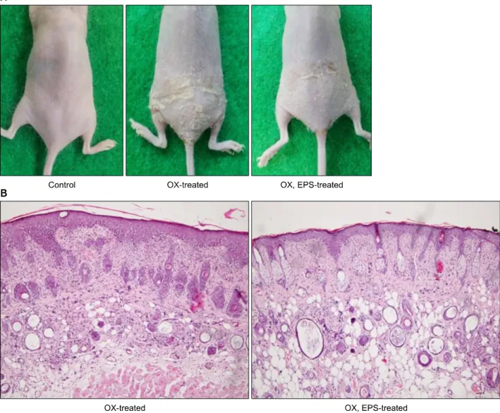
To investigate whether EPS could exert anti-inflammatory activity based on its anti-oxidant properties, the anti-in- flammatory activity of EPS was tested on mouse skin in vivo. From physical examination, we could observe in- flammatory changes, such as edema, scaling, and thicken- ing (lichenification), in the dorsal skin of OX-treated mice.
The OX-induced inflammatory changes were suppressed markedly in OX+EPS-treated mice (Fig. 1A). Semiquanti- tative scores of the three inflammatory criteria in the OX-treated group (n=4) versus the OX+EPS-treated group (n=4) were: 2.50±0.58 versus 1.50±0.58 for edema (p=0.097), 2.75±0.50 versus 1.50±0.58 for scaling (p=0.017), and 2.75±0.50 versus 1.25±0.50 for thicken- ing (p=0.005), respectively (Table 1). In the histological study, inflammatory changes in the skin, including hyper- keratosis and spongiosis in the epidermis, as well as vaso- dilation and the presence of inflammatory cell infiltrates in the dermis, were clearly observed in OX-treated mice, but were less prominent in the OX+EPS-treated mice (hematoxylin and eosin [H&E] staining, ×100; Fig. 1B).
Skin thickness, measured using a dial thickness gauge, in the OX-treated mice increased to 233.2%±22.52% versus
Fig. 1. Oxazolone (OX)-induced inflammatory changes in mouse skin are suppressed by application of 5% ethanol extract of peanut sprouts (EPS). (A) Representative pictures of untreated control mouse, OX-treated mouse, and OX+EPS-treated mouse. (B) Histological results of OX-treated and OX+EPS-treated mice are shown (H&E, ×100).
Table 1. Semi-quantitative analysis on inflammatory scores (n=4) Criteria OX-treated mice OX, EPS-treated mice p-value*
Edema 2.50±0.58 1.50±0.58 0.097
Scaling 2.75±0.50 1.50±0.58 0.017
Thickening 2.75±0.50 1.25±0.50 0.005
Values are presented as mean±standard deviation. OX: oxazolone, EPS: ethanol extract of peanut sprouts. *The degree of skin inflammation was arbitrarily graded by physical examination.
Severity of edema, scaling, and thickening was graded as 0 (none)∼3 (severe), and statistical significance was calculated between OX-treated and OX, EPS-treated mice groups.
the untreated control group. The OX-induced increase in skin thickness was less prominent in the OX+EPS-treated mice to be 201.6%±12.46% of the control group (Fig. 2).
There was no statistically significant difference between the OX-treated and the OX+EPS-treated mice groups (p=0.328, n=4).
Next, we assessed the expression levels of COX-2 and NGF-used as biomarkers for skin inflammation-in skin ex- tracts from the OX-treated mice, OX+EPS-treated mice, and untreated control mice groups (n=4 for each group).
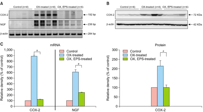
Using RT-PCR and western blot analyses, we found that mRNA and protein levels of COX-2 were upregulated in OX-treated mice, but this increase was suppressed mark- edly in OX+EPS-treated mice (Fig. 3A, B). Using RT-PCR, NGF mRNA was found to be upregulated in OX-treated mice, versus the control mice, but this was suppressed in OX+EPS-treated mice (Fig. 3A). Densitometric analysis to quantify the relative expression levels compared to the
Fig. 2. Anti-inflammatory activity of ethanol extract of peanut sprouts (EPS) reduces the oxazolone (OX)-induced increase of skin thickness in hairless mice. Skin thickness was measured with a dial thickness gauge to semi-quantify the degree of skin inflammation in vivo. Skin flaps were obtained from the dorsal skin of untreated control, OX-treated, and OX+EPS-treated mice.
Statistical significance was assessed between the OX-treated and OX+EPS-treated mice groups.
Fig. 3. Anti-inflammatory activity of ethanol extract of peanut sprouts (EPS) on oxazolone (OX)-induced skin inflammation in hairless mice. (A) In reverse transcriptase-polymerase chain reaction (RT-PCR) and (B) western blot analyses for COX-2, as well as (A) RT-PCR for nerve growth factor (NGF) mRNA expression, OX-treated mice (n=4) showed increased expression levels of the markers tested, but the increase was suppressed markedly in OX+EPS-treated mice (n=4). (C) Densitometric analysis of mRNA and protein bands of A, B was performed, and data represent mean±standard deviation of relative density of each group versus control mice without OX and EPS treatment (n=4 for each group). #p<0.01 between OX-treated and OX+EPS-treated mice groups. Control refers to the OX-untreated mice group.
control group indicated a significant difference in ex- pression levels of COX-2 and NGF between the OX-treat- ed group and the OX+EPS-treated group in a dose-de- pendent manner (p<0.01; Fig. 3C).
EPS suppresses ROS production in compound 48/80- treated HaCaT cells
To confirm our observation that EPS had an anti-in- flammatory activity in mouse skin, we developed an in vi- tro system to assess skin inflammation. Based on a report that compound 48/80 could induce ROS production in mast cells in a manner similar to allergic inflammation6, we tested whether compound 48/80 had a similar effect on HaCaT cells. In a previous study, HaCaT cells were re- ported to produce COX-2 and MMP-9 when stimulated with ultraviolet B irradiation12. In preliminary experiments using the MTT cell viability assay, it was found that 0.1∼
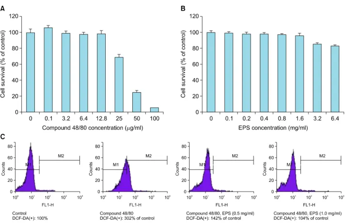
12.8 μg/ml of compound 48/80 (Fig. 4A) and 0.1∼1.6 mg/ml of EPS (Fig. 4B) were the optimum, non-toxic con- centrations for use in HeCaT cells. Therefore, all following experiments were performed using a concentration of 10 μg/ml of compound 48/80. In a FACS analysis, EPS was found to effectively scavenge the compound 48/80-pro-
Fig. 4. Anti-inflammatory activity of ethanol extract of peanut sprouts (EPS) via reactive oxygen species (ROS)-scavenging effect in compound 48/80-treated HaCaT cells. MTT assay was used to detect non-cytotoxic concentrations of (A) compound 48/80 and (B) EPS in HaCaT cells. (C) Fluorescence-activated cell sorting (FACS) analysis demonstrated that compound 48/80 induced ROS production in HaCaT cells, which was suppressed markedly by EPS treatment.
duced ROS from HaCaT cells in a dose-dependent man- ner, showing DCF-DA(+) signals to be 302% of the un- treated control in compound 48/80-treated cells, 142% of the control in compound 48/80+EPS (0.5 mg/ml)-treated cells, and 104% of the control in compound 48/80+EPS (1.0 mg/ml)-treated cells (Fig. 4C).
EPS downregulates expression levels of COX-2 and NGF in compound 48/80-treated HaCaT cells
From the above results, we confirmed that compound 48/80 stimulated HaCaT cells to produce ROS, which might, in turn, induce skin inflammation. As markers for skin inflammation, expression levels of COX-2 and NGF were measured in compound 48/80-treated HaCaT cells.
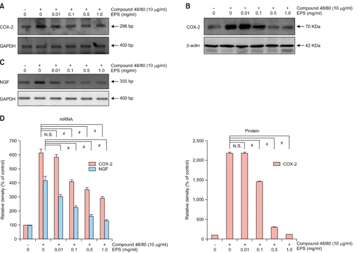
In RT-PCR and western blot analysis, mRNA and protein levels of COX2 (Fig. 5A, B) were upregulated by treatment with 10 μg/ml of compound 48/80 for 6 h. Compound 48/80-induced COX-2 expression was suppressed mark- edly by EPS treatment in a dose-dependent manner, at doses of between 0.01 and 1.0 mg/ml at the mRNA level and between doses of 0.1 and 1.0 mg/ml at the protein level. Next, we checked the expression levels of NGF, a
biomarker of neurogenic inflammation, in compound 48/80-treated HaCaT cells. In RT-PCR, NGF mRNA was upregulated by treatment with 10 μg/ml compound 48/80 for 24 h (Fig. 5C). The compound 48/80-induced NGF expression was suppressed by EPS treatment in a dose-dependent manner between doses of 0.01 and 1.0 mg/ml (Fig. 5C). Densitometric analysis showed that there was a statistically significant difference between HeCaT cells treated with compound 48/80-treated and HeCaT cells treated with compound 48/80+different concen- trations of EPS (p<0.01; Fig. 5D).
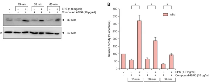
EPS upregulates IκB expression in compound 48/80- treated HaCaT cells
After demonstrating the anti-inflammatory activity of EPS in vivo and in vitro, we examined the regulatory effects of EPS by assessing IκB expression levels in HaCaT cells.
Western blot analyses indicated that EPS induced upregu- lation of IκBα, an inhibitor of NFκB, in a dose-dependent manner; this indicates that EPS exerts an anti-inflammatory effect by inhibiting NFκB transcription factor activity in HaCaT cells (Fig. 6).
Fig. 5. Inhibitory effect of ethanol extract of peanut sprouts (EPS) on compound 48/80-induced expression of cyclooxygenase-2 (COX-2) and nerve growth factor (NGF) in HaCaT cells. In (A) reverse transcriptase-polymerase chain reaction (RT-PCR) and (B) western blot analyses for COX-2, and (C) RT-PCR for NGF mRNA expression, compound 48/80 (10 μg/ml) increased expression levels of COX-2 and NGF, but this induction was suppressed markedly in compound 48/80+EPS-treated HaCaT cells in a dose-dependent manner between 0.01 and 1.0 mg/ml at the mRNA level and 0.1 and 1.0 mg/ml at the protein level. (D) Densitometric analysis for mRNA and protein bands of (A∼C) was performed, and data represent means±standard deviations of three independent experiments. #p<0.01 between compound 48/80-treated and compound 48/80+EPS-treated sample with different EPS concentration. N.S. means no significance between samples. GAPDH: glyceraldehyde 3-phosphate dehydrogenase.
DISCUSSION
In this study, we assessed whether EPS-with its potent an- ti-oxidant properties-is able to exert an antiinflammatory effect against skin inflammation. Oxidative stress, caused by overproduction of ROS and/or by insufficient scaveng- ing of anti-oxidants, is known to induce harmful biological responses in the skin, such as inflammation, aging, and cancer. Oxidative stress in the skin is thought to induce dysfunction in cell signaling pathways, triggering down- stream signaling cascades and producing inflammatory cy- tokines in skin inflammation14. This work demonstrates that EPS had potent anti-inflammatory activity in mouse skin in vivo; EPS suppressed OX-induced contact dermati- tis in hairless mice, and EPS downregulated expression levels of COX-2 and NGF in skin extracts of OX-treated
mice. The in vitro anti-inflammatory activity of EPS was examined using compound 48/80-treated HaCaT cells.
Unbalanced cellular redox status is known to cause skin inflammation, and it is also known that a variety of an- ti-oxidants are potent anti-inflammatory agents14. Although the beneficial effects of anti-oxidants such as resveratrol can be demonstrated in vitro, there is frequently little cor- relation between in vitro and in vivo activity. The incon- sistency between in vitro and in vivo systems may simply due to there being insufficient levels of active ingredients to cause pharmacological activity in the body, or in- efficient delivery of anti-oxidants. Recently, the popularity of resveratrol-containing foods and beverages has in- creased worldwide because of their antioxidant activities, and both grapes and red wine have been studied as natu- ral sources of resveratrol. The resveratrol content of grape
Fig. 6. Ethanol extract of peanut sprouts (EPS) exerts an anti-inflammatory activity by upregulating IκBα expression in compound 48/80-treated HaCaT cells. (A) In western blot analysis, compound 48/80 down-regulated IκBα expression, which was reversed by EPS treatment at a dose of 10 μg/ml in HaCaT cells. The EPS-induced upregulation of IκBα occurred in a time-dependent manner between 15 and 60 min. (B) Densitometric analysis of western blot protein bands was performed, and data represent means±standard deviations of three independent experiments. #p<0.01 between compound 48/80-treated and compound 48/80+EPS-treated samples at each time point (15, 30, and 60 min).
skin is 35.6∼170.6 μg/g, and that of red wine is 0.05∼
10.9 μg/ml in the trans-form and 0.04∼8.71 μg/ml in the cis-form15,16. Peanut sprouts have also been reported to be a source of resveratrol2,17,18. In our previous study, EPS was found to contain a relatively high concentration of trans-resveratrol (54.2 μg/ml) compared other sour- ces2,19. Besides resveratrol, the presence of other poly- phenols in EPS, such as epicatechin (EC), epicatechin gal- late, epigallocatechin, and epigallocatechin-3-gallate1, are thought to further enhance its antioxidant activity. For this study, we prepared a more concentrated form of EPS con- taining 176.75 μg/ml of trans-resveratrol-a value far high- er than the resveratrol content of red wine. To our knowl- edge, our new preparation of EPS contains the highest re- ported concentration of trans-resveratrol obtained from a natural source. Per-orally administered resveratrol has been reported to have a half-life of 8∼14 min, suggesting that resveratrol is metabolized rapidly and would not be expected to show much biological activity in the body20. In contrast, per-cutaneous application of EPS was consid- ered to be advantageous as it would not be metabolized as rapidly21.
Because epidermal KCs represent ∼90% of all epidermal cells, some mediators in epidermal KCs are likely to play important roles in the pathogenesis of skin inflammation.
For this study, we selected compound 48/80 as a non-im- munological stimulator to induce ROS-mediated skin in- flammation in HaCaT cells. In comparison with other chemicals (irritant or allergic) used in in vitro skin in- flammatory models, compound 48/80 is thought to play a
similar role to substance P (SP) in the stimulation of KCs during neurogenic inflammation. Various studies have clearly demonstrated that neurogenic inflammation plays a crucial role in the pathogenesis of skin inflammation7,9,22,23. Itch is known to be meditated primarily by histamine, and thus anti-H1 blockers are major drugs used to control itch in various skin inflammatory conditions. However, the fact that antihistamines do not alleviate itch effectively in many chronic eczemas indicates that there are other medi- ators beyond mast cell-derived histamine. Recent studies have suggested that NPs, NTs, proteases, cytokines, and opioids may also be involved in a histamine-independent itch pathway23. The itch sensation is mediated by un- myelinated, afferent C-fibers derived from the dorsal root ganglia, and NGF is the major NT to be expressed at C-fi- ber endings24. Damage to skin by scratching is an im- portant triggering factor inducing NGF expression, which, in turn, facilitates abnormal innervation of C-fibers with upregulated NGF expression in the epidermis25,26. Thus, NGF is considered to play an important role in skin in- flammation in the epidermis. For example, there is a pos- itive correlation between NGF levels in the epidermis and itch severity or objective symptoms of chronic eczema and psoriasis9,26. NGF levels in the horny layer of AD pa- tients reflect both the severity of itch and the extent of skin lesions9, suggesting that NGF may be a good marker to predict the severity of subjective symptoms (itch) and/or objective symptoms (skin eruptions) in chronic eczema.
Tumor necrosis factor-α (TNF-α) and interleukin (IL)-1 are key proinflammatory cytokines in AD to induce NGF
production, which activates T cells and thus stimulates the production of IL-427,28. Thus, we selected NGF as a suit- able marker for assessing the anti-inflammatory effect of EPS in HaCaT cells or skin extracts. In this study, NGF was found to be upregulated by compound 48/80, and the ef- fect could be blocked by EPS treatment in HaCaT cells.
This suggests that compound 48/80-stimualted HaCaT cells could be a useful new cell model for studying ROS-mediated skin inflammation. The results from our in vitro model of skin inflammation were consistent with those from the contact dermatitis model of mice in vivo, in that NGF and COX-2 were up-regulated by compound 48/80.
Compound 48/80 is commonly used to stimulate mast cells to release histamine, by immunological or non-im- munological mechanisms. Compound 48/80 was reported to play either a similar role of SP, which induces hista- mine production in mast cells, or to upregulate COX-2 ex- pression3,22,29. SP augments the production of pro-in- flammatory cytokine of IL-1 from KCs30. In a previous study, compound 48/80 up-regulated SP and NGF, and it induced ROS production from mast cells31. Interestingly, compound 48/80 was reported to induce skin in- flammation, especially neurogenic inflammation, in mice32,33. In this study, we treated HaCaT cells with com- pound 48/80, instead of SP, in order to induce ROS pro- duction and skin inflammation. In our study, compound 48/80-stimulated HaCaT cells produced ROS, which are thought to act as second messengers, and might induce skin inflammation by upregulating biomarkers such as COX-2 and NGF. The compound 48/80-induced in- flammation (via ROS) in HaCaT cells was further con- firmed by our results that EPS blocked ROS production in HaCaT cells, as it suppressed the expression levels of bio- markers of skin inflammation. The ear swelling response (ESR) has been commonly used as a non-invasive techni- que to measure inflammatory responses in intact skin in vivo34. For this study, we modified the ESR technique to allow the degree of skin inflammation to be semi-quantified.
The technique is considered to be invasive and aggressive for mice, but we can semi-quantify the degree of skin in- flammation in vivo without further manipulation. Using our new technique, we could easily detect the decrease in skin thickness in the OX+EPS-treated group versus the OXtreated group (Fig. 3).
In conclusion, EPS was found to have anti-inflammatory activity via its anti-oxidant properties in both a mouse model of OX-induced contact dermatitis and compound 48/80-treated HaCaT cells in vitro. In addition, we pro- pose a new cell model to test neurogenic inflammation of the skin by treating HaCaT cells with compound 48/80.
From these results, we expect to further develop EPS for use as a novel anti-inflammatory agent against skin in- flammation in chronic eczemas.
ACKNOWLEDGMENT
This study was supported by a grant Chonnam National University Hospital Research Institute of Clinical Medicine (CRI-11079-21).
REFERENCES
1. Sales JM, Resurreccion AV. Resveratrol in peanuts. Crit Rev Food Sci Nutr 2014;54:734-770.
2. Choi JY, Choi DI, Lee JB, Yun SJ, Lee DH, Eun JB, et al.
Ethanol extract of peanut sprout induces Nrf2 activation and expression of antioxidant and detoxifying enzymes in human dermal fibroblasts: implication for its protection against UVB-irradiated oxidative stress. Photochem Photobiol 2013;
89:453-460.
3. Gallicchio M, Rosa AC, Benetti E, Collino M, Dianzani C, Fantozzi R. Substance P-induced cyclooxygenase-2 expression in human umbilical vein endothelial cells. Br J Pharmacol 2006;147:681-689.
4. Aridor M, Rajmilevich G, Beaven MA, Sagi-Eisenberg R.
Activation of exocytosis by the heterotrimeric G protein Gi3. Science 1993;262:1569-1572.
5. Ferry X, Brehin S, Kamel R, Landry Y. G protein-dependent activation of mast cell by peptides and basic secretagogues.
Peptides 2002;23:1507-1515.
6. Okayama Y. Oxidative stress in allergic and inflammatory skin diseases. Curr Drug Targets Inflamm Allergy 2005;
4:517-519.
7. Truzzi F, Marconi A, Pincelli C. Neurotrophins in healthy and diseased skin. Dermatoendocrinol 2011;3:32-36.
8. Aubdool AA, Brain SD. Neurovascular aspects of skin neurogenic inflammation. J Investig Dermatol Symp Proc 2011;15:33-39.
9. Yamaguchi J, Aihara M, Kobayashi Y, Kambara T, Ikezawa Z. Quantitative analysis of nerve growth factor (NGF) in the atopic dermatitis and psoriasis horny layer and effect of treatment on NGF in atopic dermatitis. J Dermatol Sci 2009;53:48-54.
10. Lee JH, Piao MS, Choi JY, Yun SJ, Lee JB, Lee SC.
Up-regulation of cyclooxygenase 2 and matrix metallo- proteinases-2 and -9 in cutaneous squamous cell carcinoma:
active role of inflammation and tissue remodeling in carcinogenesis. Ann Dermatol 2013;25:145-151.
11. Lee JL, Mukhtar H, Bickers DR, Kopelovich L, Athar M.
Cyclooxygenases in the skin: pharmacological and toxi- cological implications. Toxicol Appl Pharmacol 2003;192:
294-306.
12. Anggakusuma, Yanti, Hwang JK. Effects of macelignan isolated from Myristica fragrans Houtt. on UVB-induced matrix metalloproteinase-9 and cyclooxygenase-2 in HaCaT cells. J Dermatol Sci 2010;57:114-122.
13. Man MQ, Hatano Y, Lee SH, Man M, Chang S, Feingold KR, et al. Characterization of a hapten-induced, murine model with multiple features of atopic dermatitis: structural, immunologic, and biochemical changes following single versus multiple oxazolone challenges. J Invest Dermatol 2008 128:79-86.
14. Portugal M, Barak V, Ginsburg I, Kohen R. Interplay among oxidants, antioxidants, and cytokines in skin disorders:
present status and future considerations. Biomed Phar- macother 2007;61:412-422.
15. Pascual-Martí MC, Salvador A, Chafer A, Berna A.
Supercritical fluid extraction of resveratrol from grape skin of Vitis vinifera and determination by HPLC. Talanta 2001;54:735-740.
16. Paulo L, Domingues F, Queiroz JA, Gallardo E. Development and validation of an analytical method for the deter- mination of trans- and cis-resveratrol in wine: analysis of its contents in 186 Portuguese red wines. J Agric Food Chem 2011;59:2157-2168.
17. Kang HI, Kim JY, Kwon SJ, Park KW, Kang JS, Seo KI.
Antioxidative effects of peanut sprout extracts. J Korean Soc Food Sci Nutr 2010;39:941-946.
18. Kim HJ, Kang JS, Park HR, Hwang YI. Neuroprotective effects of methanolic extracts from peanut sprouts. J Life Sci 2010;20:253-259.
19. Wang KH, Lai YH, Chang JC, Ko TF, Shyu SL, Chiou RY.
Germination of peanut kernels to enhance resveratrol biosynthesis and prepare sprouts as a functional vegetable. J Agric Food Chem 2005;53:242-246.
20. Baur JA, Sinclair DA. Therapeutic potential of resveratrol:
the in vivo evidence. Nat Rev Drug Discov 2006;5:493- 506.
21. Hung CF, Lin YK, Huang ZR, Fang JY. Delivery of resveratrol, a red wine polyphenol, from solutions and hydrogels via the skin. Biol Pharm Bull 2008;31:955-962.
22. Le Filliatre G, Sayah S, Latournerie V, Renaud JF, Finet M, Hanf R. Cyclo-oxygenase and lipoxygenase pathways in mast cell dependent-neurogenic inflammation induced by electrical stimulation of the rat saphenous nerve. Br J Pharmacol 2001;132:1581-1589.
23. Tominaga M, Takamori K. An update on peripheral me- chanisms and treatments of itch. Biol Pharm Bull 2013;
36:1241-1247.
24. Albers KM, Wright DE, Davis BM. Overexpression of nerve growth factor in epidermis of transgenic mice causes hypertrophy of the peripheral nervous system. J Neurosci 1994;14:1422-1432.
25. Kou K, Nakamura F, Aihara M, Chen H, Seto K, Komori-Yamaguchi J, et al. Decreased expression of sema- phorin-3A, a neurite-collapsing factor, is associated with itch in psoriatic skin. Acta Derm Venereol 2012;92:521-528.
26. Nakamura M, Toyoda M, Morohashi M. Pruritogenic mediators in psoriasis vulgaris: comparative evaluation of itch-associated cutaneous factors. Br J Dermatol 2003;
149:718-730.
27. Santambrogio L, Benedetti M, Chao MV, Muzaffar R, Kulig K, Gabellini N, et al. Nerve growth factor production by lymphocytes. J Immunol 1994;153:4488-4495.
28. Shi Y, Jin Y, Guo W, Chen L, Liu C, Lv X. Blockage of nerve growth factor modulates T cell responses and inhibits allergic inflammation in a mouse model of asthma. Inflamm Res 2012;61:1369-1378.
29. Jallat-Daloz I, Cognard JL, Badet JM, Regnard J. Neural- epithelial cell interplay: in vitro evidence that vagal mediators increase PGE2 production by human nasal epithelial cells. Allergy Asthma Proc 2001;22:17-23.
30. Song IS, Bunnett NW, Olerud JE, Harten B, Steinhoff M, Brown JR, et al. Substance P induction of murine kera- tinocyte PAM 212 interleukin 1 production is mediated by the neurokinin 2 receptor (NK-2R). Exp Dermatol 2000;9:
42-52.
31. Brooks AC, Whelan CJ, Purcell WM. Reactive oxygen species generation and histamine release by activated mast cells: modulation by nitric oxide synthase inhibition. Br J Pharmacol 1999;128:585-590.
32. Baolin L, Weiwei W, Ning T. Topical application of luteolin inhibits scratching behavior associated with allergic cutaneous reaction in mice. Planta Med 2005;71:424-428.
33. Takubo M, Ueda Y, Yatsuzuka R, Jiang S, Fujii Y, Kamei C.
Characteristics of scratching behavior induced by some chemical mediators in hairless mice. J Pharmacol Sci 2006;100:285-288.
34. Ikai K, Danno K, Horio T, Narumiya S. Effect of ultraviolet irradiation on mast cell-deficient W/Wv mice. J Invest Dermatol 1985;85:82-84.