pISSN 1229-5418 eISSN 2671-6623
Implantology 2020; 24(1): 40-50
https://doi.org/10.32542/implantology.202005
Received: March 5, 2020 Revised: March 22, 2020 Accepted: March 28, 2020 ORCID
Ji Hoon Jun
https://orcid.org/0000-0001-8017-2088 Kyung Chul Oh
https://orcid.org/0000-0003-4584-2597 Hong Seok Moon
https://orcid.org/0000-0001-8118-8145 Copyright © 2020. The Korean Academy of Oral &
Maxillofacial Implantology
This is an Open Access article distributed under the terms of the Creative Commons Attribution Non-Commercial License (http://creativecommons.
org/licenses/by-nc/4.0/) which permits unrestricted non-commercial use, distribution, and reproduction in any medium, provided the original work is properly cited.
OPEN ACCESS
Ⅰ. Introduction
An implant-supported hybrid prosthesis is a viable option for the rehabilitation of completely edentulous patients with resorbed alveolar ridges.
1It incorporates the features of both removable and fixed prostheses and restores lip support, compensates for oral soft tissue defects, and is functionally and psychologically beneficial for the patient. This type of prosthesis usually consists of a framework and a veneering material.
2Conventionally, the framework has been fabricated using titanium, nonprecious or precious metals, or zirconia.
Recently, polyaryletherketones (PAEKs), a group of high-performance thermoplastic polymers, have garnered increased attention as an alternative framework material.
Members of this group exhibit excellent chemical stability, low density, high strength, and
This clinical report describes a case involving a 74-year-old woman with maxillary edentulism who was successfully treated using an implant-supported hybrid prosthesis. The framework of the prosthesis was fabricated using polyetherketoneketone (PEKK), which is a high-performance polymer, and individual lithium disilicate ceramic crowns were cemented for optimal esthetics. No mechanical or biological complications were observed during a 3-year follow-up period. Further clinical studies and long-term clinical data regarding the usefulness of PEKK as a framework material for dental prostheses are necessary.Keywords: Edentulism, Framework, Implant-supported hybrid prosthesis, Polyetherketoneketone
Abstract
†These authors contributed equally to this work.
* Corresponding author: Hong Seok Moon, Department of Prosthodontics, Yonsei University College of Dentistry, 50-1 Yonsei-ro, Seodaemun-gu, Seoul 03722, Korea.
Tel: +82-2-2228-3155. Fax: +82-2-312-3598. E-mail: [email protected]
Fabricated using a Polyetherketoneketone Framework: A Case Report
Ji Hoon Jun, DDS1†, Kyung Chul Oh, DDS, PhD2†, Hong Seok Moon, DDS, MSD, PhD3*
1 Graduate student, Department of Prosthodontics, Yonsei University College of Dentistry, Seoul, Korea
2 Clinical assistant professor, Department of Prosthodontics, Yonsei University College of Dentistry, Seoul, Korea
3 Professor, Department of Prosthodontics, Yonsei University College of Dentistry, Seoul, Korea
good rigidity over a broad temperature range. Therefore, they are considered suitable materials for use in the airplane and car-manufacturing industries.
3In addition, their outstanding biocompatibility has enabled their varied applications in the medical field, such as in the fabrication of artificial joints and spinal implants.
4Polyetherketoneketone (PEKK) is a member of the PAEK family that is gaining popularity in the field of dentistry. It exhibits high compressive, flexural, and tensile strengths in addition to a remarkable wear resistance.
5Owing to its unique properties, PEKK has been recognized by several clinicians as an appropriate material for the framework fabrication of implant-supported prostheses.
4,6,7It can act as a shock-absorber, thus preventing the transfer of excessive stresses to the implant fixtures.
4Furthermore, it can be veneered with a wide spectrum of dental materials.
5,8-10PEKK is lighter than conventional materials, consequently providing increased patient comfort.
4However, few reports have documented the clinical applications of PEKK, possibly because of the short history of clinical usage in the dental field.
3,4,6,7The present clinical report documents the prosthetic rehabilitation of a patient with maxillary edentulism using an implant-supported hybrid prosthesis comprising a PEKK framework with individually luted lithium disilicate crowns.
Ⅱ. Case Report
A 74-year-old woman presented to the Department of Prosthodontics at the
○○Dental Hospital with multiple oral problems including secondary dental caries and severe mobility in multiple teeth.
Thorough clinical and radiographic examinations revealed the presence of generalized chronic moderate periodontitis; dental caries involving the maxillary left canine, left first premolar, and right second premolar; and root remnants of the maxillary right first molar (Fig. 1). Several treatment options were suggested to the patient, who rejected the option of a removable definitive prosthesis. Facial analyses were performed for evaluating the vertical dimension and facial profile, and preliminary impressions were made using irreversible hydrocolloid impression material (Aroma Fine Plus, GC Corp., Tokyo, Japan). Subsequently, a tentative treatment plan, involving rehabilitation of the maxillary arch using an implant-supported hybrid prosthesis exhibiting features of both fixed and removable prostheses, was established. In addition, multiple implant-supported fixed dental prostheses were planned for restoration of the mandibular arch.
Teeth with poor prognosis were extracted, followed by placement of provisional dentures in the
Fig. 1. Preoperative clinical photographs and panoramic radiograph. A, Facial photograph obtained in right lateral view. B, Intraoral photograph obtained in frontal view. C, Panoramic radiograph.
C
A B
maxilla and mandible. The facial profile and lip support were evaluated, and presence of adequate lip support eliminated the need of a denture flange. A soft liner (Coe-Comfort, GC Corp., Tokyo, Japan) was periodically applied on the provisional dentures during the healing period. After 1 month, preliminary impressions were repeated, followed by registration of the interarch relationship using polyvinylsiloxane bite registration material (O-Bite, DMG Dental, Englewood, NJ) at the adapted vertical dimension.
A diagnostic trial denture using prosthetic teeth (Biotone, Dentsply Sirona, York, PA) was fabricated and tried in the oral cavity for reconfirmation of a satisfactory outcome without a denture flange (Fig. 2).
The trial denture was duplicated using polyvinylsiloxane impression material (Aquasil Soft Putty, Dentsply Sirona, York, PA), and radiographic templates were fabricated using autopolymerizing clear acrylic resin (Ortho-Jet, Lang Dental Manufacturing Co., Wheeling, IL). Cone-beam computed tomography was performed with the radiographic templates placed in the patient’s mouth, and the following definitive treatment plan was established according to the findings: prosthetic rehabilitation of the maxillary arch using an implant-supported hybrid prosthesis supported by 6 implants and that of the mandibular arch using implant-supported fixed dental prostheses supported by 5 implants. The radiographic templates were modified to function as surgical templates to guide the implant surgery.
Implants (Implantium, Dentium, Seoul, Korea) were placed in the bilateral lateral incisor, first premolar,
and first molar regions of the maxilla.
Six months after implant placement, a final impression of the mandibular arch was made using polyether impression material (Impregum Penta, 3M ESPE, Minneapolis, MN). After another 6 months, an implant-level final impression of the maxillary arch was made. The impression copings were splinted using autopolymerizing acrylic resin (GC Pattern Resin, GC Corp., Tokyo, Japan), and a definitive cast was prepared using type IV dental stone (Snowrock, DK Mungyo, Gimhae, Korea). Accurate transfer of the implant fixtures in the oral cavity to the implant analogs on the definitive cast was verified using a set of jigs fabricated from autopolymerizing acrylic resin.
11The vertical dimension was determined with reference to physiologic rest position, phonetics and the provisional denture. The centric relation was registered using polyvinylsiloxane using a bimanual
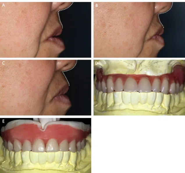
Fig. 2. Clinical evaluation of diagnostic trial denture from the lateral aspect. The dotted lines represent lip support. A, Facial appearance before insertion of the diagnostic trial denture. B, Facial appearance after insertion of the diagnostic trial denture. C, Facial appearance after insertion of the provisional denture. D, Frontal view of diagnostic trial denture without labial flange. E. Frontal view of the provisional denture.
E C A
D B
method, and the definitive cast was mounted on a semiadjustable articulator (Hanau modular articulator, Whip Mix, Louisville, KY). Subsequently, denture teeth were arranged on the maxillary record base and diagnostic wax-ups were prepared for the mandibular teeth. They were placed in the oral cavity and assessed for esthetics and occlusion (Fig. 3). The interarch relationship was additionally verified. Then, implant-supported metal-ceramic fixed dental prostheses were fabricated for the mandibular arch and cemented using methacrylate cement (Premier Implant Cement, Premier Dental Products Co., Plymouth Meeting, PA).
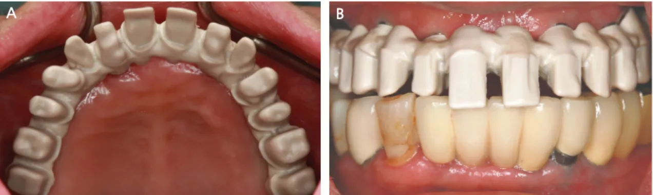
The trial arrangement was scanned using a desktop scanner (Identica Hybrid, Medit Co., Seoul, Korea) for designing the definitive prostheses using a computer-aided designing software program (exocad DentalCAD, exocad GmbH, Daramstadt, Germany). Customized titanium abutments were tried in the patient’s mouth, and accurate transfer from the definitive cast to the maxillary arch of the patient was confirmed using a verification jig (Fig. 4). The PEKK (Pekkton Ivory, Cendres+Métaux, Biel/Bienne, Switzerland) framework, which included 14 abutment tooth forms, was milled in a milling machine (Trione Z, DIO Implants, Busan, Korea). The framework was designed in one piece without palatal coverage.
Fig. 3. Clinical evaluation of full-contour trial teeth arrangement. A, Frontal view. B, Lateral view.
A B
Fig. 4. Verification procedure for maxillary abutments. A, Placement of customized titanium abutments.
B, Placement of verification jig on titanium abutments.
A B
A passive fit and favorable adaptation of the framework on the abutments in the patient’s mouth were verified with the use of powder spray (Occlude, Pascal, Bellevue, WA) and by alternatively applying figure pressure to the terminal ends of the framework
(Fig. 5). The interocclusal relationship wasregistered using polyvinylsiloxane. Individual lithium disilicate ceramic crowns (IPS e.max CAD, Ivoclar Vivadent, Schaan, Liechtenstein) were designed, milled, and temporarily cemented to the framework using zinc oxide noneugenol cement (Temp-Bond NE, Kerr, Karlsruhe, Germany).
The framework and the temporarily cemented crowns were tried in the oral cavity, and esthetics and
Fig. 5. Intraoral placement of polyetherketoneketone (Pekkton Ivory; Cendres+Métaux) framework. A, Occlusal view. B, Frontal view.
A B
Fig. 6. Postoperative clinical photographs and radiograph. A, Intraoral photograph obtained in frontal view. B, Intraoral photograph obtained in left lateral view. C, Intraoral photograph obtained in occlusal view. D, Panoramic radiograph.
C A
D B