J Korean Soc Coloproctol Vol. 22, No. 2, 2006
91
Clinical Features of Colorectal Serrated A - denomas
Hyung Joon Kim, M.D., Tae Hyo Kim, M.D., Byung Lyul Lim, M.D., Gyung Ah Jung, M.D., Hyun Jin Kim, M.D., Woon Tae Jung, M.D., Young Tae Joo, M.D.
1, SangKyung Choi, M.D.
1, Jung Hee Lee, M.D.2Departments of Internal Medicine, 1Surgery and 2Pathology, Gyeon- gsang National University College of Medicine, Jinju, Korea
Purpose: Colorectal cancer is believed to progress through
an adenoma-carcinoma sequence. However, recent eviden- ce increasingly supports the existence of an alternative rou- te for colorectal carcinogenesis through a serrated adeno- ma, which combines the architectural features of hyper- plastic polyps with the cytological features of traditional adenomas. We assessed the characteristics and the endo- scopic features of serrated adenomas and compared them with those of hyperplastic polyps and traditional adenomas in Korea. Methods: The medical records of 344 consecu- tive patients who underwent a colonoscopic biopsy or poly- pectomy from January 2003 through August 2004 at Gyeon- gsang National University Hospital were analyzed retro- spectively.Results: Serrated adenomas were seen in 12
cases (3.4%), and the most common site was the rectum (50%). Endoscopically in most cases, the serrated adeno- mas had small diameters (≤0.5 cm) and were single polyps.Morphologically, the serrated adenomas were flat and non- pedunculated. The coincidental rate of the carcinomas was 8.3%.
Conclusions: According to this study, serrated ade-
nomas are generally single, sessile adenomas with diameters less than 5 mm, and they are commonly observed in the left colon, especially in the rectum. J Korean Soc Colopro-ctol 2006;22:91-96
Key Words: Serrated adenomas, Hyperplastic polyps, Traditio- nal adenomas, Colorectal cancer
거치상 선종, 증식성 용종, 전통적 선종, 대장암 ꠏꠏꠏꠏꠏꠏꠏꠏꠏꠏꠏꠏꠏꠏꠏꠏꠏꠏꠏꠏꠏꠏꠏꠏꠏꠏꠏꠏꠏꠏꠏꠏꠏꠏꠏꠏꠏꠏꠏꠏꠏꠏꠏꠏꠏꠏꠏꠏꠏ
서 론
대장에서 발견되는 용종의 일반적인 조직학적 분류 는 신생화성 용종(neoplastic polyps)과 비 신생화성 용 종(non-neoplastic polyps)으로 구분된다. 신생화성 용종 에는 선종성 용종(adenomatous polyps)과 암성용종(car- cinomatous polyps)이 있고 비 신생화성 용종에는 증식 성 용종(hyperplastic polyps)이 대표적이고 그 외에 염 증성 용종(inflammatory polyps)과 유연성 용종(juvenile polyps), Peutz-Jegher's 용종 등이 있다.1 대부분의 대장 암은 복잡한 과정을 거쳐 선종에서 유발되는 것으로 알려져 있으나 대표적인 비 신생화성 용종인 증식성 용종은 특별한 경우를 제외하고는 암성 변화는 하지 않는 것으로 알려져 있다.2
그러나 1990년에 형태학적으로는 증식성 용종의 특 징을 가지고 있지만 조직학적으로는 전통적인 선종 (traditional adenomas)의 특성을 가진 혼합형태로 일반 적인 증식성 용종과는 다르게 암성 변화를 하는 거치 상 선종을 보고3한 이후 이에 대한 대장 내시경적, 조 직학적 특징과 분자생물학적 특성에 관하여 연구가 활발히 진행되고 있다.4,5
우리나라에서도 대장암은 식생활, 환경 변화 등의 요 인으로 인해 발생 빈도가 증가하고 있어 관심이 고조 되고 있다. 하지만 우리나라에서 악성화 경향이 보고 되고 있는 거치상 선종의 역학과 대장 내시경적 특징 등에 관한 연구는 드물다.6,7 이에 저자는 대장용종에 서 거치상 선종의 임상적, 대장 내시경 및 조직학적 특 성을 전통적 선종, 증식성 용종과 함께 비교 분석하고
대장 용종에서 거치상 선종의 임상적 특징
경상대학교 의과대학 내과학교실, 1외과학교실, 2병리학교실
김형준․김태효․임병렬․정경아․김현진․정운태․주영태1․최상경1․이정희2
접수: 2005년 8월 5일, 승인: 2006년 3월 30일 책임저자: 김태효, 660-702, 경남 진주시 칠암동 90
경상대학교 의과대학 내과학교실 Tel: 055-750-8726, Fax: 055-758-9122 E-mail: [email protected]
Received August 5, 2005, Accepted March 30, 2006
Correspondence to: Tae Hyo Kim, Department of Internal Medicine, Gyeongsang National University College of Medicine, 90 Chiram- dong, Jinju-si, Gyeongsangnam-do 660-702, Korea.
Tel: +82-55-750-8726, Fax: +82-55-758-9122 E-mail: [email protected]
자 하였다.
방 법 1) 대상
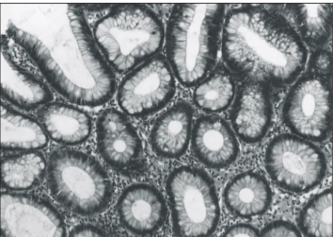
2003년 1월부터 2004년 8월까지 경상대학교병원을 방문하여 대장내시경 검사에서 용종이 발견되어 생검 및 용종절제술을 시행한 환자를 대상으로 하였다. 조직 학적 검사에서 거치상 선종(Fig. 1), 전통적 선종(Fig. 2), 증식성 용종(Fig. 3)으로 확인된 환자를 각각 3군으로 분류하여 조사하였다. 같은 기간에 대장내시경에서 용 종으로 의심되어 시행한 조직검사에서 비특이적 대장 염 및 염증성 용종으로 진단받았거나 대장 내 서로 다
른 용종성 병변이 혼합된 경우는 본 연구에서 제외하 였다.
2) 방법
대상 환자의 의무기록과 조직검사결과를 후향적으 로 조사하여 환자의 평균 연령, 성별 분포, 대장내시경 을 시행하게 된 이유와 Body Mass Index (BMI, kg/m2) 를 비교하였다.
용종의 위치는 비장 굴곡을 기준으로 하여 맹장과 상행 결장 및 횡행 결장을 우측으로, 하행 결장과 에스 자 결장 및 직장을 좌측으로 구분하였고 용종이 비장 굴곡에 위치한 경우는 우측으로 분류하였다. 크기에 따른 구분은 0.5 cm 미만, 0.5∼1 cm, 1∼2 cm, 2 cm 이상으로 나누었다. 개수는 6개 이하인 경우 각각 조 사하였고 6개 이상은 다발성으로 분류하였다. 용종의 일반적인 형태분류 방법인 Yamada 분류법8을 사용하 여 융기의 정도와 목(stalk)의 유무에 따라서 제I형에서 IV형까지 구분하였다. 각각의 군에서 이형성(dyspla- sia) 정도와 선암종의 동반여부를 재차 확인하였다.
통계적 분석은 SPSS (SPSS for window, Release 10.05) 를 이용하여 chi-square test를 시행하였고, P값이 0.05 이하일 때 통계적으로 유의한 것으로 판정하였다.
결 과 1) 대상 환자의 특징
대장내시경을 시행한 1,782명 중 용종이 확인되어 생검 및 용종절제술을 시행받은 환자는 495명(27.8%) 이었다. 이 중 비특이적 대장염(102명), 염증성 용종 Fig. 1. Microscopic finding of hyperplastic polyps. Crypts of
hyperplastic polyps are noted (H&E stain, ×100).
Fig. 2. Microscopic finding of serrated adenomas. Colonic crypts look like saw-tooth as the case of hyperplastic polyps, but lined by adenomatous epitheliums like tubular adenomas (H&E stain, ×100).
Fig. 3. Microscopic finding of traditional adenomas. Crypts of adenomas are noted (H&E stain, ×100).
(10명), 대장 내 서로 다른 용종성 병변이 혼합된 경우 (39명)를 제외한 344명 환자를 대상으로 하였다. 대상 환자 중에서 거치상 선종군 12명(3.4%), 증식성 용종 군 63명(18.4%), 전통적 선종군 269명(78.2%)이었고 평 균 연령은 증식성 용종군에서 57.2세, 전통적 선종군 60.4세, 거치상 선종군이 64.2세였으며 통계적으로 유 의하지는 않았다(P>0.05). 남자 대 여자의 비율은 3군 모두에서 남자의 비율이 높았다. BMI는 3군 간에 차이 가 없었다(Table 1). 대장내시경을 시행한 이유는 하복 부통증(29%), 혈변(20%), 변비(11%), 설사(10%)였고, 무증상도 9%를 차지하였다(Table 1).
2) 대장내시경적 특징
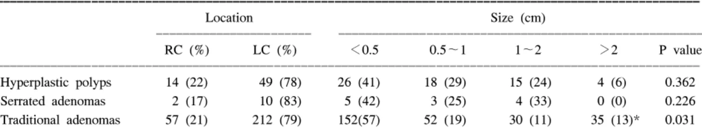
대장내시경에서 용종의 위치는 3군 모두 좌측 대장 에 호발하였으며 특히 거치상 선종군의 경우는 에스 자 결장과 직장에서 주로 발견되었다. 용종의 크기는 3군에서 모두 1 cm 이하가 대부분이었지만, 전통적 선
종군의 경우는 2 cm 이상의 크기가 큰 용종의 비율이 13%로 다른 군에 비해서 높았다(P<0.05)(Table 2). 용 종의 개수는 거치상 선종군과 증식성 용종군의 경우 단일 용종(75%, 50.7%)이 많았지만, 전통적 선종군의 경우 6개 이상의 다발성 용종이 13.3%로 통계적으로 유의하게 높은 빈도를 보였다(P<0.05). Yamada 분류 법에 의한 형태학적 분류로는 3군에서 모두 표면 무경 형인 제I형이 흔하였으며 II, III, IV형은 비슷한 분포를 보였다(Table 3).
3) 조직학적 특징
조직학적으로 선암성 병변과 고도 이형성이 동반된 경우는 전통적 선종군에서 각각 41예(15.2%), 34예 (12.6%)였고, 거치상 선종군에서 1예(8.3%)의 선암성 병변이 동반된 것이 확인되었으며 증식성 용종군에서 는 관찰되지 않았다(Table 4).
Table 1. Clinical characteristics of hyperplastic polyps, serrated adenomas and traditional adenomas
ꠚꠚꠚꠚꠚꠚꠚꠚꠚꠚꠚꠚꠚꠚꠚꠚꠚꠚꠚꠚꠚꠚꠚꠚꠚꠚꠚꠚꠚꠚꠚꠚꠚꠚꠚꠚꠚꠚꠚꠚꠚꠚꠚꠚꠚꠚꠚꠚꠚꠚꠚꠚꠚꠚꠚꠚꠚꠚꠚꠚꠚꠚꠚꠚꠚꠚꠚꠚꠚꠚꠚꠚꠚꠚꠚꠚꠚꠚꠚꠚꠚꠚꠚꠚꠚꠚꠚꠚꠚꠚꠚꠚꠚꠚꠚꠚꠚꠚꠚꠚꠚꠚꠚꠚ Hyperplastic polyps Serrated adenomas Traditional adenomas P value ꠏꠏꠏꠏꠏꠏꠏꠏꠏꠏꠏꠏꠏꠏꠏꠏꠏꠏꠏꠏꠏꠏꠏꠏꠏꠏꠏꠏꠏꠏꠏꠏꠏꠏꠏꠏꠏꠏꠏꠏꠏꠏꠏꠏꠏꠏꠏꠏꠏꠏꠏꠏꠏꠏꠏꠏꠏꠏꠏꠏꠏꠏꠏꠏꠏꠏꠏꠏꠏꠏꠏꠏꠏꠏꠏꠏꠏꠏꠏꠏꠏꠏꠏꠏꠏꠏꠏꠏꠏꠏꠏꠏꠏꠏꠏꠏꠏꠏꠏꠏꠏꠏꠏꠏ
Number (%) 63 (18.4) 12 (3.4) 269 (78.2)
Age (years) 57.2±17.7 64.2±9.7 60.4±13.6 0.320
Male (%) 33 (52.4) 10 (75) 177 (66) 0.146
Female (%) 30 (47.6) 2 (25) 92 (34) 0.158
BMI (kg/m2) 23.2±3.6 22.6±2.6 22.35±3.0 0.075
ꠏꠏꠏꠏꠏꠏꠏꠏꠏꠏꠏꠏꠏꠏꠏꠏꠏꠏꠏꠏꠏꠏꠏꠏꠏꠏꠏꠏꠏꠏꠏꠏꠏꠏꠏꠏꠏꠏꠏꠏꠏꠏꠏꠏꠏꠏꠏꠏꠏꠏꠏꠏꠏꠏꠏꠏꠏꠏꠏꠏꠏꠏꠏꠏꠏꠏꠏꠏꠏꠏꠏꠏꠏꠏꠏꠏꠏꠏꠏꠏꠏꠏꠏꠏꠏꠏꠏꠏꠏꠏꠏꠏꠏꠏꠏꠏꠏꠏꠏꠏꠏꠏꠏꠏ Cause*
ꠏꠏꠏꠏꠏꠏꠏꠏꠏꠏꠏꠏꠏꠏꠏꠏꠏꠏꠏꠏꠏꠏꠏꠏꠏꠏꠏꠏꠏꠏꠏꠏꠏꠏꠏꠏꠏꠏꠏꠏꠏꠏꠏꠏꠏꠏꠏꠏꠏꠏꠏꠏꠏꠏꠏꠏꠏꠏꠏꠏꠏꠏꠏꠏꠏꠏꠏꠏꠏꠏꠏꠏꠏꠏꠏꠏꠏꠏꠏꠏꠏꠏꠏꠏꠏꠏꠏꠏꠏꠏꠏꠏꠏꠏꠏꠏꠏꠏꠏꠏꠏꠏꠏꠏ
Abdominal pain (%) 19 (30) 2 (17) 78 (29) 0.53
Diarrhea (%) 4 (6) 1 (8) 30 (11) 0.27
Constipation (%) 13 (21) 1 (8) 24 (9) 0.09
Hematochezia (%) 17 (27) 2 (17) 50 (19) 0.76
Asymptomatic (%) 10 (16) 6 (50) 16 (6) 0.75
ꠏꠏꠏꠏꠏꠏꠏꠏꠏꠏꠏꠏꠏꠏꠏꠏꠏꠏꠏꠏꠏꠏꠏꠏꠏꠏꠏꠏꠏꠏꠏꠏꠏꠏꠏꠏꠏꠏꠏꠏꠏꠏꠏꠏꠏꠏꠏꠏꠏꠏꠏꠏꠏꠏꠏꠏꠏꠏꠏꠏꠏꠏꠏꠏꠏꠏꠏꠏꠏꠏꠏꠏꠏꠏꠏꠏꠏꠏꠏꠏꠏꠏꠏꠏꠏꠏꠏꠏꠏꠏꠏꠏꠏꠏꠏꠏꠏꠏꠏꠏꠏꠏꠏꠏ
*cause of undergoing colonoscopy.
Table 2. Location and size of hyperplastic polyps, serrated adenomas and traditional adenomas
ꠚꠚꠚꠚꠚꠚꠚꠚꠚꠚꠚꠚꠚꠚꠚꠚꠚꠚꠚꠚꠚꠚꠚꠚꠚꠚꠚꠚꠚꠚꠚꠚꠚꠚꠚꠚꠚꠚꠚꠚꠚꠚꠚꠚꠚꠚꠚꠚꠚꠚꠚꠚꠚꠚꠚꠚꠚꠚꠚꠚꠚꠚꠚꠚꠚꠚꠚꠚꠚꠚꠚꠚꠚꠚꠚꠚꠚꠚꠚꠚꠚꠚꠚꠚꠚꠚꠚꠚꠚꠚꠚꠚꠚꠚꠚꠚꠚꠚꠚꠚꠚꠚꠚꠚ
Location Size (cm)
ꠏꠏꠏꠏꠏꠏꠏꠏꠏꠏꠏꠏꠏꠏꠏꠏꠏꠏꠏꠏꠏꠏꠏ ꠏꠏꠏꠏꠏꠏꠏꠏꠏꠏꠏꠏꠏꠏꠏꠏꠏꠏꠏꠏꠏꠏꠏꠏꠏꠏꠏꠏꠏꠏꠏꠏꠏꠏꠏꠏꠏꠏꠏꠏꠏꠏꠏꠏꠏꠏꠏꠏꠏꠏꠏꠏꠏꠏ
RC (%) LC (%) <0.5 0.5∼1 1∼2 >2 P value
ꠏꠏꠏꠏꠏꠏꠏꠏꠏꠏꠏꠏꠏꠏꠏꠏꠏꠏꠏꠏꠏꠏꠏꠏꠏꠏꠏꠏꠏꠏꠏꠏꠏꠏꠏꠏꠏꠏꠏꠏꠏꠏꠏꠏꠏꠏꠏꠏꠏꠏꠏꠏꠏꠏꠏꠏꠏꠏꠏꠏꠏꠏꠏꠏꠏꠏꠏꠏꠏꠏꠏꠏꠏꠏꠏꠏꠏꠏꠏꠏꠏꠏꠏꠏꠏꠏꠏꠏꠏꠏꠏꠏꠏꠏꠏꠏꠏꠏꠏꠏꠏꠏꠏꠏ Hyperplastic polyps 14 (22) 49 (78) 26 (41) 18 (29) 15 (24) 4 (6) 0.362 Serrated adenomas 2 (17) 10 (83) 5 (42) 3 (25) 4 (33) 0 (0) 0.226 Traditional adenomas 57 (21) 212 (79) 152(57) 52 (19) 30 (11) 35 (13)* 0.031 ꠏꠏꠏꠏꠏꠏꠏꠏꠏꠏꠏꠏꠏꠏꠏꠏꠏꠏꠏꠏꠏꠏꠏꠏꠏꠏꠏꠏꠏꠏꠏꠏꠏꠏꠏꠏꠏꠏꠏꠏꠏꠏꠏꠏꠏꠏꠏꠏꠏꠏꠏꠏꠏꠏꠏꠏꠏꠏꠏꠏꠏꠏꠏꠏꠏꠏꠏꠏꠏꠏꠏꠏꠏꠏꠏꠏꠏꠏꠏꠏꠏꠏꠏꠏꠏꠏꠏꠏꠏꠏꠏꠏꠏꠏꠏꠏꠏꠏꠏꠏꠏꠏꠏꠏ RC = right colon; LC = left colon. *P<0.05.
고 찰
대장 내의 증식성 용종은 일반적으로 양성으로 알 려졌으나 증식성과 선종성이 혼합된 형태인 거치상 선종에서 대장암의 발생이 보고되면서3,4 이러한 증식 성과 선종성 용종의 혼합형이 주목을 받게 되었다. 거 치상 선종은 과증식성 용종과 유사한 선증식에 선종 성 변화가 동반된 대장의 상피성 종양으로 병리학적 으로 소와의 내강이 톱니모양 형태이며 각각의 상피 세포가 증식성 및 선종성 상피를 동시에 가지고 있 다.4,5
대장암의 암성 변화에 대한 일반적인 가설은 선종- 암종 순서이다. 암종화 과정은 정상 상피조직의 비정 상적 메틸화(abnormal methylation)와 APC 유전자 돌연 변이 등에 의한 종양시작(tumor initiation) 과정에서 K-ras 돌연변이, p53 deletion, 18q deletion 등에 의한 종 양증진(tumor promotion) 과정이 선종의 암종변화의 가 설로 제시되고 있다.9,10
그러나 거치상 선종의 암성 변화는 대장암 발생의 일반적인 가설인 선종-암종 순서 외에 또 다른 암성화 경로(carcinogenesis pathway)가 있다는 증거로 거치상 선종에 대한 연구는 임상적으로 의미가 있다고 하겠 다. 최근의 몇몇 보고11-13에 의하면 과거 암성 변화를 하지 않는 것으로 알려진 증식성 용종에서도 암성 변 화 과정에서 보이는 K-ras 돌연변이와 현미위성체불안 정(microsatellite instability)이 관찰된다. 또한 암성 변 화의 증거가 있는 거치상 선종과의 형태학적 특징 및 뮤신(mucin) 생성5에서의 유사성으로 인해 증식성 용 종에서 거치상 선종을 걸쳐 대장암(colonic carcinoma) 이 발생한다는 가설이 대장암의 또 다른 암성변화 경 로(The serrated pathway of colorectal tumorigenesis)로 제시되고 있다. 또한 최근 면역조직화학염색법의 발달 로 인해 거치상 선종의 조직학적, 생물학적 성상에 대 한 연구와 대장암의 발생에 있어서의 역할에 대해서 도 활발한 연구가 진행 중이다.12,14,15
또한, 증식성 용종에 비해 거치상 선종에서 고도 이 형성 변이의 가능성이 많다는 것이 알려졌으며, 거치 Table 3. Numbers and morphologic feautures in hyperplastic polyps, serrated adenomas and traditional adenomas ꠚꠚꠚꠚꠚꠚꠚꠚꠚꠚꠚꠚꠚꠚꠚꠚꠚꠚꠚꠚꠚꠚꠚꠚꠚꠚꠚꠚꠚꠚꠚꠚꠚꠚꠚꠚꠚꠚꠚꠚꠚꠚꠚꠚꠚꠚꠚꠚꠚꠚꠚꠚꠚꠚꠚꠚꠚꠚꠚꠚꠚꠚꠚꠚꠚꠚꠚꠚꠚꠚꠚꠚꠚꠚꠚꠚꠚꠚꠚꠚꠚꠚꠚꠚꠚꠚꠚꠚꠚꠚꠚꠚꠚꠚꠚꠚꠚꠚꠚꠚꠚꠚꠚꠚ
Hyperplastic polyps Serrated adenomas Traditional adenomas
P value
(n=63) (n=12) (n=269)
ꠏꠏꠏꠏꠏꠏꠏꠏꠏꠏꠏꠏꠏꠏꠏꠏꠏꠏꠏꠏꠏꠏꠏꠏꠏꠏꠏꠏꠏꠏꠏꠏꠏꠏꠏꠏꠏꠏꠏꠏꠏꠏꠏꠏꠏꠏꠏꠏꠏꠏꠏꠏꠏꠏꠏꠏꠏꠏꠏꠏꠏꠏꠏꠏꠏꠏꠏꠏꠏꠏꠏꠏꠏꠏꠏꠏꠏꠏꠏꠏꠏꠏꠏꠏꠏꠏꠏꠏꠏꠏꠏꠏꠏꠏꠏꠏꠏꠏꠏꠏꠏꠏꠏꠏ Number (%)
1 30 (47.6) 10 (83.3) 107 (39.8) 0.067
2 8 (12.6) 1 (8.3) 50 (18.6) 0.253
3 15 (23.8) 0 (0) 38 (14) 0.724
4 3 (4.8) 0 (0) 20 (7.4) 0.726
5 2 (3.3) 0 (0) 19 (7) 0.422
≥ 6 5 (7.9) 1 (8.3) 35 (13) 0.021
Yamada type (%)
I 32 (50.8) 6 (50.0) 136 (50.6) 0.082
II 20 (31.7) 2 (16.6) 37 (13.8) 0.350
III 7 (11.1) 2 (16.6) 66 (24.5) 0.256
IV 4 (6.4) 2 (16.6) 30 (11.1) 0.854
ꠏꠏꠏꠏꠏꠏꠏꠏꠏꠏꠏꠏꠏꠏꠏꠏꠏꠏꠏꠏꠏꠏꠏꠏꠏꠏꠏꠏꠏꠏꠏꠏꠏꠏꠏꠏꠏꠏꠏꠏꠏꠏꠏꠏꠏꠏꠏꠏꠏꠏꠏꠏꠏꠏꠏꠏꠏꠏꠏꠏꠏꠏꠏꠏꠏꠏꠏꠏꠏꠏꠏꠏꠏꠏꠏꠏꠏꠏꠏꠏꠏꠏꠏꠏꠏꠏꠏꠏꠏꠏꠏꠏꠏꠏꠏꠏꠏꠏꠏꠏꠏꠏꠏꠏ
Table 4. Dysplasia and carcinoma rate of hyperplastic polyps, serrated adenomas and traditional adenomas ꠚꠚꠚꠚꠚꠚꠚꠚꠚꠚꠚꠚꠚꠚꠚꠚꠚꠚꠚꠚꠚꠚꠚꠚꠚꠚꠚꠚꠚꠚꠚꠚꠚꠚꠚꠚꠚꠚꠚꠚꠚꠚꠚꠚꠚꠚꠚꠚꠚꠚꠚꠚꠚꠚꠚꠚꠚꠚꠚꠚꠚꠚꠚꠚꠚꠚꠚꠚꠚꠚꠚꠚꠚꠚꠚꠚꠚꠚꠚꠚꠚꠚꠚꠚꠚꠚꠚꠚꠚꠚꠚꠚꠚꠚꠚꠚꠚꠚꠚꠚꠚꠚꠚꠚ
HGD (%) Carcinoma (%) Total P value
ꠏꠏꠏꠏꠏꠏꠏꠏꠏꠏꠏꠏꠏꠏꠏꠏꠏꠏꠏꠏꠏꠏꠏꠏꠏꠏꠏꠏꠏꠏꠏꠏꠏꠏꠏꠏꠏꠏꠏꠏꠏꠏꠏꠏꠏꠏꠏꠏꠏꠏꠏꠏꠏꠏꠏꠏꠏꠏꠏꠏꠏꠏꠏꠏꠏꠏꠏꠏꠏꠏꠏꠏꠏꠏꠏꠏꠏꠏꠏꠏꠏꠏꠏꠏꠏꠏꠏꠏꠏꠏꠏꠏꠏꠏꠏꠏꠏꠏꠏꠏꠏꠏꠏꠏ
Hyperplastic polyps 0 (0) 0 (0) 0 0.523
Serrated adenomas 0 (0) 1 (8.3) 1 0.264
Traditional adenomas 34 (12.6) 41 (15.2) 75 0.028
ꠏꠏꠏꠏꠏꠏꠏꠏꠏꠏꠏꠏꠏꠏꠏꠏꠏꠏꠏꠏꠏꠏꠏꠏꠏꠏꠏꠏꠏꠏꠏꠏꠏꠏꠏꠏꠏꠏꠏꠏꠏꠏꠏꠏꠏꠏꠏꠏꠏꠏꠏꠏꠏꠏꠏꠏꠏꠏꠏꠏꠏꠏꠏꠏꠏꠏꠏꠏꠏꠏꠏꠏꠏꠏꠏꠏꠏꠏꠏꠏꠏꠏꠏꠏꠏꠏꠏꠏꠏꠏꠏꠏꠏꠏꠏꠏꠏꠏꠏꠏꠏꠏꠏꠏ HGD = high grade dysplasia.
상 선종의 개수가 증가할수록 선암종의 발생이 유의 하게 증가된다. 따라서 선암종으로 진행되지 않는 증 식성 용종과의 감별이 임상적으로 중요하다. 본 연구 에서 용종의 빈도는 일반적으로 알려진4 대장내시경 에서 용종의 발견 빈도 30%와 유사한 결과였다. 거치 상 선종의 발생률은 보고에 따라 차이가 심하여 0.005%에서 29%까지 다양한데 3,16,17 이는 대장내시경 을 시행하는 의사의 숙련도와 병리의사 간의 판독 차 이 등에 의한 것으로 추정된다. 본 연구에서는 대장내 시경 검사를 통하여 조직학적으로 확진된 거치상 선 종은 344명의 환자 중 12명(3.4%)이었다.
초기 연구들의 보고에서는 거치상 선종의 크기가 5 mm 이하인 경우 17.3%, 6∼10 mm인 경우 61.8%, 1 cm 이상인 경우가 20.9%였으나3 이후 Jaramillo 등16의 보 고에서는 5 mm 이하가 72%이고 1 cm 이상은 3.7%였 다. 본 연구에서도 5 mm 이하가 62.5%로 대부분을 차 지하여 거치상 선종의 크기는 최근 보고와 유사하게 나타났다. 이는 작은 용종에서 조기 선암종이 발생한 다는 인식과 내시경 기기의 발달로 과거에는 그냥 지 나칠 수도 있던 작은 병변을 더욱 더 유심히 관찰하고 조직검사까지 시행하는 결과라고 여겨진다. 대장 내시 경에서 보이는 용종의 형태는 표면무경형이 6예(50%), 융기무경형 2예(16.7%)로 표면융기형과 융기무경형이 대부분을 차지하여 다른 문헌과 유사한 결과를 보여
주었다.16,17 거치상 선종의 호발위치는 주로 좌측 대장,
특히 에스자 결장과 직장이었고 증식성 용종과 전통 적 선종도 좌측 대장에 빈발하였다. 대장내시경 시행 때에 용종들의 일반적 특징을 염두해 두면 임상적으 로 도움이 될 수 있을 것이다.
악성화의 빈도가 8∼13%로 알려져 있지만 유의성 을 갖는 적절한 증례의 빈도는 미미하며 본 연구에서 는 1예에서 선암종이 병발해 있었다. 기존 연구보고와 큰 차이가 없을 것으로 생각되지만 대상군의 수가 적 어 객관적으로 비교할 수 없어 보다 더 많은 대상자를 통하여 확인이 필요하리라 생각된다.
본 연구에서 대장 내 서로 다른 용종성 병변이 혼합 된 39예는 대상군에서 제외하였다. 거치상 선종에 전 통적 선종과 증식성 용종이 혼합된 예는 각각 6명 (15.4%), 5명(12.8%)이었고 전통적 선종과 증식성 용종 이 혼합된 예는 28명(71.8%)이었다. 이런 혼합형 용종 을 제외한 것은 용종 전체보다는 각 개인별로 용종의 특징을 확인하고 통계처리 시에 있을 수 있는 오류를 줄이기 위해서이다. 그러나 혼합형의 환자에서 거치상 선종이 상대적으로 많이 포함되어 있어 이런 혼합형
에 대한 보다 더 많은 연구가 필요하리라 생각된다. 또 한 거치상 선종은 전통적 선종에 비하여 발생빈도가 낮지만 악성화의 또 다른 경로로 추정되므로 이에 대 한 대규모 국내 연구와 악성화 경향에 대한 보다 많은 연구가 필요하겠다.
결 론
거치상 선종의 크기와 형태는 5 mm 이하의 작고 표 면융기형과 융기무경형이 대부분을 차지한다. 호발위 치로는 좌측 대장의 에스자 결장과 직장에서 가장 호발 하며 전통적 선종처럼 악성화 경향(malignant potentials) 에 대한 보고가 있어 주의 깊게 추적 관찰을 해야 하며 특히, 작고 편평한 용종에 대한 더욱 세심한 조직 생검 이 필요하겠다. 추후 더욱 많은 환자를 대상으로 한 거 치상 선종의 임상적, 내시경적 특징과 암성 변화와 관 련하여 면역조직화학염색법을 이용한 유전자 발현이 상 등에 관한 활발한 연구가 필요할 것으로 생각한다.
REFERENCES
1. Enterline HT. Polyps and cancer of the large bowel. Curr Top Pathol 1976;63:95-141.
2. Estrada RG, Spjut HJ. Hyperplastic polyps of the large bo- wel. Am J Surg Pathol 1980;4:127-33.
3. Longacre TA, Fenoglio-Preiser CM. Mixed hyperplastic ade- nomatous polyps/serrated adenomas. A distinct form of colo- rectal neoplasia. Am J Surg Pathol 1990;14:524-37.
4. Makinen MJ, George SM, Jernvall P, Makela J, Vihko P, Karttunen TJ. Colorectal carcinoma associated with serrated adenoma-prevalence, histological features, and prognosis. J Pathol 2001;193:286-94.
5. Jass JR. Serrated adenoma of the colorectum: a lesion with teeth. Am J Pathol 2003;162:705-8.
6. 김상균, 이문성, 김진오, 천갑진, 정인섭, 천영국. 거치상 선 종의 조직학적 및 대장내시경적 특징과 p53 단백의 과발현.
대한소화기학회지 2002;39:350-5.
7. 유민규, 김영호, 손희정, 이풍렬, 김재준, 백승운. 거치상 선 종과 전통적 선종의 악성화 경향에 대한 비교연구. 대한소 화기내시경학회지 2004;29:358.
8. Yamada T, Flkutomi H. Protruding lesions of the stomach.
I To CHYOU (Japanese) 1966;1:145-50.
9. Vogelstein B, Fearon ER, Hamilton SR, Kem SE, Preisinger AC, Leppert M, et al. Genetic alterations during colorectal- tumor development. N Engl J Med 1988;319:525-32.
10. Jacoby RF, Marshall DJ, Kailas S, Schlack S, Harms B, Lo- ve R. Genetic instability associated with adenoma to carcino- ma progression in hereditary nonpolyposis colon cancer.
Gastroenterology 1995;109:73-82.
11. Jass JR, Young J, Leggett BA. Hyperplastic polyps and DNA microsatellite unstable cancers of the colorectum. Histopatho- logy 2000;37:295-301.
12. Chan TL, Zhao W, Leung SY, Yuen ST. BRAF and KRAS mutation in colorectal hyperplastic polyps and serrated ade- nomas. Cancer Res 2003;63:4878-81.
13. Rashid A, Houlihan PS, Booker S, Petersen GH, Giardiello FM, Hamilton SR. Phenotypic and molecular characteristics of hyperplastic polyposis. Gastroenterology 2000;119:323- 32.
14. Rubio CA, Rodensjo M. p53 overexpression in flat serrated adenomas and flat tubular adenomas of the colorectal mu-
cosa. J Cancer Res Clin Oncol 1995;121:571-6.
15. Sawyer EJ, Cerar A, Hanby AM, Gorman P, Arends M, Tal- bot IC. Molecular characteristics of serrated adenomas of the colorectum. Gut 2002;51:200-6.
16. Jaramillo E, Watanabe M, Rubio C, Sleza KP. Small colo- rectal serrated adenomas: endoscopic findings. Endoscopy 1997;29:1-3.
17. Matsumoto T, Mizuno M, Shimizu M, Manabe T, Iida M, Fujishima M. Serrated adenoma of the colorectum: colono- scopic and histologic features. Gastrointest Endosc 1999;49:
736-42.