POVL after Spine Surgery / 287 Asian Spine Journal Vol. 6, No. 4, pp 287~290, 2012 http://dx.doi.org/10.4184/asj.2012.6.4.287
Copyright Ⓒ 2012 by Korean Society of Spine Surgery
This is an Open Access article distributed under the terms of the Creative Commons Attribution Non-Commercial License (http://creativecommons.org/licenses/by-nc/3.0/) which permits unrestricted non-commercial use, distribution, and reproduction in any medium, provided the original work is properly cited.
Asian Spine Journal • pISSN 1976-1902 eISSN 1976-7846
Received May 23, 2011: Revised Jun 24, 2011; Accepted Jun 30, 2011
Corresponding author: Sujit Kumar Tripathy, MS, DNB, Dip SICOT, MNAMS Clinical fellow, Department of Orthopaedics, Friarage Hospital,
Northallerton, DL6 1JG, United Kingdom
Tel: +44-7423388617, E-mail: sujitortho@yahoo.co.in
Cortical Blindness Following Spinal Surgery: Very Rare Cause of Perioperative Vision Loss
Vijay Goni1, Sujit Kumar Tripathy1,2, Tarun Goyal1,3, Tajir Tamuk1, Bijnya Birajita Panda4, Shashidhar BK1
1Department of Orthopaedics, Postgraduate Institute of Medical Education and Research, Chandigarh, India
2Department of Orthopaedics, Friarage Hospital, Northallerton, United Kingdom
3Department of Orthopaedics, Freeman Hopstal, Newcastel upon Tyne, United Kingdom
4Department of Opthalmology, SCB Medical College, Cuttack, India
A 38-year-old man was operated with posterior spinal decompression and pedicle screw instrumentation for his L2 fracture with incomplete neurological deficit. In the recovery, he complained of blindness in both eyes after twelve hours. Computed tomographic scan and magnetic resonance angiography revealed bilateral occi pital lobe infarcts. He remained permanently blind even after three years follow-up. Though rare, perioperative vision loss is a potential complication following spine surgery in prone position. We report a rare occurrence of cortical blindness following lumbar spine surgery.
Key Words: Blindness, Prone, Surgery, Spinal injuries, Postoperative vision loss
Introduction
Vision loss is a very rare but devastating complication of nonocular surgeries, and reported incidence is 0.003%
to 0.0008% in the general surgical population [1]. The risk of perioperative vision loss (POVL) has been commonly noted after cardiac and spinal surgeries. The incidence as reported in literature is 8.64/10,000 for cardiac surgeries and 3.09/10,000 for spinal fusions [2].
Most cases of perioperative vision loss following spine surgery are mentioned as case reports in literature [3-8].
The specific pathogenesis of POVL remains elusive in most cases, with much controversy surrounding patient and surgi- cal risk factors. Important causes of POVL include ischemic optic neuropathy (ION), retinal vascular occlusion (RVO) and cortical blindness. Among these major causes, cortical blindness is the rarest cause of POVL [7-9]. Myers et al.
[7] reported only three cases of cortical ischemia leading to
blindness while reviewing 37 patients of POVL after spine surgery. Because of the rarity of occurrence and as most of the data are retrospective, it is difficult to establish definite cause-effect relationship for the cortical blindness. In this article we report a case of POVL secondary to cortical isch- aemia in a 38-year-old man following lumbar spinal fusion without any predisposing factors for vaso-occlusive disease.
Case Report
A 38-year-old manual labourer was brought to the emer- gency after a high energy motor vehicle accident. He was hemodynamically stable at the time of admission. On pri- mary and secondary survey a lumbar spine fracture possibil- ity was suspected. Radiographic evaluation confirmed our diagnosis and X-ray showed fracture of the second lumbar vertebra (Fig. 1A). Neurologically he had diminished power in both lower limbs (motor power of grade 3 or less around
288 / ASJ: Vol. 6, No. 4, 2012
hip, knee and ankle joints). He had complete loss of bladder and bowel sensations, but no sensory impairment was elic- ited in the lower limbs.
His past medical history was not suggestive of any neuro- logical problems, vision problems, diabetes, hypertension, coronary artery disease, deep vein thrombosis, peripheral vascular disease, collagen vascular disorder or previous chest or heart problems. He was a non smoker and a social drinker. His body mass index was 32.4.
Magnetic resonance imaging of the lumbar spine was per- formed to better delineate the severity of spinal cord injury and compression (Fig. 1B). He was operated after 72 hours of injury. Surgery was carried out with posterior decompres- sion and fusion using pedicle screw instrumentation in the prone position under general anaesthesia (Fig. 2). Constant monitoring of the arterial blood pressure was performed during the surgery. Total duration of surgery was 105 min- utes. Perioperative blood loss was 420 ml. Systolic blood pressure throughout the surgery was in the range of 90 to 110 mm Hg. The oxygen saturation as measured by pulse oxymeter was above 98% at all times. There was no other anaesthetic or surgical complication intraoperatively. In the recovery, patient complained of complete loss of vision after twelve hours. He was evaluated by an ophthalmolo- gist, neurologist and a cardiologist. His ocular examination revealed complete bilateral loss of vision with preservation of papillary and corneal reflexes and normal ocular move- ments. Intraocular pressure and fundus examination were within normal limits. There was no new neurological deficit occurring after surgery and his cerebellar functions were intact. His postoperative haemoglobin was 10.6 g and he did not require any blood transfusion in the perioperative period.
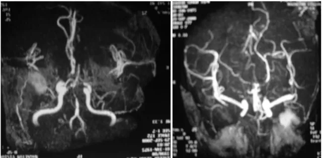
The computed tomographic scan and magnetic resonance angiography of the brain revealed infarcts in bilateral oc- cipital lobes (Fig. 3). Electrocardiography and echocardiog- raphy evaluation for underlying cardiac problem did not reveal any cardiac source of emboli. He was treated by the ophthalmologist, but no recovery in vision was observed even after three years follow-up.
Discussion
The article by Berg et al. [10] was surprising to the spine surgeons where they mentioned that the incidence of POVL following ocular surgeries is much lower than that seen in nonocular surgeries. Incidence estimates for POVL after
nonocular surgery range from 0.013% for all surgeries up to 0.2% following spine surgery. Ischemic optic neuropathy is the most common cause of POVL accounting for more than 81%, followed by retinal artery thrombosis. Cortical blind- ness is the rarest cause of POVL with a handful of cases in the literature [11].
Work up of a patient with perioperative visual loss in- volves consideration of anatomy of the visual pathway.
Anterior ischemic optic neuropathy and retinal vascular oc- clusion reveal remarkable changes on fundus examination, but no such changes are observed in posterior ischemic op- tic neuropathy (PION). The diagnosis in PION can be made
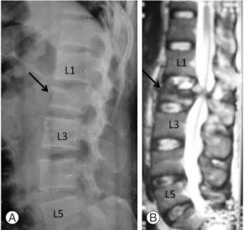
Fig. 1. Radiograph (A) and magnetic resonance imaging (B) of the lumbar spine showing fracture of the L2 vertebra (ar- row).
A B
Fig. 2. Radiogarphs (antero-posterior and lateral views) after posterior spinal decompression and pedicle screw fixa- tion for L2 vertebra fracture.
A B
POVL after Spine Surgery / 289 by contrast enhancement seen in the optic nerve on ocular
magnetic resonance imaging. Cortical blindness is diag- nosed by looking for the ischemic changes in the occipital lobe on intracranial imaging. In our case, the diagnosis was made by cortical occipital changes seen on magnetic reso- nance imaging with the absence of fundoscopic examina- tion findings.
Despite numerous efforts and explanations, pathogenesis of POVL is still elusive. Various aetiologies’ such as fall in systemic blood pressure, anaemia, direct ocular compres- sion, hypercoagulable states, embolism, increased venous pressure, prone positioning during surgery and increased cerebrospinal fluid pressure have been elucidated but none has proved so far [12-15]. Two important factors in cortical blindness are generalised cerebral hypoperfusion and em- bolism. It has been suggested that more than one factor may be working in any patient making this a multifactorial event [7,10]. Pathogenesis in cardiac surgery is relatively easy to explain. Embolisation may take place due to cardiac and great vessel manipulation, atrial or ventricular fibrillation [7].
The source of emboli in spinal surgery is difficult to explain.
It is also unclear why this phenomenon is commoner in sur- geries carried out in the prone position. Direct pressure on eye, raised intraocular pressure or vascular congestion does not explain cortical infarcts, but may explain other causes of vision loss such as ION and RVO. Intraoperative hypoten- sion, hypoxia, blood loss and anaemia are contributory fac- tors, but are not found in this patient. Further, they should classically affect the watershed areas of blood supply in the brain which innervate the proximal muscles of upper and
lower limb. Also, it has been shown that the use of deliber- ate hypotensive anaesthesia during spine surgery does not increase chances of POVL [7]. Huber and Grob [8] sug- gested that abnormal posture of the neck when the patient is being positioned prone for surgery could be a contributory factor for reducing perfusion in the vertebra-basilar area manifesting as stroke. This is purely hypothetical thought and cannot be definitively proved. It has been recommended to keep the neck at the level of heart or above in a neutral forward position at the time of surgery to avoid chances of hypoperfusion due to vertebra-basilar compression. The bilateral infarction as in the present case does not support the hypothesis. Review of POVL following general surger- ies include many procedure carried out in the lower limb in prone position [7,16]. Irrespective of the types of surgeries, the prone position itself is a predisposing factor for POVL.
Shen et al. [2] found some important finding on periopera- tive visual loss following spinal surgeries. Incidences were higher for age less than 18 or more than 65 years, male gender, anaemia, and posterior approach. Many cases occur in patients who have no identified preoperative risk factors, although hypertension, smoking, diabetes, and vascular dis- ease appear to lead to increased risk. None of these risk fac- tors was present in this patient. He had no cardio-pulmonary comorbidities and he had no fluctuation in hemodynamic status in the perioperative period as well. Considering these situations, other than the prone position we could not find any predisposing condition in our case.
Most of patients with cortical blindness have a partial vi- sion loss. They often have other associated symptoms such
Fig. 3. Magnetic resonance angiography of brain reveals bilateral infarction of occipital lobe.
290 / ASJ: Vol. 6, No. 4, 2012
as cerebellar signs and other focal neurological deficits, de- pending upon the area of infarct. This patient had purely vi- sion loss with no other deficit. There is no definite treatment for cortical blindness. No drugs including steroids have shown to reduce morbidity in these cases and most postop- erative visual deficits do not show significant improvement with time. There is a hope of some improvement in initial months, but once the window time period has passed there is no hope of any further improvement.
This case report warns the spine surgeon about such fatal complication following spine surgery in prone position. Ex- treme cautions at every step should be taken to prevent the development of perioperative visual loss. Despite these pre- cautions, some cases may still land up with visual loss and the patient should be explained about the grievous situation well before the surgical procedure.
REFERENCES
1. Warner ME, Warner MA, Garrity JA, MacKenzie RA, Warner DO. The frequency of perioperative vision loss.
Anesth Analg 2001;93:1417-21.
2. Shen Y, Drum M, Roth S. The prevalence of periopera- tive visual loss in the United States: a 10-year study from 1996 to 2005 of spinal, orthopedic, cardiac, and general surgery. Anesth Analg 2009;109:1534-45.
3. Grossman W, Ward WT. Central retinal artery occlu- sion after scoliosis surgery with a horseshoe headrest.
Case report and literature review. Spine (Phila Pa 1976) 1993;18:1226-8.
4. Hoski JJ, Eismont FJ, Green BA. Blindness as a com- plication of intraoperative positioning. A case report. J Bone Joint Surg Am 1993;75:1231-2.
5. Katzman SS, Moschonas CG, Dzioba RB. Amaurosis
secondary to massive blood loss after lumbar spine sur- gery. Spine (Phila Pa 1976) 1994;19:468-9.
6. Lee AG. Ischemic optic neuropathy following lumbar spine surgery: case report. J Neurosurg 1995;83:348-9.
7. Myers MA, Hamilton SR, Bogosian AJ, Smith CH, Wagner TA. Visual loss as a complication of spine surgery: a review of 37 cases. Spine (Phila Pa 1976) 1997;22:1325-9.
8. Huber JF, Grob D. Bilateral cortical blindness after lumbar spine surgery: a case report. Spine (Phila Pa 1976) 1998;23:1807-9.
9. Newman NJ. Perioperative visual loss after nonocular surgeries. Am J Ophthalmol 2008;145:604-10.
10. Berg KT, Harrison AR, Lee MS. Perioperative visual loss in ocular and nonocular surgery. Clin Ophthalmol 2010;4:531-46.
11. Kamming D, Clarke S. Postoperative visual loss fol- lowing prone spinal surgery. Br J Anaesth 2005;95:257- 60.
12. Remigio D, Wertenbaker C. Post-operative bilateral vi- sion loss. Surv Ophthalmol 2000;44:426-32.
13. Stevens WR, Glazer PA, Kelley SD, Lietman TM, Bradford DS. Ophthalmic complications after spinal surgery. Spine (Phila Pa 1976) 1997;22:1319-24.
14. Stambough JL, Dolan D, Werner R, Godfrey E. Oph- thalmologic complications associated with prone positioning in spine surgery. J Am Acad Orthop Surg 2007;15:156-65.
15. Roth S. Perioperative visual loss: what do we know, what can we do? Br J Anaesth 2009;103:i31-40.
16. Williams EL, Hart WM Jr, Tempelhoff R. Postop- erative ischemic optic neuropathy. Anesth Analg 1995;80:1018-29.