Effect of liners and primers on tensile bond strength between zirconia and resin-based luting agent
Eun-Hye Jo, Yoon-Hyuk Huh, Kyung-Ho Ko, Chan-Jin Park, Lee-Ra Cho*
Department of Prosthodontics and Research Institute of Oral Science, College of Dentistry, Gangneung-Wonju National University, Gangneung, Republic of Korea
PURPOSE. The effect of silica-based glass-ceramic liners on the tensile bond strength between zirconia and resin-based luting agent was evaluated and compared with the effect of 10-methacryloyloxydecyl dihydrogen phosphate (MDP)-containing primers. MATERIALS AND METHODS. Titanium abutments and zirconia crowns (n
= 60) were fabricated, and the adhesive surfaces of the specimens were treated by airborne-particle abrasion. The specimens were divided into 5 groups based on surface treatment: a control group, 2 primer groups (MP:
Monobond Plus; ZP: Z Prime Plus), and 2 liner groups (PL: P-containing Liner; PFL: P-free Liner). All specimens were cemented with self-adhesive resin-based luting agent. After 24-hour water storage and thermocycling (5,000 cycles, 5°C/55°C), the tensile bond strength was measured using a universal testing machine. Failure mode analysis and elemental analysis on the bonding interface were performed. The data were analyzed using Kruskal-Wallis test, Dunn’s post hoc test, and Fisher’s exact test. RESULTS. The liner groups and primer groups showed significantly higher tensile bond strengths than that of the control group (P<.05). PFL showed a significantly higher tensile bond strength than the primer groups (P<.05). The percentage of mixed failure was higher in the primer groups than in the control group (P<.001), and all the specimens showed mixed failure in the liner groups (P<.001). A chemical reaction area was observed at the bonding interface between zirconia and liner. CONCLUSION. The application of liner significantly increased the tensile bond strength between zirconia and resin-based luting agent. PFL was more effective than MDP-containing primers in improving the tensile bond strength with the resin-based luting agent. [J Adv Prosthodont 2018;10:374-80]
KEYWORDS: Resin-based luting agent; Methacryloyloxydecyl dihydrogen phosphate (MDP); Liner; Tensile bond strength; Zirconia
INTRODUCTION
Yttria-stabilized tetragonal zirconia polycrystal (Y-TZP) has
been widely used as a material for prostheses owing to its biocompatibility and excellent mechanical properties.1 Zirconia prostheses have shown a wide range of survival rate (81 - 100%) in 5-year observation, which was reported to be associated with mechanical complications such as veneer chipping and loss of retention.2-4 Loss of retention occurs more frequently in Y-TZP than in porcelain-fused metal prostheses.5 Ortorp et al.3 monitored monolithic zirco- nia prostheses for 5 years and insisted that loss of retention accounted for the highest percentage of total complications (30%). Most of those prostheses were bonded using resin- based luting agent.
All-ceramic prostheses can form stable chemical bonds with resin-based luting agent through hydrofluoric acid and silane applications.6 However, such surface processing is not possible in zirconia due to its polycrystalline structure.7 For this reason, researchers have explored various surface treat-
Corresponding author:
Lee-Ra Cho
Department of Prosthodontics and Research Institute of Oral Science, College of Dentistry, Gangneung-Wonju National University 7 Jukheon-gil, Gangneung, Gangwondo 25457, Republic of Korea Tel. +82336403153: e-mail, [email protected]
Received March 6, 2018 / Last Revision July 2, 2018 / Accepted August 13, 2018
© 2018 The Korean Academy of Prosthodontics
This is an Open Access article distributed under the terms of the Creative Commons Attribution Non-Commercial License (http://creativecommons.
org/licenses/by-nc/3.0) which permits unrestricted non-commercial use, distribution, and reproduction in any medium, provided the original work is properly cited.
This work was supported by the Cooperative Research Fund (CR1502) of Gangneung-Wonju National University Dental Hospital.
ment methods to improve the bond strength between zirco- nia and resin-based luting agent, such as airborne-particle abrasion (APA), laser treatment, elective infiltration etching, silica coating, and functional monomer application.8 For lut- ing of zirconia prostheses, APA followed by 10-methacry- loyloxydecyl dihydrogen phosphate (MDP) or silane applica- tion is the most common treatment.9-11
MDP, a type of acid phosphate monomer, was originally developed to increase the bond strength between metal and resin-based luting agent, and is now used in zirconia pros- thesis.12 It forms phosphate-oxygen-zirconium bonds with zirconia.13 To obtain a sufficient bond strength, MDP should be accompanied with APA treatment.1 However, APA can cause microcracks on zirconia surface, which lead to frac- ture or damage on the zirconia margin.14,15
Tribochemical silica coating has been reported to effec- tively improve the bond strength in high-strength ceram- ics.16-18 Micromechanical retention and chemical bonding with silica particles can be achieved through tribochemical silica coating.11,19 However, these effects may be reduced in zirconia, which has a high surface hardness.20 Matinlinna et al.,20 through energy-dispersive X-ray spectroscopy (EDS) analysis, reported that tribochemical treatment of zirconia surface induced insufficient silica coating. It has also been claimed that silica does not chemically bind to zirconia sur- faces.21 The effect of silica coating can be improved by increasing the blasting pressure during tribochemical silica coating.22 However, the possibility of zirconia surface dam- age cannot be ruled out.23
Researchers have proposed another technique in which silica is added to zirconia surfaces instead of the subtraction techniques.24-26 Several studies reported that chemical bond- ing occurred at the bonding interface between zirconia and ceramics.27-29 Lung et al.25 proposed a sol-gel method for sili- ca-based ceramics. However, this technique resulted in a sig- nificantly low shear bond strength with resin-based luting agent compared with that obtained after APA, and the coat-
ing was unstable after thermocycling. Kitayama et al.24 pro- posed an internal coating system (INT), in which the inner surface of zirconia prostheses is partially or fully covered with silica-based ceramics through high-temperature firing.
A significant increase in bond strength with resin-based lut- ing agent and compensation of the large internal gap within the zirconia prostheses were achieved with INT.26 However, few studies have compared the INT with different surface processing methods, and have assessed the stability of the coating after thermocycling.
In this study, the effects of silica-based glass-ceramic liners and MDP-containing primers on the tensile bond strength between zirconia and resin-based luting agent fol- lowing thermocycling were investigated. The null hypothesis was that different surface treatment techniques do not have any effect on the tensile bond strength between zirconia and resin-based luting agent and on the failure mode.
MATERIALS AND METHODS
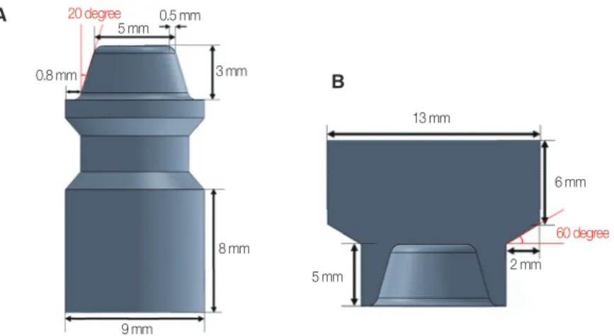
Sixty titanium abutments and zirconia crowns (12 per group) were fabricated from titanium blocks (commercially pure titanium, grade V) and pre-sintered block (Zirtooth, Hass, Gangneung, Korea) with CAD/CAM program (hyperMiLL, Samwoohyper, Seongnam, Korea). To mini- mize the effects of mechanical retention, the convergence angle of the titanium abutment was set to 20 degrees on one side, making a total of 40 degrees (Fig. 1). The titanium abutments and intaglio of zirconia crowns were subjected to APA (Basic master, Renfert, Hilzingen, Germany) by ver- tically spraying alumina (Al2O3) particles from a distance of 10 mm at 0.25 MPa. Specimens were divided into 5 groups depending on the applied surface treatment: control group (APA), MP group (APA+Monobond Plus), ZP group (APA+Z Prime Plus), PL group (APA+P-containing Liner), and PFL group (APA+P-free Liner) (Table 1).
The internal gap between the titanium abutment and zir-
Table 1. Materials used
Material Brand name Lot No. Main composition Manufacturer
Zirconia Zirtooth S41FIB901 Zirconium dioxide, hafnium dioxide, yttrium trioxide,
inorganic pigment, organic binder HASS
Resin-based luting agent RelyX Unicem 3328586
Glass powder, initiator, silica, substituted pyrimidine, calcium hydroxide, peroxy compound, pigment, methacrylate phosphoric ester, dimethacrylate, acetate, stabilizer
3M ESPE
Hydrofluoric acid gel Porcelain etchant 1700004012 4% Hydrofluoric acid Bisco Inc
Silane coupling agent Monobond S U17000 3-(methacyrloxy)propyltrimethoxysilane, water, ethanol Ivoclar Vivadent Primer Monobond Plus (MP) V33449 3-(trimethoxysilyl)propyl methacrylate, sulphide
methacrylate, methacrylated phosphoric acid ester Ivoclar vivadent Primer Z Prime Plus (ZP) 1600002409 Organophosphate monomer, carboxylic acid monomer Bisco Inc Liner P-containing Liner (PL) EO1D03 Silicon dioxide, lithium oxide, aluminum oxide,
phosphorus pentoxide, etc. HASS
Liner P-free Liner (PFL) EO1D11 Silicon dioxide, lithium oxide, aluminum oxide, etc. HASS
conia crown was determined by measuring the mean liner thickness in 10 additional zirconia crowns that were cut using an Accutom-50 cutting machine (Struers, Ballerup, Denmark) (Fig. 2). An internal gap of 20 μm for cement was set in the control and primer groups, while the mean liner thickness (15 μm) was added for the liner group. A spray-type liner was sprayed from a distance of 15 - 20 mm for 2 - 3 seconds and fired for 1.5 minutes at 950°C in a porcelain furnace (Austromat D4, Dekema, Freilassing, Germany). The zirconia crowns were bonded to the titanium abutments using the self-adhesive RelyX Unicem (3M ESPE, Seefeld, Germany). In the liner group, the intaglio of zirco- nia crown was treated with 4% hydrofluoric acid (Porcelain etchant, Bisco Inc., Schaumburg, IL, USA) for 5 minutes and uniformly coated with silane (Monobond S, Ivoclar Vivadent, Liechtenstein, Germany) for 60 seconds. The bonding interface between the titanium abutment and zirco- nia specimen was observed at ×10,000 magnification using a scanning electron microscope (SEM, Quanta FEG 250, FEI, Hillsboro, OR, USA). For the liner group, an EDS (Apollo XP, EDAX, Mahwah, NJ, USA) connected to the SEM was used for line scan analysis to observe the elemen- tal distribution between the liner and zirconia.
All specimens were stored in distilled water at 37°C for 24 hours, and then thermocycling was performed for 5,000 cycles (5°C and 55°C, 2 seconds dwelling time; Thermal cyclic tester, R&B Inc., Daejeon, Korea). A tensile force was applied in the axial direction at a crosshead speed of 1.0 mm/min using a universal testing machine (Model 5982, Instron, Norwood, MA, USA). After the tensile bond strength test, the outer surface of the titanium abutment and the intaglio of the zirconia crown were examined at × 300 magnification using an optical microscope (VHX S550E, Keyence, Itasca, TX, USA). Failure modes were cat- egorized into adhesive failure, cohesive failure, and mixed failure. If a mixed failure was observed, the specimen was examined at × 100 and × 200 magnifications under a SEM
to confirm the failure mode.
The Kruskal-Wallis test and Dunn’s post hoc test (IBM SPSS 23.0, IBM, Armonk, NY, USA, α = .05) were used to analyze the significant differences among the tensile bond strengths achieved by the different surface treatment tech- niques. Two-sided P values < .05 were accepted as statisti- cally significant. Further, Fisher’s exact test was used to determine the significant differences in the failure mode among the groups.
Fig. 1. Titanium abutment and zirconia crown. (A) Schematic diagram of titanium abutment, (B) Schematic diagram of zirconia crown.
13 mm
6 mm
5 mm 2 mm 8 mm
9 mm
3 mm 0.5 mm 5 mm
0.8 mm 20 degree
60 degree
A
B
Fig. 2. Liner thickness measurement (optical microscope;
original magnification: × 500). (A) Measurement sites, (B) Optical microscopic image to measure occlusal space, (C) Optical microscopic image to measure axial space.
L, Liner; Z, Zirconia.
A
B C
RESULTS
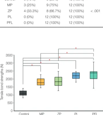
All the experimental groups showed significantly higher ten- sile bond strengths than that of the control group (P < .05).
The tensile bond strengths of the PFL group was signifi- cantly higher than those of the ZP and MP groups (Fig. 3, P
< .05).
In the failure mode analysis, significant differences in the failure modes were observed according to the surface treatment (Table 2, P < .001 with Fisher’s exact test). Cohesive failure was not observed in any specimens. In the control group, adhesive failures were more frequently observed than mixed failures, whereas in the primer groups, mixed failures were more common than adhesive failures. The MP and ZP groups showed similar frequencies of adhesive failures. In both the liner groups (PL and PFL), all specimens presented mixed failures. When closely observed using a SEM, cohe- sive failures were observed mostly in the resin-based luting agent layers in the occlusal surface, and adhesive failures were observed between the zirconia crown and resin-based luting agent in the axial surface. Similar failure modes were observed in the occlusal surface of the liner group.
However, in the axial surface, adhesive failures between the
resin-based luting agent and liner were usually observed, whereas adhesive failure between the liner and zirconia was not observed. In addition, it was seen that the zirconia sur- faces were covered with the liner (Fig. 4).
EDS was used to perform line scanning to determine the elemental distribution of the specimens’ components (Fig. 5). In the liner group, the intensities of zirconium (Zr) and silicon (Si) ion distributions in a chemical reaction layer changed. Diffusion of Zr into the liner and of Si, main components of the liner, into zirconia were observed. The interdiffusion zone was approximately 1 - 2 μm thick (Fig.
5).
Table 2. Distribution number (%) of failure modes Group Adhesive
failure Mixed failure Total P value
Control 9 (75%) 3 (25%) 12 (100%)
< .001
MP 3 (25%) 9 (75%) 12 (100%)
ZP 4 (33.3%) 8 (66.7%) 12 (100%)
PL 0 (0%) 12 (100%) 12 (100%)
PFL 0 (0%) 12 (100%) 12 (100%)
Fig. 4. Scanning electron microscopic (SEM) images of inner surface of zirconia crown after tensile bond test.
Black arrow indicates cohesive failure in resin-based luting agent. White arrow indicates adhesive failure between zirconia and resin-based luting agent or liner and resin-based luting agent. (A) MP (occlusal surface), (B) MP (axial surface), (C) ZP (occlusal surface), (D) ZP (axial surface), (E) PL (occlusal surface), (F) PL (axial surface), (G) PFL (occlusal surface), and (H) PFL (axial surface). (A, C, E, G: original magnification ×200; B, D, F, H: original magnification ×100). R, Resin-based luting agent; Z, Zirconia; L, Liner.
A B
C D
E F
G H
Fig. 3. Box plot for comparison of tensile bond strengths of different groups.
* Indicate statistically significant difference (P < .05).
Control MP ZP PL PFL 3500
3000 2500 2000 1500 1000 500 0
Tensile bond strengths (N)
DISCUSSION
Based on the measurement results, the null hypotheses of this study were rejected. The primer treatment and liner treatment significantly increased the tensile bond strength between zirconia and resin-based luting agent.
The MP and ZP groups both include MDP. The MP group additionally includes sulfide methacrylate and 3-(tri- methoxysilyl) propyl methacrylate (3-MPS) monomers, which are silane monomers, while the ZP group additionally includes carboxylic acid monomer. In this study, there was no significant difference between the tensile bond strengths of the MP and ZP groups. MDP may significantly affect the bonding behavior of zirconia.
The biggest compositional difference between the PFL and PL groups was the presence of phosphorous pentoxide (P2O5). P2O5 acts as a nucleating agent to induce the forma- tion of bonds between components.30 Therefore, a definite crystal structure can be formed in the presence of P2O5. As the crystal structure becomes clearer, the strength of the liner increases; however, the wettability decreases. This explains the significantly higher tensile bond strength of the PFL group than those of the primer groups, despite of no significant difference between the PL group and the primer groups.
Liner treatment increased the tensile bond strength more effectively than the primer treatment did. This was also confirmed in the failure mode analysis. In the failure mode analysis, the liner groups showed less adhesive failures than the primer groups did, which indicates that more stable and stronger bonds were formed between the zirconia, lin- er, and resin-based luting agent. Very few adhesive failures were observed between zirconia and liner, and most zirco- nia surfaces were covered with the liner. It appears that chemical reactions occurred during the liner firing pro- cess.27-29 Such results are consistent with previous findings.
It was reported that the interdiffusion zone within the bond- ing interface between zirconia and ceramic was observed in EDS analysis.27,28 Durand et al.29 suggested that the interdif- fusion zone is 2 μm in thickness; this value is close to the thickness of the interdiffusion zone observed in this study.
Since the liners are crystallized glass-ceramics with silica as the main component, they can form stable siloxane bonds with resin-based luting agent through hydrofluoric acid and silane treatments, which were applied in the conventional ceramic bonding procedures.
In this study, all the bonding surfaces between the titani- um abutments and zirconia crowns were subjected to APA.
There has been a controversy regarding the optimal blasting pressure and particle size for producing efficient bond strengths Fig. 5. Line scan analysis (original magnification: ×10,000) of bonding interface between zirconia and liner. Chemical reaction area and diffusion of elements (Si: silicon, Zr: zirconium) observed across the interface. (A) PL, (B) PFL.
Z, Zirconia; L, Liner.
A
B
×10000
×10000
100
50
0
100
50
0
wt (%)wt (%)
2.19 4.38 6.58 Distance (µm)
2.38 4.76 7.15 Distance (µm)
and durability of zirconia. In this study, 50-μm alumina particles were sprayed at 0.25 MPa onto the intaglio of zirconia crown, which was suggested in a previous study.23 The control, prim- er, and liner groups were all treated under the same condi- tions to minimize confounding effects.
Both the primer and liner groups showed higher tensile bond strengths than that of the control group after thermo- cycling. Studies that investigate the stability of liners after thermocycling procedure are rare, and there is a controversy regarding the stability of primers during thermocycling.15 The magnitude of the effects of thermocycling can vary depending on the exposure of the bonding interface. In this study, only the margin area was exposed to water. Furthermore, because the zirconia crowns were relatively thick, they may have been less susceptible to the effects of thermocycling.
However, the liner may effectively resist thermocycling in which a 1 - 2 μm thick chemical reaction layer was formed in the bonding interface between the zirconia and liner.
There are various methods of liner treatment including dipping and spraying. Although Lee et al.26 recommended the use of dipping method, it is a complicated process since it involves the mixing of powder and liquid. In addition, this method leads to a thick accumulation of liner around the line angle from the occlusal to the axial intaglio of the zirconia crown. In this study, the liners produced in the form of a spray were sprayed onto the intaglio of the zirco- nia crowns. The spraying method is simple and results in comparatively uniform distribution of particles. However, it is technically sensitive as the quantity of sprayed particles, and consequently, the liner thickness can be changed depending on the spraying distance and time.
Further, the tensile bond strength with resin-based lut- ing agent significantly increased for the PFL group com- pared with the primer groups. However, the liner applica- tion has several limitations. First, the liner treatment is infe- rior in terms of convenience. Liners require high-tempera- ture firing following application and must be treated with hydrofluoric acid and silane for bonding. Second, primer treatment can be performed following internal adjustment of ceramics in the chairside, and the liner layer can disap- pear during internal adjustment. Third, complete seating may be difficult to achieve if the thickness of the liner is larger than the internal gap of the prostheses previously set.
Therefore, further studies are needed to simplify the liner treatment processes and to find methods to sensitively adjust the liner thickness. However, since zirconia prostheses typi- cally have large and various internal gaps compared to other prostheses, the liners may compensate the misfit.24
CONCLUSION
In this study, the inner surfaces of zirconia crowns were treated with primers or silica-based glass ceramic liners, and their tensile bond strengths with resin-based luting agent were measured. Primer treatment led to an increase in the tensile bond strength regardless of the primer types com- pared with the control group, and led to an increase in the
percentage of mixed failures. Liner treatment led to a signif- icant increase in the tensile bond strength regardless of liner composition compared with the control group, and all spec- imens showed mixed failures. In addition, the liners without phosphorous compounds exhibited the highest tensile bond strength. Chemical reaction layers and 1 - 2 μm thick inter- diffusion zone were observed at the bonding interface between zirconia and liner.
ACKNOWLEDgEMENTS
The authors thank HASS Corporation for their materials used in this study.
ORCID
Eun-Hye Jo https://orcid.org/0000-0001-8375-2563 Yoon-Hyuk Huh https://orcid.org/0000-0003-4072-5199 Kyung-Ho Ko https://orcid.org/0000-0002-1260-8844 Chan-Jin Park https://orcid.org/0000-0003-4734-214X Lee-Ra Cho https://orcid.org/0000-0003-3989-2870 REFERENCES
1. Yi YA, Ahn JS, Park YJ, Jun SH, Lee IB, Cho BH, Son HH, Seo DG. The effect of sandblasting and different primers on shear bond strength between yttria-tetragonal zirconia poly- crystal ceramic and a self-adhesive resin cement. Oper Dent 2015;40:63-71.
2. Schmitter M, Mussotter K, Rammelsberg P, Gabbert O, Ohlmann B. Clinical performance of long-span zirconia frameworks for fixed dental prostheses: 5-year results. J Oral Rehabil 2012;39:552-7.
3. Ortorp A, Kihl ML, Carlsson GE. A 5-year retrospective study of survival of zirconia single crowns fitted in a private clinical setting. J Dent 2012;40:527-30.
4. Rinke S, Gersdorff N, Lange K, Roediger M. Prospective evaluation of zirconia posterior fixed partial dentures: 7-year clinical results. Int J Prosthodont 2013;26:164-71.
5. Heintze SD, Rousson V. Survival of zirconia- and metal-sup- ported fixed dental prostheses: a systematic review. Int J Prosthodont 2010;23:493-502.
6. Kato H, Matsumura H, Ide T, Atsuta M. Improved bonding of adhesive resin to sintered porcelain with the combination of acid etching and a two-liquid silane conditioner. J Oral Rehabil 2001;28:102-8.
7. Cavalcanti AN, Foxton RM, Watson TF, Oliveira MT, Giannini M, Marchi GM. Bond strength of resin cements to a zirconia ceramic with different surface treatments. Oper Dent 2009;34:280-7.
8. Tzanakakis EG, Tzoutzas IG, Koidis PT. Is there a potential for durable adhesion to zirconia restorations? A systematic review. J Prosthet Dent 2016;115:9-19.
9. Casucci A, Osorio E, Osorio R, Monticelli F, Toledano M, Mazzitelli C, Ferrari M. Influence of different surface treat- ments on surface zirconia frameworks. J Dent 2009;37:891-7.
10. Ntala P, Chen X, Niggli J, Cattell M. Development and testing
of multi-phase glazes for adhesive bonding to zirconia sub- strates. J Dent 2010;38:773-81.
11. Chen C, Chen G, Xie H, Dai W, Zhang F. Nanosilica coating for bonding improvements to zirconia. Int J Nanomedicine 2013;8:4053-62.
12. Papia E, Larsson C, du Toit M, Vult von Steyern P. Bonding between oxide ceramics and adhesive cement systems: a sys- tematic review. J Biomed Mater Res B Appl Biomater 2014;
102:395-413.
13. Chen L, Suh BI, Brown D, Chen X. Bonding of primed zir- conia ceramics: evidence of chemical bonding and improved bond strengths. Am J Dent 2012;25:103-8.
14. Kosmac T, Oblak C, Jevnikar P, Funduk N, Marion L.
Strength and reliability of surface treated Y-TZP dental ce- ramics. J Biomed Mater Res 2000;53:304-13.
15. Blatz MB, Sadan A, Martin J, Lang B. In vitro evaluation of shear bond strengths of resin to densely-sintered high-purity zirconium-oxide ceramic after long-term storage and thermal cycling. J Prosthet Dent 2004;91:356-62.
16. Kern M, Thompson VP. Bonding to glass infiltrated alumina ceramic: adhesive methods and their durability. J Prosthet Dent 1995;73:240-9.
17. Awliya W, Odén A, Yaman P, Dennison JB, Razzoog ME.
Shear bond strength of a resin cement to densely sintered high-purity alumina with various surface conditions. Acta Odontol Scand 1998;56:9-13.
18. Blixt M, Adamczak E, Lindén LA, Odén A, Arvidson K.
Bonding to densely sintered alumina surfaces: effect of sand- blasting and silica coating on shear bond strength of luting cements. Int J Prosthodont 2000;13:221-6.
19. Khan AA, Al Kheraif AA, Jamaluddin S, Elsharawy M, Divakar DD. Recent trends in surface treatment methods for bonding composite cement to zirconia: A reveiw. J Adhes Dent 2017;19:7-19.
20. Matinlinna JP, Heikkinen T, Ozcan M, Lassila LV, Vallittu PK.
Evaluation of resin adhesion to zirconia ceramic using some organosilanes. Dent Mater 2006;22:824-31.
21. Chen L, Suh BI, Kim J, Tay FR. Evaluation of silica-coating techniques for zirconia bonding. Am J Dent 2011;24:79-84.
22. Heikkinen TT, Lassila LV, Matinlinna JP, Vallittu PK. Effect of operating air pressure on tribochemical silica-coating. Acta Odontol Scand 2007;65:241-8.
23. Yang B, Barloi A, Kern M. Influence of air-abrasion on zirco- nia ceramic bonding using an adhesive composite resin. Dent Mater 2010;26:44-50.
24. Kitayama S, Nikaido T, Maruoka R, Zhu L, Ikeda M, Watanabe A, Foxton RM, Miura H, Tagami J. Effect of an in- ternal coating technique on tensile bond strengths of resin cements to zirconia ceramics. Dent Mater J 2009;28:446-53.
25. Lung CY, Kukk E, Matinlinna JP. The effect of silica-coating by sol-gel process on resin-zirconia bonding. Dent Mater J 2013;32:165-72.
26. Lee ES, Huh YH, Park CJ, Cho LR. Effect of silica-contain- ing glasseceramic liner treatment on zirconia coping reten- tion. J Prosthet Dent 2018 Jun 28. pii: S0022-3913(18)30001- 5. doi: 10.1016/j.prosdent.2017.12.005. [Epub ahead of print].
27. Aboushelib MN, Kleverlaan CJ, Feilzer AJ. Microtensile bond strength of different components of core veneered all-ceram- ic restorations. Part II: Zirconia veneering ceramics. Dent Mater 2006;22:857-63.
28. Kawai Y, Uo M, Watari F. Microstructure evaluation of the interface between dental zirconia ceramics and veneering por- celain. Nano Biomed 2010;2:31-6.
29. Durand JC, Jacquot B, Salehi H, Fages M, Margerit J, Cuisinier FJ. Confocal Raman microscopic analysis of the zir- conia/feldspathic ceramic interface. Dent Mater 2012;28:661- 71.
30. Holand W, Beall GH. Glass ceramic technology. 2nd. Hoboken;
John Wiley & Sons; 2012. p. 32-45.