大韓放射線뽑學會誌 Vol. 21, No. 2. 1985
- Abstract-
子宮頭部潤의 放射線治據 後의 骨盤脫內 變化의 電算化斷層擺影 所見
高神醫科大學 放射線科學敎室 禹 永 勳 · 金 湖 俊·田 炳 熙
혈明大學校 醫科大學放射線科學敎室 徐 修 之
Computed Tomographic Findings in Pelvic Cavity after Radiation Therapy for Carcinoma of Cervix
Young Hoon Woo
,
M.D.,
Ho Joon Kim,
M.D.,
Byung Hee Chun,
M.D.Oepartment of Oiagnostic Radiologι Kosin Medical Col/ege
,
Busan,
Korea500 Jhi 5uh
,
M.D.Oepartment of Radiology
,
School of Medicine,
Keimyung Universitχ Oaegμ KoreaFrom J비Y 1
,
1981 to August 31,
1984,
59 patients who had radiation therapy for carcinoma of cervix had CT scanning at Department of Diagnostic Radiology, Kosin Medical College.The authors analysed the CT fin
d.
ings of the patients in regard to the recurrence of the disease and postradia.tion changes
The results are as followings;
1. The incidence of recurrence was most common in advanced stage over IIb.
2. Changes in pelvic cavity were as followings;
; Widening of presacral space Increased perirectal fat space
; Symmetrical thickening of perirectal fascia Fibrous connection between sacrum and rectum
; Anterior connection between rectum and perirectal fascia
; Increased bowel wall thickness
Increased blad.der wall thickness with trabeculations
7796
7896 8196 9796 9296 4796 4496 5196 3. In most patients who had CT scanning within 3 months after radiation therapy
,
CT did not demonstratepostradiation changes characterized by an increased pelvic fibrous and fatty tissue.
4. In 10 patients who had postoperative radiation therapy, 8 patients show increased bowel wall thickness.
이 논문은 1985년 2월 28일에 접수하여 1985년 3 월 15일에 채돼되었음.
- 334-
5. 30 patients with recurrent carcinoma of cervix were as followings;
Pelvic tumor recurrence
Parametrial and side wall extension Pelvic and paraaortic Iymphadenopathy Hydronephrosis
Bladder involvement
뼈찌 m 씨 級 W 생 였
Lumbar spine involvement 10%
And 1 patient shows distant metastasis to paraaortic Iymph node
,
1 patient to lumbar spine,
and 1 patient to liver without recurrent tumor mass in pelvic cavity.6. 2 patients showing mass without other sign in the pelvic cavity were unable to be differentiated between irradiated uterus and recurrent tumor.
1. 絡 論 %흉愚者中 放射線 治續 또는 手術과 m用治擔後 추적 검 사로써 CT 을 시행한 59 例를 대상으로 하였£며 放射 放射線治續 또는 手術과 m用治展 後 再發된 경우의 線治續는 4MeV線型加速機를 使用히여 全 骨盤에 前 약 半數에서는 骨盤용內에 再發된 睡塊를 볼 수 있으며,
遠隔轉移는 主로 뼈, 府 및 없모節등에 나타난다. 이 러 한 局所再發과 遠隔轉移를 얄기 위해서는 骨盤內談 및 뼈部題影, 經靜服뽑굶造影術, 바리움 灌陽法, 뽑스캔.~
巴管造影術, 超音波談斷法, 電算化斷層嚴影 等이 있다.
骨盤內談으로서 는 放射線治覆 後에 오는 骨盤많의 硬 化로 病變 및 病變範圍測定의 失敗率이 20% 정도라는 報告가 있고 1) CT 와 相互 보완적으로 使用하는 超흡 波談斷法도 骨盤tK의 硬化, 骨盤骨과 陽內호氣 等으로 因한 影像의 障害가 많아 그談斷能力에限界가있다?’ 3, 4)
이 들에 比해 骨盤업의 CT 는 骨盤용內器官들이 兩測 이 對照的이 거 나 中心에 놓이 게 되 어 比較觀察이 容易하 고 또한 풍부한 8읍防組織에 依해 各 器官틀의 境界가 뚜 렷 하며, 骨盤骨때 문에 各 器官들의 位置變動이 거의 없 고 呼吸運動에 依한 影像의 障害가 거의 없기 때문에 다 른 部位의 CT 보다 많은 利點이 있다고 할 수 있다 5)
또한 CT 는 再發인 경우 睡塊의 存在有無, 正確한 容 積, 周圍嚴器의 홉潤 等을 正確히 알 수 있을 뿐만 아니 라 放射線治續 後에 骨盤홈內變化의 特徵的인 所見을 보 인다.
이에 著者들은 1981年 7 月 1 日 부터 1984年 8 月 31 日 까지, 추적검사에서 CT 를 利用한 59例를 分析, 檢討하 여 相當한 結果를 얻 었기에 文敵考察과 함께 報告하고 자한다.
2.
對象 및 方法 對 象1981 年 7 月 I 日부터 1984年 8月 31 日까지, 子宮題部
- 335-
後 2 門 혹은 4 門對向 照射하였으며, 흩內治續는 Flet- cher- Suit-Delclos 또는 Henschke Afterloading appl- icator 를 使用하고 Cs-137 밀 봉소선원으로 하였으며, 흩 內治魔을 할 수 없는 境遇는 外部에 서 追加量을 照射하 였다.
方 法
使用한 CT 는 Varian-360-3 이었으며 像은 256 x 256 Matrix 로 構成되며 各 節片의 두께는 10mm이고 Scan
time 은 3 쟁、이 다.
,훌者을 {fp 몽A位로 하여 會陰部로 부터 上方으로 1D-v13 mm 間隔으로 兩測뽑魔의 下部까지 擺影하였으며 境遇에 따라서
JJf
및 뼈部도 題影하였 다.陽管과 睡塊를 區別하기 짧하여 擺影 1 乃至 2 時間 前에 10% Gastrografin 500 rrù 를 經口 授與하여 題홈 內造影을 하였으며. 65% Angiografin 100ml 를 靜服內 潤注하여 造影增꿇을 시키므로 血管과 增大된 ~밍節을 區別하였고, 病황에 따라 適切한 window setting 을 調 節하여 適當한 影像을 얻 었 다.
轉移에 依한 빼巴節增大는 兩뼈O 陽骨血管周圍의 非對 稱性 樣狀 및 찌*밍節의 크기가 1. 5cm 以上인 6) 境遇 또 는 빼밍節中心部애 底密度가 보이는 혈遇 等을 判定基 準으로 하였으며, 骨盤慶으로의 룹犯與否는 睡塊와 周 圍節肉사이의 正常 9읍防組織消失 및 睡塊周圍節肉을 結合 시 키는 不規則한 軟組織의 線狀漫潤을 보이는 境遇 等으
로 하였다 7 , B).
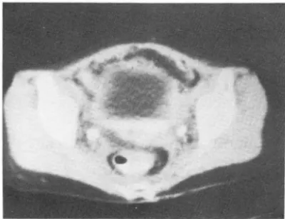
CT scan demonstrate widening of presacral space due to increased fat. The
increasedperirectal fat space
issurro
undedby thickened perirectal fascia , creating
“halo"
effeαFig.
1.年都別分布는 全@빼 서 5 야t 群이 30 例(53%)이고, 再 發한 例에서는 50 代群이 14 例 (57%) 로써 가장 많은 頻 度를 보였다
(Table] ).
Table
ß에서 나타난 바와 같이 병기별 分布는 ßb 以 上의 進行된 例가 41例(7 2.2%) 이었으며, 進行된 例가 많은 理由는 낮은 病期에서는 手術에 만 依存하는 例가 많았기 때문이다. 再發된 30例中 ßb 以上의 進行된 病 期는 23名 (81%) 이었다.放射線治續後의 骨盤홈內 變化들은
(Table 3)
1) 薦骨前호間의 據張이78%(Fig.
1)2)
直陽周圍R읍防組織의 增加81%(Fig.l) 3)
直陽周圍觸維組織의 對稱的ßE原97%(Fig. 1) 4)
薦骨과 直陽間 織維組織結合92%(Fig. 2)
5) 直陽과 直陽周圍繼維組織間의 前方繼維性結슴48
%(Fig. 3)
6)
陽ßE훔44%(Fig. 4)
7)
勝ID\;뚫의 ßE~흥51%(Fig.
5)를 나타내었고, 直陽 周圍脫防組織과 直陽周圍鐵維組織의 對稱的 ßEJ훔로 因 한 • Halo effect" (Fig. 1) 가 大多數에서 나타났다.이들中 가장 많은 例에서 나타낸 直陽周圍織維組織의
*훌
3.
成Table
1. Age distríbution of carcinoma of cervix Age
20-29 30-39 40
-49 50-59 60-69
Recurrence Total
Fibrotic
∞ nnectionbetween sacrum and rectum
“Halo" effect by increase in perirectal fat and fibrous tissue is also seen.
Fig.2.
1 4 6 M 5 1
4
n
% 9
59 30
Table 3. CT findings after radiation therapy for carcinoma of cervix
Table 2. Stage distribution of carcinoma of cervix
78%
81%
97%
92%
44%
48%
51
%Widening of presacral space
Increased perirectal fat space
Symmetrical thickening of perirectal fascia Fibrous connection between sacrum and rectum
Anterior connection between rectum and perirectal fascia
Increased bladder wall thickness with trabeculations
Increased bowel wall thickness 6.
7 --
--
1i
?ι 끼j
A?
5.
Recurrence
30
nU
이ι 〈」O。A약 (、)
/0
Total No. of patient
--‘
j i4 nxu nU 7t ro
---
59 Stage
Ia
Ib
IIa
IIb
IIIa
IIIb
IV對稱的R巴原, 直陽周圍8읍防組織의 t홈加, 또 薦骨前호間의 陽사이에서 睡塊를 보인 覆遇가 27 例이 었으며 이들은 隨張등이 며 이 들 세 가지 變化를 나타내 지 않은 例들은 大部分 험等한 固形睡塊로써 나타났고
(Fig. 6)
때 로는 거의가 放射線治擾 後 짧은 期間內에 CT를 施行한 境 睡塊中心에 壞死를 同伴한 不均等密度를 나타냈다 遇들이었다.즉 直陽周圍組織의 對稱的nE~륨를 나타내지 않은 2 例 中 I 例는 1 個月以內에, 다른 例는 6 個月以內에 CT를 施行했고, 薦骨前호間의 據張이 나타나지 않은 13 例中
7 例는 3 個月 以內에 施行한 境遇이며, 直陽周圍R읍防組 織의 t홈加를 볼 수 없었던 12例中 7 例가 3 個月以內에
CT를施行했었다
手術을 받은 10 例中 8 例에서 陽뼈享를 나타냈고, 또 이 들에 서 陽服原가 深하게 나타났다
再發한 30 例
(Table 4)
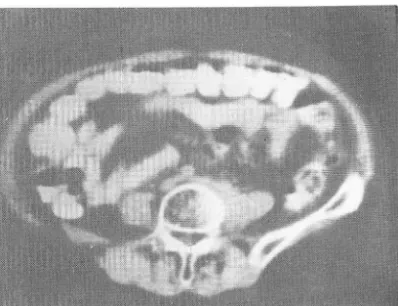
에 서 는 骨盤폼中央部, 勝脫과 直Fig. 3. Rectum and perirectal fascia are
∞ nnectedanterio
r1y by fibrous tissue
.Fig.4. CT scan reveals marked thickening of visible bowel loops in pelvic cavity causing narrowing of lumen. There is slight dilatation of bowel
100
ps in pelvic
ca띠ty.(Fig . 7).
骨盤測慶으로의 파급은 16例이 었으며 大部分 睡塊와
Table 4. CT findings of recurrent carcinoma of cervix after radiation therapy
J. Pelvic tumor recurrence
2. Parametrial and side wall extension 3. Pelvic and paraaortic Iymphadenopathy 4. Hydronephrosis
5. Bladder involvement 6. Lumbar spine involvement
Fig. 5. Smooth thickening of bladder wall.
90%
53%
40%
23%
23%
10%
Fig.6. Recurrent carcinoma of cervix after irradiation
appears as homogenous mass between bladder
and rectum. There is also postradiation effect.
憐接된 內測閒銷ij'fj
(m. obturator I nternus) ,
尾骨節(m.
coccygeus) ,
또는 햇狀節 (m.
pyriformis) 과 連結되는 不規則한 軟組織의 線狀홉潤(Fig. 8)
이 거 나 또는 節周 圍暗防組織이 消失되고 節組織과 융뽀合되는 充寶性睡塊 로써 나타났다(Fig. 9).
骨盤용內 /#.모節t홈大는
12
171J에서 보였으며, 大部分 睡塊로써 陽骨血管周圍에서 發見되었으여(Fig.
10) ,題 部大動服周圍빼巴節轉移는 6171J이었고 이中 1 例는 骨 盤脫內 睡塊의 陰影없이 題部大動服周圍 빼모節增大만 을 나타냈다 (Fig.11).
水賢효이 7 例로써 오른쪽
4171J.
왼쪽 2 例,양쪽 1 例 이었 다(Fig. 12).
勝뾰의 홉犯은 7 例로써 睡塊에 依한 外因性壓浪과 正
Fig. 7. The mass between bladder and rect um shows inhomogenous density suggesting necrosis and arr m cer
V1X.Fig. 9. CT scan reveais tumor extension to right pelvic side wall by relatively homogenous mass.
There is metastasis to pelvic lymph nodes
Fig. 10. Common iliac lymph nodes are enlarged.
Fig. 8. The large conglomerated recurrent tumor Fig. 1
1.Extensive retroperitoneal lymph node metasta- extends to pelvic side wall with irregular sis obliterates the borders of aorta and inferior
linear strands. vena cava.
常 6읍防組織의 消失 및 勝 &t률좋의 不規則↑生을 나타냈다.
CFig.
13).體堆波及이 3 例를 보였£며 이 中 1 例는 꽉盤뾰內 睡塊의 出現없이 제 5 顆推部位에서 左댐‘” 體節의 局所 ßE j享플 나타내는 遠隔빼移의 所見을 냐타냈다 (Fig.
14).
1例에서 骨盤용內에 再發완 睡塊의 陰影과 더불어 Cul -de-Sac 에 W훗體가 고여 있음을 볼 수 있었다 CFig.15).
略形많을 骨盤뾰內에 가진例도 냐타났다
CFig.
16).그러 나 再發된 睡場의 j룹潤性 睡塊와 放射線治續後의 繼維形成과의 區分이 어려운 혈遇도 2 ØlJ가 있었다.
4.
考 察子宮題部햄의 放射線治爾 後에는 解副學的으로 6읍防
Fig. 12.
CTscan at
levelof kidneys shows bilateral
hydronephrosisand extensive retroperitoneal
lymph node metastasis.Fig. 13.
Recurrent tumor invadesposterior
wallof bladde
r.Fig. 14. At
level ofL5 vertebral body , focal enlarge-
mentof
leftpsoas mus
cIe with ob
Iiteration of
fatbetween vertebral body and psoas
muscIeis indicative of distant metastasis.
Fig. 15.
Fluid shadow is
in cul-de-sac, which is posterior to recurrent mass.
Fig. 16.
Incidental finding of teratoma.組織과 繼維組織이 t홉加한다. 直 陽周圍 織維組織은 힐常 모습을 볼 수 있 다 17)
對稱的으로 薦骨前호間에서 兩測으로 增加된 直陽周圍 再發된 {7U의 約 半이 骨盤엎內에서 일어났다는 報告 8읍防組織을 싸면서 나타내 며 9), 直陽과 앞쪽으로 織維 가 있£나 1) 저자들의 境遇는 30 例 中 27 例에서 骨盤 性結슴을 形成하거나 薦骨과 直陽間의 織維性結合組織 용內에서 再發되었고, 이와 더불어 遠隔轉移를 나타내지 을 보이기도 한다. 않은때는放射線照射後 織維注增꺼홉을 일£킨 子宮인지 再
이것은 正常A에서는 없거나 있다고 해도 겨우 알아 發된 R重塊陰影인지 區別하기 어렵다19) 著者들의 境遇 볼 程度이 다. 도 子宮麗內 睡塊陰影만 보인 8 例 中 2 i71J에 서 區分하
이런 繼維性變化는 骨盤뾰의 手術을 무척 어렵게하나 기 어려웠다.
血被供給을 줄여서 放射線治續의 셨果을 높인다 6, 10) • 骨盤測뿔S로의 파급은 碼塊와 周圍骨盤節組織을連結 薦骨前호間의 據張은 lcm 以上으로 나타내고 이는題 하는 軟組織과 同等密度의 不規則한 線狀홈潤이거나 節 題, 慣處性大陽잦, 性病性빼巴肉풍睡, 骨盤脫助睡효l 結 周圍R읍防組織을 消失시키고 節組織과 밟合되는 固型鍾 核, 梅毒, 쿠싱씨病 等에서도 나타날 수 있으나 放射線
에 依한 變化에서 볼 수 있는 直陽周圍織維組織의 對稱 的 R巴原가 나타나지 않는點으로 區分된다 11)
子宮과 題은 放射線治爾의 破壞的 했果로 부터 回生 할 수 있는相當한 能力을 가지고 있다 12)
勝ID't은 휠좋이 두꺼워지고 크기가 줄며 勝ID't周圍R읍防組 織과 織維組織이 增加하나 %跳自體의 {$展性과 勝뾰周 圍組織의 個A에 따론 差異가 많아 區分이 어 렵 다 10)
勝뾰도 比較的 放射線에 대한 저항성이 강하나, 後뿔에 治짧되지 않는 慣陽의 發達로 드물게 勝뾰麗훨를 形成
한다 13)
直陽은 解헤學的으로 睡塊와 가까이 있고 比較的 固 定되어 있으며, 또 比較的 放射線에 敏感하여 그變化가 잦아서 慶이 두꺼워지고 口쩔은 좁아지며 뼈展性이 줄
어든다 10
,
14) •小陽은 勝뾰이 나 直陽보다는 敏感하나 局所的인 子宮
塊로 나타나는데 20) 著者을의 例에서도 同-한 所見을 보였다.
子宮題部經의 轉移는 #巴管을 通하여 이 루어지며 가 장 먼저 轉移되는 #巴節은 閒銷#巴節을 包含한 外部
陽骨#巴節과 內部陽骨빼밍節이 며 1,21,22 ,23), 其他 脫陽
骨#巴節, 大動服周圍빼巴節 및 鼠앓部1#모節의 順으로
轉移된 다 24 ,25) ..
CT 는 子宮題部病의 骨盤홈內 #밍節 파급의 談斷에 있어서 1#밍管照影術에 比하여 多少 옷미치고 있으나
CT 의 ó~斷的 優位性은 骨盤용內 睡塊를 直接 確認할 수 있다는 點과 廳塊의 容짧을 얘U定할 수 있으며 #밍 管照影術上 보이지 않는 #모節 및後題題뺨#모節의 增 大를 알 수 있고, 骨盤얘I}뿔£로의 파급 및 憐接魔器로 의 훔犯 與否를 쉽게 및j定할 수 있다는 點이다 5,6,17,26,
27,28,29,30,31,32)
그러 나 CT는 轉移된 #巴節이 正常 또는 輕微한 增 띤홉部痛의 放射線治續에 包含되는 容積이 比較的 적고 또 大를 보이는 혈遇에 談斷이 不可能하여 27,33,34) 增大된 正常의 補動運動에 依해 固有의 保護가 있다 그러나 骨 1#밍節이 轉移로 因한 것인지 反應性增植요로 因한 것인 盤手術 後에 는 放射線照、射野內에 서 小陽의 固定때 문에 지 區分이 안된 다는 點이 短點이 다 6)
이 러한 保護를 잃어 버린다. 放射線治爾 後에 나타나는 水뽑효은 織維性 變化때문 著者들의 例에서도 骨盤手術을 받은 境遇는 慢性不可 인지 再發된 睡塊때문에 오는 康管閒銷때문인지를 區分 遊的 小陽陽害의 發生率이 上昇했다. 이때 勝뼈을 樓張 하기 어렵다는 報줌가 있으나 35{ 輸행管은 放射線에 振 시키거나 題없位로 해서 小陽을 放射線照射野에서 轉置 抗性이 크므로, 放射線治續 後의 輸행管 閒銷의 發生은 시킬 수 있다 15) 거의 睡陽의 再發때문이다 6, 36)
gp
陽陽害의 程度는 放射線量과 그 照射野에 依해, 또 g용ID't은 .l.t較的 放射線에 振抗하나 後慶에 週場의 發達 動 !w 硬化fíE. 樓康病, 高血塵, 많往題部手術, 骨盤感짧fíE 이 可能하여 드물게 勝ID't麗製로 말탈한다 13)等의 血管狀態나 陽運動에 依해 左右된다 l
‘’
15,16). 睡塊의 勝 ID't ìl호及은 後慶을 따라 外因性睡展, 正常R읍防 放射線治標後의 子宮題部 1홉의 再發은 2 年內에 잘 얼 組織의 消失, 勝뼈활의 不規則性 및 勝跳內에 睡塊의 存 어난다 17,18) 在로써 나타난다 19)CT 에 依해 再發된 睡塊의 陰影, 子宮周圍結合組織과 顆堆波及은 遠隔轉移을 意味하며 6) 한쪽의 骨破壞나 骨盤뻐뿔으로의 파급, 骨盤 및 題部大動服 周圍#巴節 周圍 體推의 硬化fíE( Selerosis)을 나타내고, 가끔은 體 轉移, 水뽑효의 閒銷部位를 CT로서 찾음으로써 再發된 推間版의 ~9움을 나타낸 다. 한쪽 波及일 쩔遇에 는 왼쪽
이 많은데 이것은 왼쪽의 8흥部大動Il!æ周國#巴節이 體推 에 더 가까이 있기때문이다. 한쪽편에만 오는 境遇, 둘 혹은 그 以上의 體 f훌에 周圍 軟組織 l國塊와 함께 體唯波 及이 나타나는 例가 보고되었는데, 이것은血行性보다는 빼巴節의 波及。l 體唯波及을 일으키 는 原因이 되 기 때 문
이 마 37
,
38) ..5.
結 論著者들은 子宮趙部提으로 放射線治續를 받은 後 追없;
檢훌를 위해 高神醫大 放射線科學 敎室에서 1981年 7月 1 日부터 1984年 8 月 31 日 까지 CT를 利用한 59 例를 分 析한 結果 다음과 같은 結論을 얻을 수 있었다.
1) 再發된 例에 서 病期別 分布는
ßb
以上의 進行된 例 가 많았다 77%2) 放射線治續 後의 骨盤뾰內 變化을은 薦骨前호間의
據張이 78%. 直陽周圍R읍防組織의 增加가 81%. 直陽周
圍鐵維組織의 對擺성 6巴훔가 97%. 薦骨과 直陽間 鐵維
組織結合이 92%. 直陽과 直陽周圍織維組織間의 前方繼
維住結合이 48%. 陽ßE훔 44%. 勝뾰慶의 ßl"ft콩-가 51
%를 나타내었다.
3) 放射線治續後 特徵的 所見인 6읍助組織과 織維組織
의 t曾加를 나타내 지 않은 大健의 例가 放射線照、射後 3 個月 以內의 짧은 期間안에 CT를 施行한 境遇였다.
4) 手術을 받은 後 放射線治續를 施行한 10 例中 8例
에 서 陽 R巴j훔가 나타났다.
5) 再發한 30 f11빼서는 勝脫과 直陽사이의 睡塊를 보 인 境遇가 90%. 子宮周圍結合組織과 骨盤測뚫으로 波
及 53%. 骨盤 및 題部大動服周圍빼巴節轉移 40%. 水
쩔훈 23%. 睡塊의 勝Ib't波及 23%. g훌推波及이 10%를 보였 3 며, 骨盤홈內 睡塊의 陰影없이 題部大動服周圍#
巴節로의 遠隔轉移, 睡堆轉移. "꾸轉移를 일으킨 l71J가 1 例썩 있었다.
6) 다른 所見이 없이 骨盤엎內에 睡塊陰影만을 보임
으로써 放射線治續後의 鐵維性增꺼훌을 보이는 子宮인지, 再發된 睡塊인지를 區分하기 어려운 境遇도 2 例를보였
다
REFERENCES
1. Parsons L. sommers 5.C: Cynecalogγ Philadelphia
,
Saunders
,
1300-1420,
19782. Carter B. L., Kahn P.C, Wolper 5.M. et al: Unusual pelvic
masses; A comparison of computed tomographic scan- ning and ultrasophgray. Rad. 121:383-390
,
1976 3. Sukov R.j., 5cardine P.T., 5ample W.F. et al: Computedtomography and transabdominal ultrasound in the evalua- tion of the prostate. j. of CAT 1:281-289
,
1977.4. Walsh j
.w.,
Resenfield A.T.,
ja야 CC et al: Prospective comparison of ultraseund and computed tomography in the evaluation of gynecologic masses. A}R 131:955-960,
1978.
5. Levitt R.G., 5agel 5.5., et al: Computed tomography of the pelvis. Seminars in Roent. 13:193-200, 1978.
6. Ginaldi 5.
,
Wallace 5. et al: Carcinoma of the Cervix; Lym- phangiography and computed tomography. A}R 136:1087-1091, 1981.7. Husband j.E.
,
Hodson N.j.,
Parsons C A.: The use of com- puted tomography in reéurrent rectal tumors. Rad 134:677-682, 19808. Walsh j.W., Amendela M .A., Konerding K.F. et al: Com- puted tomographic detection of pelvic and inguinallym- phnode metastasis from primary & recurrent pelvic malign낀nt disease. Rad. 137:157-166, 1980
9. Eckhardt G., Werner L., Rainer W.: The perirectal fascia, morphology and use in staging of rectal carcinoma. Rad 149‘241-246, Oct. 1983
10. Doubleday L. C Bemardino M.E.: CT findings in the perirec- tal area following radiation therapy. j. of CA T 4(딩:634-638.
Oct. 1980
11. Edling NP
,
Eklöf 0: The retrorectal soft-tissue space in ulcerative colitis. A roentgen-diagnostic study.
Acta Radiol (5tockh) 80:949-953,
1963.12. Wang CC Principles of radiation therapy of gynecologic cancer. In: Cusberg 5A. Frick H
c.,
eds. Corscaden's gynecelogic cancer. Baltimere: Williams & Wilkins,
69-82,
1978
13. Aron B5, 5chlesinger A.: Complications of radiation therapy: the gemitourinary tract. 5emin. Roent. 9:65-74, 1974
14. jack E. Meyer: Radiography of the distal colon and rec- tum after irradiation of carcinoma of the cervix. A}R 136:691-699
,
April 1981.15. Green N.
,
Iba G.,
5mith W.: Measures to minimize sma /l intesting injury in the irradiation pelvis. Cancer 35:1633-1640,
197516. Maruyama Y., Van Nagel RR, Utley J., Vider ML, parker j.C: Radiation and small bowel complications in cervical carcinoma therap
y.
Rad. 112:699-703, 1974- 341-
17. james W. Walsh et al‘ Recurrent carcinoma of the cer- 28. Price j.M., Davidson A. j.: Computed tomography in the vix: CT diagnosis; AjR 136:117-122 jan. 1981. evaluation of the suspected carcinomatous prostate. Uro 18. 5alitha Reddy, Myung-sook Lee, F.R. Hendrickson: Pat- Rad. 1:39-42, 1979.
tern 01 recurrences in endometrial carcinoma and their 29. Redman H.C.: Computed tomography of the pe/vis. Rad management. Rad. 133:737-740. Dec. 1979‘ Clin. North am. 15:441-448, 1977
19. james W. Walsh, Dean R. Goplerud: Prospective com- 30. 5tanley R.j., 5agel 5.5., Fair W.R.: Computed Tomography parison between clinical and CT staging in primary cer- of the genitourinary tract. j. Urol. 119:780-782, 1978 vical carcinoma: AjR 137:997-1003, Nov. 1981. 31. Walsh j.W.: Comparison of u/trasound and computed 20. 5pellman M.C., Castellino R.A., Ray G.B. et al: An eva/ua- Tomography in the evaluation of pe/vic masses. Clin. Diag
tion of Iymphography in localized carcinoma of the pro- Ultrasound 2:229-242
,
1979.state. Rad. 125:637-644, 1977‘ 32. Walsh j.W., Amendola M.A., Konerding K.F. et al: Com- 21. Cherry
c.
P., Williamson B., 5heedy P.F. et al: Computed puted tomographic detection of pe/vic and inguinallym- tomography of the retroperitonea/space. Rad. c/in. North phnode metastasis from primary and recurrent pe/vic Am. 15:337-390, 1977. malignant disease. Rad. 137:157-166, 198022. Henriksen E.: The Iymphatic spread of carcinoma of the 33. Lee j.K.T.
,
5tanley R.j.,
5agel 5.5. et al: Accuracy of com- cervix and of the body of the uterus: A study 420 necrop- puted tomography in detecting intraabdomina/ and pe/vic sies. Am. j. Ob. Cy. 58:924-942, 1949. adenopathy in /ymphoma. AjR 131:311-315.23. Holzaeplel j.H., Ezell H.E ’ Sites of metastases of uterine 34. Schaner E.G., Head G.L., Deppman j.L. et al: Computed carcinoma. Am. j. Ob. Cy. 69:1027-1038, 1955. tomography in the diagnosis, stagin
g,
and management 24. HiU A.M.: The pathology and treatment of malignant glan- of abdomina/ Iymphoma. j. CAT 1:176-180,
1977dular metastases of the /ateral pe/vic wall. Br. j. Ob. C
y.
35. Cunningham JJ, Fuks ZY, Castellino RA: Radiographic67:717-723, 1960. manifestations of carcinoma of the cervix and complica
25. Mitani Y., Fujii j.l., Miyamura M. et al: Lymph node tions of its treatment. Rad. Clin. Nor. Am. 12:93-108, 1974.
metastases of carcinoma of the uterine cervix. Am. j. Ob. 36. Slater j.M., Fletcher GH: Ureteral strictures after radia- Cy. 84:515-522, 1962. tion therapy for carcinoma of the uterine cervix. AjR 26. Brizel H.e., Livingston P.A., Grayson E.V.: Radiotherapeutic 111:259-272, 1971
app
![Fig. 1. 年都別分布는 全@빼 서 5 야t 群이 30 例(53%)이고, 再發한 例에서는 50 代群이 14 例 (57%) 로써 가장 많은 頻度를 보였다 (Table] )](https://thumb-ap.123doks.com/thumbv2/123dokinfo/5313554.163694/3.918.480.880.123.433/Fig이고한에서는代群이로써가장많은頻度를보였.webp)