I. 서론
세균내독소 (bacterial endotoxin)는 그람음성 세균 의 세포벽 외막에 존재하는 lipopolysaccharide (LPS) 성분으로, 염증의 유발을 비롯한 다양한 생물학적 작 용을 가지고 있으며, 구강 내에서는 치태, 타액, 치은 삼출액, 염증치은조직, 치석, 그리고 치주질환에 이 환된 백악질 등에 세균내독소가 존재한다1-3). Gibbons 등4)은 치은열구 내에 세균내독소를 방출할 수 있는 다수의 그람음성 세균이 존재함을 보고하였 고, 치주질환에 있어 특정 병원체로서의 다양한 그람 음성 세균들에 대한 관심이 증대됨에 따라, 이들 세 균의 세균내독소의 구조 및 독성을 규명하기 위한 몇몇 연구도 있었다5-7).
Prevotella intermedia와 Prevotella nigrescens 는 그람음성 간균으로 착색을 형성하는 혐기성 세 균이다. P. nigrescens는 P. intermedia와 밀접한 연관을 가지고 있으며, 생화학적 특성 등에 의거하 여 Shah와 Gharbia8)에 의해Prevotella종 내의 별 도의 새로운 종으로 분류된 바 있고, type strain은 ATCC 33563이다. P. intermedia와 P. nigrescens 는 용혈 및 혈구응집 작용9), 효소활성10), 그리고
beta-lactamase 생성 및 항균제에 대한 감수성11-13) 등에 있어 상이함이 보고된 바 있고, 이 두가지 균 종은 SDS-PAGE와 PCR 등의 방법에 의해 구분 될 수 있다14-18).
P.intermedia는 치주질환의 주요 병인 균주 중의 하나로서, 성인성치주염 환자의 치주낭 내에서 우세 하게 존재한다19-21). 또한, 이 균주는 급성괴사성궤양 성치은염22)과 임신성치은염23) 환자의 치은연하에서 빈번하게 검출된다. P. nigrescens는 치은염24)과 성 인성치주염25,26)환자의 치은연하에서 빈번하게 검출 되는 병인균주이다. 이 균주는 근관감염14,27)과 임플 랜트주위염28)과도 연관이 있는 것으로 알려져 있다.
P. intermedia와P. nigrescens는 염증성 치주질환 등 구강 내 감염에 있어 주요 병인균주임에도 불구 하고 이들 균주의 독성 및 조직파괴 기전에 관하여 는 많은 것이 알려져 있지는 않다. 본 연구에서는P. intermedia와P. nigrescens의 세균내독소의 각종 생 물학적 활성을 비교 분석하고자 한다. 이는 치주질 환과 치수질환 등 구강 내 감염에 있어 이들 균주와 그들의 세균내독소의 역할을 규명하는데 있어 중요 한 의의가 있으리라 사료된다.
Prevotella intermedia 와 Prevotella nigrescens 의 세균내독소에 대한 연구
- 화학적 분석 및 면역생물학적 활성 평가 -
김성조
부산대학교 치과대학 치주과학교실
대한치주과학회지 : Vol. 34, No. 2, 2004
“이 논문은 2001년도 한국학술진흥재단의 지원에 의하여 연구되었음”(KRF-2001-041-F00251)
교신저자 : 김성조, 부산광역시 서구 아미동 1-10, 부산대학교 치과대학 치주과학교실, 우편번호 : 602-739
II. 연구대상 및 방법
1. 균주 및 배양조건Prevotella intermedia ATCC 25611, Prevotella nigrescens ATCC 33563, 그 리 고 Prevotella nigrescens ATCC 25261을 연구대상으로 하였다. 이 들 균주를 통법에 따라, 1 μg/ml menadione과 5 μ g/ml hemin을 포함하고 있는, enriched trypticase soy agar 또는 GAM broth (Nissui, Tokyo, Japan)를 이 용 하 여 37℃ 의 혐 기 성 조 건 (10% H2/10%
CO2/80% N2)에서 배양하였다. 액체배지에서 24시간 배양한 early stationary phase의 균주를 4℃에서 12,000 × g로 20분간 원심분리하여 회수하고, phos- phate-buffered saline (PBS, pH 7.2)로 3회 세척한 후 동결건조 하였다.
2. 세균내독소 (lipopolysaccharide; LPS)의 분리
Westphal과 Jann29)의 hot phenol-water 방법에 의 거하여 LPS를 동결건조한 균주로부터 추출하였다.
간략히 소개하면, 균주를 소독된 증류수에 녹인 후 90 % phenol을 가하여 68℃에서 20분간 2회 추출하 고 냉각한 후, 7,000 ×g에서 15분간 원심분리하여 aqueous phase를 수집하고, 4℃에서 증류수로 철저 히 투석하였다. 투석 후 105,000 ×g에서 3시간 원 심분리하여 동결건조한 crude LPS를 0.1 M Tris (pH 8.0)에 녹인 DNase (25 μg/ml; Sigma Chemical, St.
Louis, MO, USA)와 RNase (25 μg/ml; Sigma)로 37℃
에서 밤새 배양하여 핵산을 제거하였으며, pro- teinase K (50 μg/ml)를 첨가하여 60℃에서 1시간 가 열하고 37℃에서 밤새 배양하여 오염된 단백질을 제 거하였다. 순수분리한 LPS의 단백질 함량은, Markwell 등30)의 방법에 의해 측정한 바에 의하면, 0.1 % 미만이었다. Sodium dodecyl sulfate (SDS)- polyacrylamide gel에 과량의 분리한 LPS를 가하여 전기영동한 후 Coomassie blue로 염색한 결과 단백 질 밴 드 는 보 이 지 않 았 다 (자 료 제 시 않 함 ).
Salmonella typhimurium LPS (phenol extract)는 Sigma Chemical Co. (St. Louis, Mo, USA)에서 구입 하였다.
3. KDO 및 단백질 함량의 분석
2-keto-3-deoxyoctonic acid (KDO) 함 량 은 Karkhanis 등31)의 방법에 의해 결정하였으며, 단백질 의 정량은 Markwell 등30)의 방법에 의하였다.
4. B cell mitogenicity의 측정
in vitro에서 BALB/C 마우스의 비장세포에 대한 LPS의 mitogenicity를 측정하였다. 간략히 소개하 면, 6-8주의 자성 BALB/C 마우스의 비장세포 (well 당 100 μl에 5×10⁵세포수)를 96-well microculture plate에서 다양한 농도의 LPS (0.1 μg, 1 μg, 그리고 10 μg/ml)와 함께 37℃의 5% CO2배양기에서 72시 간 배양 후, MTT assay를 이용하여 세포증식을 측 정하였다. 이를 위해, 세포 배양 후 0.5 mg/ml 농도 가 되 도 록 3-(4,5-dimethylthiazol-2-yl)-2,5- diphenyltetrazolium bromide (MTT)를 가하여 37
℃의 5% CO2배양기에서 2시간 배양한 후 상층액 을 제거하고, 세포를 dimethyl sulfoxide (DMSO)에 녹였다. 그 후 Spectra Max 250 ELISA Reader (Molecular Devices, USA)를 이용하여 570 nm에서 의 흡광도를 측정하여 MTT가 formazan으로 환원 된 정도를 평가하였다. 결과는 대조군에 대한 백분 율로 표시하였다.
5. RAW264.7 세포의 배양
마우스의 macrophage-like cell line인 RAW264.7 (American Type Culture Collection, Rockville, MD.) 을 37℃의 5% CO2/95% air 배양기에서 10% [v/v]
heat-inactivated fetal bovine serum (FBS), 100U/ml penicillin, 100μg/ml streptomycin, 10 mM HEPES, 2 mM L-glutamine, 0.2% NaHCO3, 그리고 1 mM sodi- um pyruvate이 포함된 Dulbecco's modified Eagle's
medium (DMEM)으로 배양하였다. Confluence 상태 에서 배지와 비부착세포를 제거하고 새로운 배지를 가하여 24시간 배양 후, rubber policeman으로 세포 를 모아, 3회 수세하고 생존세포의 수를 세었다.
Well 당 1×10⁶의 세포수로 24 well에 분주한 후, 2시 간 이상 배양하여 plate에 세포가 부착되도록 하였 다. 그 후 다양한 농도의 LPS를 가하여 지정된 시간 동안 배양하고, 상층액을 수집하여 NO assay 및 TNF-αassay를 위해 -70℃에 보관하였다.
6. Nitric oxide (NO) assay
NO의 농도는 배양 상층액 내의 nitrite (NO2-) 농도 를 측정하여 결정하였다32). 간략히 소개하면, 96- well flat-bottomed microtiter plate에서 배양 상층액 100 μl와 동일 부피의 Griess reagent (1% sulfanil- amide, 0.1% naphthylethylene diamine dihydrochlo- ride, and 2.5% phosphoric acid) (Sigma)를 혼합한 후 실온에서 10분간 방치하고, Spectra Max 250 ELISA Reader (Molecular Devices, USA)를 이용하여 540 nm에서 흡광도를 측정하였다. 배양액으로 연속 희석한 NaNO2(Sigma)로 제작한 표준 곡선으로부터 nitrite의 농도를 결정하였다.
7. Immunoblot analysis for inducible nitric oxide synthase (iNOS)
세포를 sample buffer (50 mM Tris-HCl, pH 6.8, 2% SDS, 20% glycerol, and 10% 2-mercaptoethanol) 에서 가열하여 whole cell lysate을 조제하였다. Cell lysate 내의 단백질들을 SDS-PAGE에 의해 분리하고, nitrocellulose paper에 transfer하였다. 5% skim milk (in PBS-Tween-20)에서 1시간 동안 membrane을 blocking한 후 anti-iNOS antibody와 배양하였다. 그 후 PBS-Tween-20으로 3회 세척하고, secondary anti- body와 30분 배양한 후, enhanced chemilumines- cence detection system (ECL) (Amersham Pharmacia Biotech, USA)을 이용하여 antibody-spe- cific protein을 관찰하였다.
8. TNF-αassay
Mouse TNF-α ELISA set (BD Biosciences Pharmingen, San Diego, CA)을 이용하여 배양액 상 층액 내의 TNF-α 농도를 결정하였다. 제조자의 지 시에 의거하여, monoclonal antibody로 microtiter plate를 coating하고, quantitative solid-phase sand- wich enzyme immunoassay를 시행하여, 배양액 ml 당 TNF-α의 양을 결정하였다.
9. Reverse transcription-polymerase chain reaction과 PCR product의 분석
세포 (2×10⁷cells/dish)를 100 mm tissue culture dish에 넣고, 1 μg/ml의 LPS를 가하여 지정된 시간 동안 배양하였다. 배양 후 세포를 PBS로 2회 수세하 고 원심분리 하여 세포를 회수하였다. 제조자의 지시 에 따라 RNeasy Mini Kit (Qiagen, Valencia, CA, USA) 을 활용하여 total RNA를 분리하였다. AccuPower RT/PCR Premix kit (Bioneer, Korea)과 thermal cycler (GeneAmp PCR system 2400; PE Applied Biosystems, USA)를 이용하여, 추출한 RNA로부터 cDNA를 합성하고, reverse transcription-polymerase chain reaction (RT-PCR)을 수행하여 cDNA를 증폭하 였다. Internal control로는 β-actin을 활용하였다. 동 일 시험관 내에서 iNOS 또는 TNF-α와 β-actin에 특 이성을 갖는 primer를 이용하여 cDNA를 PCR 증폭하 였다. Nonsaturating PCR condition을 위한 cycle 수 는 예비실험을 통해 결정하였다. iNOS와 TNF-α를 위한 PCR 증폭은 95℃에서 1 분간, 62℃에서 1 분간, 그리고 72℃에서 1 분간 35 cycle과 94℃에서 1 분간, 62℃에서 1 분간, 그리고 72℃에서 3 분간 35 cycle로 각각 수행되었다. 사용된 oligonucleotide primer는 다음과 같다: iNOS, 5'-TCACTGGGACAGCACA- GAAT-3、(sense) and 5'-TGTGTCTGCAGATGTGCT- GA-3、(antisense) (corresponding to positions 348- 367 and 857-838, respectively, of the published mouse iNOS mRNA sequence), yielding a 510-bp product; TNF-α, 5、-GTGACAAGCCTGTAGCCCA-3、
(sense) and 5、-AAAGTAGACCTGCCCGGAC-3、(anti- sense) (corresponding to positions 419-437 and 846- 828, respectively, of the published mouse TNF-α mRNA sequence), yielding a 428-bp product; β- actin, 5'-TCCTTCGTTGCCGGTCCACA-3' (sense) and 5'-CGTCTCCGGAGTCCATCACA-3' (antisense) (cor- responding to positions 44-63 and 553-534, respec- tively, of the published mouse actin mRNA sequence), yielding a 508-bp product. PCR-amplified product를 ethidium bromide를 포함하고 있는 1.5%
agarose gel에서 전기영동하여 자외선 하에서 관찰하 였다.
III. 연구결과
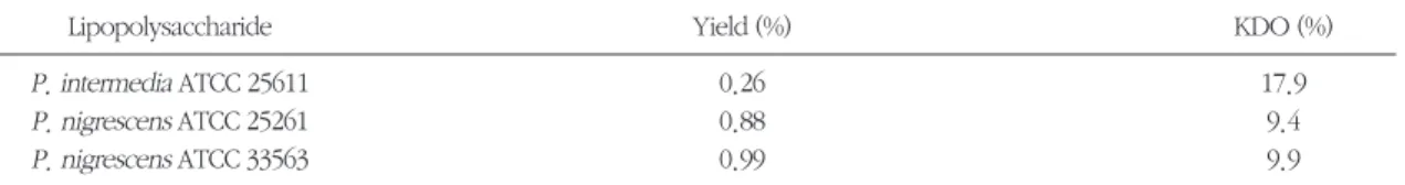
1. LPS의 KDO 및 단백질 함량LPS의 수율은 동결건조한 세포를 기준으로 하여 0.26%에서 0.99%에 이르렀고, P. nigrescens가 P. intermedia에 비해 높은 수율을 보였다 (Table 1). 순 수분리한 LPS의 KDO 함량은 9.4%에서 17.9%에 달 하였으며, P. intermedia LPS가P. nigrescens LPS에 비해 KDO 함량이 더 많았다 (Table 1). LPS의 단백 질 함량은 3종의 균주 모두에서 0.1% 미만이었다 (자료제시 않함).
2. B cell mitogenic activity
BALB/C 마우스의 비장세포에 대한P. intermedia 및P. nigrescensLPS의 증식효과가 Figure 1에 제시 되어 있다. P. intermedia와P. nigrescens의 LPS는 대 조군에 비해 현저한 세포증식을 유발하였으며, 저농 도에서는P. nigrescensATCC 33563 LPS에 의한 세 포증식이 가장 현저하였다.
Table 1. Yield and KDO content of LPSs from Prevotella intermedia and Prevotella nigrescens
Lipopolysaccharide Yield (%) KDO (%)
P. intermedia ATCC 25611 0.26 17.9
P. nigrescens ATCC 25261 0.88 9.4
P. nigrescens ATCC 33563 0.99 9.9
Figure 1. Mitogenic activity of LPS on spleen cells of BALB/C mouse. 5×10⁵prepared cells in 100μl per well were cultured in microculture plates with various concentration of each LPS. S. typhimurium LPS were used as the positive controls. The results are expressed as the mean ± standard deviation of four culture wells.
LPS (μg/ml) 0.1
% of Control
P. intermedia ATCC 25611 P. nigrescens ATCC 33563 P. nigrescens ATCC 25261 S. typhimurium
500 450 400 350 300 250 200 150 100 50
0 1 10
3. NO 생성
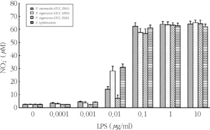
다양한 농도의 LPS를 RAW264.7 세포에 가하고 나 서 24시간 후 배양 상층액 내의 nitrite 농도를 측정하 였다. 3종의 P. intermedia 및 P. nigrescens LPS는 0.1 ng/ml-10 μg/ml에 걸쳐 RAW264.7 세포로부터 NO의 분비를 유발하였다 (Figure 2). LPS에 의한 자 극이 없이도 약 3 μM의 nitrite가 RAW264.7 세포로 부터 분비되었다. 10 ng/ml의 농도에서부터 유의한 농도의 NO가 분비되었으며, 10 μg/ml에서 최대의 NO 생성 (약 65 μM)을 보였다. 양성 대조군으로 사
용된S. typhimuriumLPS의 활성은 최소 자극용량과 최대 NO 생산량의 측면에서 P. intermedia 및 P. nigrescens LPS와 유사하였다. 다만, 10 ng/ml의 농 도에서, S. typhimuriumLPS와P. nigrescens ATCC 33563 LPS의 활성이 현저히 높았다.
RAW264.7 세포에 1 μg/ml의 각종 LPS를 가하고, 시간의 경과에 따른 NO 생성을 평가하였다 (Figure 3). P. intermedia및P. nigrescens LPS는, 4시간의 정 체기 후, 8시간에서 48시간까지 NO 생성을 지속적 으로 증가시켰다. S. typhimuriumLPS도 24시간까지 지속적으로 NO 생성을 증가시켰다.
Figure 2. Dose response of NO production by RAW264.7 cells stimulated with LPS isolated from P. interme- dia or P. nigrescens. S. typhimurium LPS was used as a control. Cells were incubated with increas- ing concentrations of LPS and supernatants were removed after 24 h and assayed for NO. The results are means ± standard deviation of four experiments.
Figure 3. Time course of NO production by RAW264.7 cells stimulated with LPS isolated from P. intermedia or P. nigrescens. Cells were incubated with 1 μg/ml of LPS. Other details as in Figure 2.
0 NO2-(μM)
P. intermedia ATCC 25611 P. nigrescens ATCC 33563 P. nigrescens ATCC 25261 S. typhimurium
80 70 60 50 40 30 20 10
0 0.0001 0.001 0.01
LPS (μg/ml)
0.1 1 10
0 NO2-(μM)
80 70 60 50 40 30 20 10
0 4 8
Time (hr)
12 24 48
Control P. intermedia ATCC 25611 P. nigrescens ATCC 33563 P. nigrescens ATCC 25261 S. typhimurium
4. iNOS 단백질과 mRNA의 발현
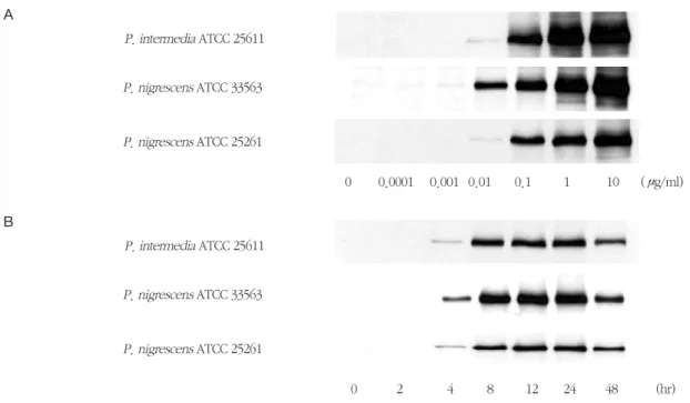
RAW264.7 세포에서의 P. intermedia 및 P. nigrescens LPS에 의한 NO 생성이 iNOS 단백질의 발 현에 의한 것인지를 확인하기 위하여, P. intermedia 및P. nigrescens LPS가 iNOS 단백질의 발현에 미치 는 영향을 평가하였다. 이들 LPS로 자극된 세포에서 는 iNOS에 대한 특이 항체와 반응하는 약 130 kDa의 단백질이 발현되었다 (Figure 4). RAW264.7 세포를 다양한 농도의 LPS에 노출시켰을 때, iNOS의 발현이 농도 의존적으로 증가하였다 (Figure 4A). iNOS 단 백질은 1 ng/ml의 LPS에서부터 발현되기 시작하여, 10 μg/ml에서 최대의 발현을 보였다. Figure 4B에는 1 μg/ml의P. intermedia및P. nigrescens LPS를 가 한 후 시간의 경과에 따른 iNOS 단백질 발현 양상을 보여주고 있다. iNOS 단백질은 4시간에서부터 발현
되기 시작하여 24시간에 최대의 발현을 보였다.
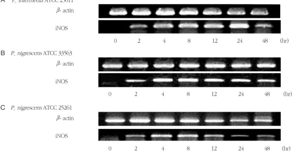
P. intermedia및P. nigrescens LPS가 iNOS 전사와 iNOS mRNA의 발현에 미치는 영향을 RT-PCR에 의 해 확인하였다. RAW264.7 세포를 1 μg/ml의 LPS에 노출시켰을 때 iNOS mRNA가 발현되었다 (Figure 5). P. intermedia ATCC 25611과P. nigrescens ATCC 33563 LPS는 2시간에서 24시간에 걸쳐 지속적으로 iNOS mRNA의 발현을 증가시켰다 (Figure 5A와 B).
P. nigrescens ATCC 25261 LPS는 8시간에 최대의 iNOS mRNA 발현을 보였다 (Figure 5C). LPS를 가하 지 않은 RAW264.7 세포는 iNOS mRNA를 발현하지 않았다.
5. Polymyxin B가 NO 생성에 미치는 영향 Polymyxin B는 LPS의 lipid A domain에 결합하여
Figure 4. Dose response (A) and time course (B) of iNOS protein expression in RAW264.7 cells stimulated with LPS isolated from P. intermedia or P. nigrescens. iNOS protein synthesis was measured by immunoblot analysis of cell lysates using iNOS-specific antibody. A representative immunoblot from two separate experiments with similar results is shown. (A) Cells were incubated with different con- centrations of LPS from P. intermedia or P. nigrescens for 24 h. (B) Cells were incubated in the pres- ence of 1 μg/ml of LPS fromP. intermedia or P. nigrescens for different times.
P. intermedia ATCC 25611 A
P. nigrescens ATCC 33563
P. nigrescens ATCC 25261
0 0.0001 0.001 0.01 0.1 1 10 (μg/ml)
P. intermedia ATCC 25611 B
P. nigrescens ATCC 33563
P. nigrescens ATCC 25261
0 2 4 8 12 24 48 (hr)
LPS의 생물학적 활성을 중화할 수 있다. Polymyxin B가P. intermedia및P. nigrescens LPS에 의한 NO 형성에 미치는 영향을 평가하였다. Polymyxin B는
P. nigrescens LPS에 의한 RAW264.7 세포에서의 NO 형성을 완전히 억제하였다 (Figure 6). 그러나, P. intermedia LPS에 의한 NO 생성은 polymyxin B에 Figure 5. Time course of iNOS mRNA expression in RAW264.7 cells stimulated with LPS isolated from P. inter- media or P. nigrescens. Cells were incubated in the presence of 1 μg/ml of LPS from P. intermedia or P. nigrescens for different time periods. See Materials and methods for further details. The PCR bands on a gel photograph in one of two separate experiments yielding similar results are shown.
Figure 6. Influences of polymyxin B (PB) on the NO-inducing activity of LPS isolated from P. intermedia or P.
nigrescens. 1 μg/ml of LPS from P. intermedia or P. nigrescens was incubated with the indicated concentrations of PB at 37°C for 60 min and then subjected to cultures with RAW264.7 cells.
Supernatants were removed after 24 h and assayed for NO. S. typhimurium LPS was used as a con- trol. The results are means ± standard deviation of four experiments.
β- actin P. intermedia ATCC 25611
P. nigrescens ATCC 33563
P. nigrescens ATCC 25261 iNOS A
0 2 4 8 12 24 48 (hr)
β- actin iNOS B
0 2 4 8 12 24 48 (hr)
β- actin iNOS C
0 2 4 8 12 24 48 (hr)
P. intermedia ATCC 25611 P. nigrescens ATCC 33563 P. nigrescens ATCC 25261 S. typhimurium
0 NO2-(μM)
80 70 60 50 40 30 20 10
0 2.5 5 10 20 40
PB (μg/ml)
의해 단지 부분적으로 억제되었다 6. TNF-α생성
다양한 농도의 LPS를 RAW264.7 세포에 가하고 24 시간 후에 TNF-α농도를 측정하였다. P. intermedia 및P. nigrescens LPS는 0.1 ng/ml-10 μg/ml에 걸쳐 RAW264.7 세포로부터 TNF-α의 분비를 유발하였다 (Figure 7). LPS에 의한 자극이 없이도 약 0.7 ng/ml 의 TNF-α가 RAW264.7 세포로부터 분비되었다. P.
intermedia및P. nigrescens LPS는 10 ng/ml의 농도 에서부터 유의한 농도의 TNF-α의 분비를 유발하였 으며, 10 μg/ml에서 최대의 TNF-α생성 (약 6 ng/ml) 을 보였다. 양성 대조군으로 사용된S. typhimurium LPS의 활성은 최소 자극용량과 최대 TNF-α생산량의 측면에서P. intermedia및P. nigrescens LPS와 유사 하였다. 다만, 10 ng/ml의 농도에서, S. typhimurium LPS의 활성이 현저히 높았고, P. nigrescens ATCC 25261 LPS의 활성이 현저히 낮았다.
RAW264.7 세포에 1 μg/ml의 각종 LPS를 가하고, Figure 7. Dose response of TNF-αproduction by RAW264.7 cells stimulated with LPS isolated from P. inter- media or P. nigrescens. S. typhimurium LPS was used as a control. Cells were incubated with increasing concentrations of LPS and supernatants were removed after 24 h and assayed for TNF-α.
The results are means ± standard deviation of four experiments.
Figure 8. Time course of TNF-αproduction by RAW264.7 cells stimulated with LPS isolated from P. intermedia or P. nigrescens. Cells were incubated with 1 μg/ml of LPS. Other details as in Figure 7.
P. intermedia ATCC 25611 P. nigrescens ATCC 33563 P. nigrescens ATCC 25261 S. typhimurium
0 7 6 5 4 3 2 1
0 0.0001 0.001 0.01 0.1 1 10
LPS (μg/ml)
TNF-α(ng/ml)
Time (hr) 0
TNF-α(ng/ml)
6 5 4 3 2 1
0 4 8 12 24 48
Control P. intermedia ATCC 25611 P. nigrescens ATCC 33563 P. nigrescens ATCC 25261 S. typhimurium
시간의 경과에 따른 TNF-α생성을 평가하였다 (Figure 8). P. intermedia및P. nigrescens LPS는 4시 간 경과 후 현저한 TNF-α생성을 유발하였고, 그 후 48시간까지는 TNF-α생성이 현저히 증대되지는 않 았다. S. typhimuriumLPS에 의한 TNF-α생성도 유 사한 양상을 보였다.
7. TNF-αmRNA의 발현
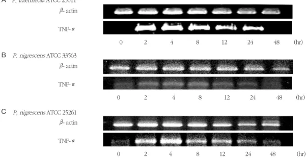
P. intermedia 및 P. nigrescens LPS가 TNF-α mRNA의 발현에 미치는 영향을 RT-PCR에 의해 확인 하였다. RAW264.7 세포를 1 μg/ml의 LPS에 노출시 켰을 때 TNF-αmRNA가 발현되었다 (Figure 9). P. intermedia ATCC 25611과P. nigrescens ATCC 33563 LPS는 2시간에서 8시간에 걸쳐 지속적으로 TNF-α mRNA의 발현을 증가시켰다 (Figure 9A와 B). P. nigrescens ATCC 25261 LPS는 4시간에 최대 의 TNF-α mRNA 발현을 보였다 (Figure 9C). LPS를 가하지 않은 RAW264.7 세포는 TNF-α mRNA를 발
현하지 않았다.
IV. 총괄 및 고찰
본 연구는P. intermedia와P. nigrescensLPS의 각 종 생물학적 활성을 분석하여, 치주질환과 치수질환 등 구강 내 감염에 있어서의 이들 균주의 역할을 규 명하기 위해 수행되었다. KDO는P. intermedia와P. nigrescensLPS에서 9.4%에서 17.9%의 함량으로 존 재하였으며, 이는 이들 균주가 온전한 LPS를 소유하 고 있음을 제시하고 있다. P. intermedia는 P. nigrescens에 비해 매우 강력한 KDO 시그날을 가지 고 있는 것으로 보여진다. LPS에 함유되어 있는 단백 질에 의해 LPS의 각종 생물학적 활성이 증진될 가능 성을 배제하기 위하여, 본 연구에서는 LPS를 pro- teinase K로 처리하였으며, 순수분리한 LPS의 단백질 함량은 0.1% 미만이었다. 이는 본 연구에서의 LPS의 각종 생물학적 활성이 전적으로 LPS에 의한 것임을 의미한다.
Figure 9. Time course of TNF-α mRNA expression in RAW264.7 cells stimulated with LPS isolated from P.
intermedia or P. nigrescens. Cells were incubated in the presence of 1 μg/ml of LPS from P. interme- dia or P. nigrescens for different time periods. See Materials and methods for further details. The PCR bands on a gel photograph in one of two separate experiments yielding similar results are shown.
β- actin TNF-α A
0 2 4 8 12 24 48 (hr)
β- actin TNF-α B
0 2 4 8 12 24 48 (hr)
β- actin TNF-α C
0 2 4 8 12 24 48 (hr)
P. intermedia ATCC 25611
P. nigrescens ATCC 33563
P. nigrescens ATCC 25261
대부분이 B-세포로 이루어져 있는 비장세포에 대 해P. intermedia 및P. nigrescens LPS가 현저한 증식 효과를 가지고 있음을 본 연구는 제시하고 있다. B- 세포 및 형질세포가 활성기 치주질환 병소에 우세하 게 존재하는 것으로 알려져 있는 바33), 이들 균주의 LPS가 치주질환의 병리에 있어 주요한 역할을 할 가 능성이 있다.
NO는 새로운 유형의 염증 매개물질로 주목을 받 고 있다34). NOS 활성과 NO 생성을 억제함으로써 골 관절염, 사구체 신염, 그리고 대장염 등 염증성 질환 의 진행과 심도를 억제할 수 있음이 밝혀진 바도 있
다35,36). NO는 다른 염증성 질환에서와 마찬가지로
염증성 치주질환에 있어서도 중요한 역할을 하는 것 으로 여겨진다. 치주질환 시 NO의 생성이 증가하며
37), Actinobacillus actinomycetemcomitans의 LPS가 대식세포에서 현저하게 NO 형성을 유발함이 보고된
바 있다38,39). 또한, 만성치주염 환자의 치은조직 내에
는, 건강한 치주조직에 비해, iNOS 단백질과 mRNA 가 고농도로 존재한다40-43). 치주조직 내에서의 iNOS 의 주 공급원으로는 대식세포, 다형핵 백혈구, 섬유 아세포, 그리고 혈관 내피세포를 들 수 있다40-43).
이에 본 연구에서는 치주질환과 근관감염 등에 있 어 주요 병인균주 중의 하나인 P. intermedia와 P. nigrescens의 LPS가 마우스의 macrophage-like cell line인 RAW264.7에서의 NO 형성과 iNOS 발현에 미 치는 영향을 평가하였다. P. intermedia 및 P. nigrescens LPS가 대식세포에서 NO 생성을 유발할 수 있 는 능 력 은 양 성 대 조 군 으 로 활 용 한 S. typhimurium LPS와 유사하였다. NO 형성에 있어 iNOS가 촉매효소로 작용하므로, 본 연구에서는 P. intermedia 및 P. nigrescens LPS가 iNOS 단백질과 mRNA의 발현에 미치는 영향을 평가하였으며, iNOS 단백질과 mRNA의 발현이 immunoblotting과 RT- PCR에 의해 각각 증명되었다. P. intermedia 및 P. nigrescens LPS는 주로 전사 수준에서 iNOS 발현을 유발하는 것으로 여겨진다. P. intermedia 및 P. nigrescens LPS가, interferon-γ등에 의한 부가적인 자극이 없이도, 마우스 대식세포에서 iNOS의 발현을 유발하여 NO 형성을 초래할 수 있음이 본 연구에서
최초로 제시되었다. 한편, P. intermedia LPS는P. nigrescens LPS에 비해 polymyxin B에 대해 덜 민감 한 것으로 여겨진다.
NO는, 직접적인 영향에 의하거나 염증성 싸이토 카인의 생성을 조절하여, 치주질환의 발병과 진행 그 리고 그로 인한 골소실에 있어 중요할 수 있다44). P. intermedia 및P. nigrescens LPS가 NO 생성을 유도 하는 자세한 기전은 추후 밝혀져야 할 것이다.
TNF-α, IL-1β, IL-6, 그리고 IL-8 등의 염증성 싸이토 카인은 염증성 치주질환에 있어 중요한 역할을 하는 것으로 알려져 있다45,46). 염증에 이환된 치주조직 내 에서 이들 싸이토카인이 국소적으로 생성될 수 있 다. P. intermedia 및P. nigrescens LPS는 RAW264.7 세포에서 TNF-α의 생성을 유발하였다. 이들 LPS가 대식세포에서 TNF-α의 생성을 유발할 수 있는 능력 은 S. typhimurium LPS와 유사한 것으로 여겨진다.
또한, 이들 LPS는 주로 전사 수준에서 TNF-α의 발현 을 유발하는 것으로 여겨진다
전반적으로P. intermediaLPS와P. nigrescensLPS 는 제반 생물학적 활성에 있어 유사하였다. 종합하 여 보면, P. intermedia및P. nigrescens LPS는 B-세 포의 증식을 초래하며, 대식세포에서 iNOS의 발현에 의한 NO 생성을 유발하고, 숙주의 싸이토카인 네트 웍을 활성화하여 TNF-α등의 염증 매개물질의 생성 을 촉진함으로써, 치주질환과 치수질환 등 구강 내 감염에 중요한 역할을 하는 것으로 사료된다.
V. 결론
본 연구는P. intermedia와P. nigrescensLPS의 각 종 생물학적 활성을 분석하여, 치주질환과 치수질환 등 구강 내 감염에 있어서의 이들 균주의 역할을 규 명하기 위해 수행되었다. 순수분리한P. intermedia 및 P. nigrescens LPS의 KDO 함량은 9.4%에서 17.9%에 달하였으며, P. intermedia LPS에서 KDO 함량이 더 많았다. P. intermedia 및 P. nigrescens LPS는 비장세포에 대해 현저한 증식효과를 보였다.
P. intermedia 및P. nigrescens LPS는 마우스 대식세 포에서, interferon-γ등에 의한 부가적인 자극이 없
이도, iNOS의 발현에 의한 NO 생성과 TNF-α의 생 성을 각각 유발할 수 있음이 본 연구에서 최초로 제 시되었다. 전반적으로 P. intermedia LPS와 P. nigrescensLPS는 제반 생물학적 활성에 있어 유사하 였다. 종합하여 보면, P. intermedia및P. nigrescens LPS는 B-세포의 증식을 초래하며, 대식세포에서 iNOS의 발현에 의한 NO 생성을 유발하고, 숙주의 싸이토카인 네트웍을 활성화하여 TNF-α등의 염증 매개물질의 생성을 촉진함으로써, 치주질환과 치수 질환 등 구강 내 감염에 중요한 역할을 하는 것으로 사료된다.
VI. 참고문헌
1. Maidwell-Smith MA, Wilson M, Kieser JB.
Lipopolysaccharide (endotoxin) from periodon- tally involved teeth. J Clin Periodontol 1987;14:453-456.
2. Shapiro L, Lodato FM Jr, Courant PR, Stallard RE.
Endotoxin determination in gingival inflamma- tion. J Periodontol 1972;43:591-596.
3. Simon BI, Goldman HM, Ruben MP, Baker E.
The role of endotoxin in periodontal disease. I.
A reproducible, quantitative method for deter- mining the amount of endotoxin in human gin- gival exudate. J Periodontol 1969;40:695-701.
4. Gibbons RJ, Socransky SS, Sawyer S, Kapsimalis B, MacDonald JB. The microbiota of the gingival crevice area of man. II. The predominant cul- tivable organisms. Arch Oral Biol 1963;8:281- 289.
5. Koga T, Nishihara T, Fujiwara T et al.
Biochemical and immunobiological properties of lipopolysaccharide (LPS) from Bacteroides gingivalis and comparison with LPS from Escherichia coli. Infect Immun 1985;47:638-647.
6. Bramanti TE, Wong GG, Weintraub ST, Holt SC.
Chemical characterization and biological proper- ties of lipopolysaccharide from Bacteriodes gin-
givalisstrains W50, W83, and ATCC 33277. Oral Microbiol Immunol 1989;4:183-192.
7. Kim SJ, Kato T, Naito Y et al. B-cell mitogenicity and IL-1 beta production of lipopolysaccharides from various Capnocytophaga strains. Bull Tokyo Dent Coll 1994;35: 79-83
8. Shah HN, Gharbia SE. Biochemical and chemi- cal studies on strains designated Prevotella inter- media and proposal of a new pigmented species, Prevotella nigrescenssp. nov. Int J Syst Bacteriol 1992;42:542-546.
9. Okamoto M, Maeda N, Kondo K, Leung KP.
Hemolytic and hemagglutinating activities of Prevotella intermediaand Prevotella nigrescens. FEMS Microbiol Lett 1999;178:299-304.
10. Stubbs S, Lewis MA, Waddington RJ, Embery G.
Hydrolytic and depolymerising enzyme activity of Prevotella intermedia and Prevotella nigrescens. Oral Dis 1996;2:272-278.
11. Matto J, Asikainen S, Vaisanen ML et al. Beta-lac- tamase production in Prevotella intermedia, Prevotella nigrescens, and Prevotella pallens genotypes and in vitro susceptibilities to selected antimicrobial agents. Antimicrob Agents Chemother 1999;43: 2383-2388.
12. Andres MT, Chung WO, Roberts MC, Fierro JF.
Antimicrobial susceptibilities of Porphyromonas gingivalis, Prevotella intermedia, and Prevotella nigrescensspp. isolated in Spain. Antimicrob Agents Chemother 1998;42:3022-3023.
13. Bernal LA, Guillot E, Paquet C, Mouton C. Beta- Lactamase-producing strains in the species Prevotella intermediaand Prevotella nigrescens. Oral Microbiol Immunol 1998;13:36-40.
14. Baumgartner JC, Bae KS, Xia T, Whitt J, David LL. Sodium dodecyl sulfate-polyacrylamide gel electrophoresis and polymerase chain reaction for differentiation of Prevotella intermedia and Prevotella nigrescens. J Endod 1999;25:324-328.
15. Premaraj T, Kato N, Fukui K, Kato H, Watanabe K. Use of PCR and sodium dodecyl sulfate-poly- acrylamide gel electrophoresis techniques for differentiation of Prevotella intermedia sensu stricto and Prevotella nigrescens. J Clin Microbiol 1999;37:1057-1061.
16. Guillot E, Mouton C. PCR-DNA probe assays for identification and detection of Prevotella inter- mediasensu stricto and Prevotella nigrescens. J Clin Microbiol 1997;35:1876-1882.
17. Conrads G, Pelz K, Hughes B, Seyfarth I, Devine DA. Optimized oligonucleotides for the differen- tiation of Prevotella intermedia and Prevotella nigrescens. Oral Microbiol Immunol 1997;12:117-120.
18. Matto J, Saarela M, von Troil-Linden B et al.
Similarity of salivary and subgingival Prevotella intermediaand Prevotella nigrescensisolates by arbitrarily primed polymerase chain reaction.
Oral Microbiol Immunol 1996;11:395-401.
19. Socransky SS, Haffajee AD. The bacterial etiolo- gy of destructive periodontal disease: current concepts. J Periodontol 1992;63:322-331.
20. Slots J, Bragd L, Wikstrom M, Dahlen G. The occurrence of Actinobacillus actinomycetem- comitans, Bacteroides gingivalisand Bacteroides intermediusin destructive periodontal disease in adults. J Clin Periodontol 1986;13:570-577.
21. Tanner ACR, Haffer C, Bratthall GT, Visconti RA, Socransky SS. A study of the bacteria associated with advancing periodontitis in man. J Clin Periodontol 1979;6:278-307.
22. Chung CP, Nisengard RJ, Slots J, Genco RJ.
Bacterial IgG and IgM antibody titers in acute necrotizing ulcerative gingivitis. J Periodontol 1983;54:557-562.
23. Kornman KS, Loesche WJ. The subgingival microbial flora during pregnancy. J Periodont Res 1980;15:111-122.
24. Lie MA, van der Weijden GA, Timmerman MF et al. Occurrence of Prevotella intermedia and Prevotella nigrescens in relation to gingival health. J Clin Periodontol 2001;28:189-193.
25. Ximenez-Fyvie LA, Haffajee AD, Socransky SS.
Microbial composition of supra and subgingival plaque in subjects with adult periodontitis. J Clin Periodontol 2000;27:722-732.
26. Teanpaisan R, Douglas CW, Walsh TF.
Characterization of black-pigmented anaerobes isolated from diseased and healthy periodontal sites. J Periodont Res 1995;30:245-251.
27. Bae KS, Baumgartner JC, Shearer TR. David LL.
Occurrence of Prevotella nigrescens and Prevotella intermediain infections of endodontic origin. J Endod 1997; 23:620-623.
28. Salcetti JM, Moriarty JD, Cooper LF. et al. The clinical, microbial, and host response characteris- tics of the failing implant. Int J Oral Maxillofac Implants 1997;12:32-42.
29. Westphal O, Jann K. Bacterial lipopolysaccha- rides: extraction with phenol-water and further applications of the procedure., p. 83-91. In RL Whistler (ed), Methods in carbohydrate chem- istry. Academic Press, Inc, New York, NY. 1965.
30. Markwell MA, Haas SM, Bieber LL, Tolbert NE.
A modification of the Lowry procedure to sim- plify protein determination in membrane and lipoprotein samples. Anal Biochem 1978;87:206- 210.
31. Karkhanis YD, Zeltner J, Jackson JJ, Carlo DJ. A new and improved microassay to determine 2- keto-3-deoxyoctonate in lipopolysaccharide of gram-negative bacteria. Anal Biochem 1978;85:595-601.
32. Green LC, Wagner DA, Glogowski J et al.
Analysis of nitrate, nitrite, and [15N]nitrate in bio- logical fluids. Anal Biochem 1982;126,131-138.
33. Seymour GJ, Greenspan JS. The phenotypic
characterization of lymphocyte populations in established human periodontal disease. J Periodnt Res 1979;4:39-46.
34. Moncada S, Palmer RMJ, Higgs EA. Nitric oxide:
physiology, pathology, and pharmacology.
Pharmacol Rev 1991;43:109-142.
35. Southey A, Tanaka S, Murakami T et al.
Pathophysiological role of nitric oxide in rat experimental colitis. Int J Immunopharmacol 1997;19:669-676.
36. Weinberg JB, Granger DL, Pisetsky DS et al. The role of nitric oxide in the pathogenesis of spon- taneous murine autoimmune disease: increased nitric oxide production and nitric oxide synthase expression in MRL-lpr/lpr mice, and reduction of spontaneous glomerulonephritis and arthritis by orally administered NG-monomethyl-L-arginine.
J Exp Med 1994;179:651-660.
37. Matejka M, Partyka L, Ulm C, Solar P, Sinzinger H. Nitric oxide synthesis is increased in peri- odontal disease. J Periodont Res 33:517- 518,1998.
38. Blix IJ, Helgeland K. LPS from Actinobacillus actinomycetemcomitansand production of nitric oxide in murine macrophages J774. Eur J Oral Sci 1998;106:576-581.
39. Sosroseno W, Barid I, Herminajeng E, Susilowati H. Nitric oxide production by a murine macrophage cell line (RAW264.7) stimulated with lipopolysaccharide from Actinobacillus
actinomycetemcomitans. Oral Microbiol Immunol 2002;17:72-78.
40. Batista AC, Silva TA, Chun JH, Lara VS. Nitric oxide synthesis and severity of human periodon- tal disease. Oral Diseases 2002;8:254-260.
41. Hirose M, Ishihara K, Saito A net al. Expression of cytokines and inducible nitric oxide synthase in inflamed gingival tissue. J Periodontol 2001;72:590-597.
42. Kendall HK, Haase HR, Li H, Xiao Y, Bartold PM. Nitric oxide synthase type-II is synthesized by human gingival tissue and cultured human gingival fibroblasts. J Periodont Res 2000;35:194- 200.
43. Lappin DF, Kjeldsen M, Sander L, Kinane DF.
Inducible nitric oxide synthase expression in periodontitis. J Periodont Res 2000;35:369-373.
44. Brennan PA, Thomas GJ, Langdon JD. The role of nitric oxide in oral diseases. Arch Oral Biol 2003; 48: 93-100.
45. Honig J, Rordorf-Adam C, Siegmund C, Wiedemann W, Erard F. Increased interleukin-1 beta concentration in gingival tissue from peri- odontitis patients. J Periodont Res 1989;24:362- 367.
46. Stashenko P, Jandinski JJ, Fujiyioshi P, Rynar J, Socranski SS. Tissue levels of bone resorptive cytokines in periodontal disease. J Periodontol 1991;62:504-509.
-Abstract-
Chemical and Immunobiological Characterization of Lipopolysaccharides
from Prevotella intermedia and Prevotella nigrescens
Sung-Jo Kim
Department of Periodontology, College of Dentistry, Pusan National University
The purpose of this study was to assess some biological activities of lipopolysaccharides (LPSs) from P. inter- mediaand P. nigrescens. LPS was prepared by the standard hot phenol-water method. NO production was assayed by measuring the accumulation of nitrite in culture supernatants. TNF-α production was determined by enzyme-linked immunosorbent assay. Western blot analysis of iNOS and analysis of reverse transcription (RT)-PCR products were carried out. LPS from P. intermediademonstrated higher KDO content than those from two stains of P. nigrescens. LPSs from P. intermediaand P. nigrescenswere mitogenic for spleen cells of BALB/C mouse. The present study clearly shows that LPSs from P. intermediaand P. nigrescens fully induced iNOS expression and NO production in RAW264.7 cells in the absence of other stimuli. Moreover, LPSs from P. intermediaand P. nigrescens clearly induced TNF-αproduction in RAW264.7 cells. The biological activities of LPS from P. intermedia was found to be comparable to those of P. nigrescens LPS. The ability of LPSs from P. intermediaand P. nigrescens to promote the production of NO and TNF-αmay be important in the patho- genesis of inflammatory periodontal disease.
Key words: Prevotella intermedia, Prevotella nigrescens, Lipopoplysaccharide, B-cell mitogenicity, Nitric oxide, iNOS, TNF-α