대한소화기학회지 2005;46:447-454
ꠏꠏꠏꠏꠏꠏꠏꠏꠏꠏꠏꠏꠏꠏꠏꠏꠏꠏꠏꠏꠏꠏꠏꠏꠏꠏꠏꠏꠏꠏꠏꠏꠏꠏ 접수: 2005년 8월 8일, 승인: 2005년 11월 23일
연락처: 김현수, 220-701, 강원도 원주시 일산동 162번지 연세대학교 원주의과대학 내과학교실 소화기내과 Tel: (033) 741-1229, Fax: (033) 741-1228
E-mail: hskim@wonju.yonsei.ac.kr
Mallory-Weiss 증후군 환자에서 재출혈의 예측인자
연세대학교 원주의과대학 내과학교실, 의과대학 내과학교실*
김재우․김현수․변종원․원찬식․지명관․박용순․백순구․권상옥․이동기*
Predictive Factors of Recurrent Bleeding in Mallory-Weiss Syndrome
Jae Woo Kim, M.D., Hyun Soo Kim, M.D., Jong Won Byun, M.D., Chan Sik Won, M.D., Myeong Gwan Jee, M.D., Yong Soon Park, M.D., Soon Koo Baik, M.D.,
Sang Ok Kwon, M.D., and Dong Ki Lee, M.D.*
Department of Internal Medicine, Yonsei University Wonju College of Medicine, Wonju;
Department of Internal Medicine, Yonsei University College of Medicine*, Seoul, Korea
Background/Aims: Although the majority of patients with Mallory-Weiss syndrome (MWS) have a benign course, MWS patients with recurrent bleeding have an unfavorable outcome and require intensive care. Therefore, this study was carried out to identify the risk factors for recurrent bleeding in MWS patients. Methods: The medical records of patients with MWS between January 1999 and December 2003, were reviewed retrospectively. Demo- graphics, initial clinical and laboratory parameters, and endoscopic findings of the patients with and without recur- rent bleeding were compared and the potential risk factors predicting recurrent bleeding in MWS were evaluated.
Results: A total of one hundred and fifty-nine patients (22 women, 137 men, mean age 48.1 years old) were enrolled in the study. Recurrent bleeding was observed in 17 patients (10.7%). Those patients with recurrent bleeding showed higher frequency for the presence of shock at initial manifestation, combined liver cirrhosis and endoscopic findings of active bleeding, lower hemoglobin level and platelet count, higher amount of transfusions and epinephrine-mixed fluid injections, and longer hospital stay than those patients without recurrent bleeding.
Significant risk factors predicting the recurrent bleeding in MWS were the presence of shock at initial manifestation (OR 3.71, 95% CI 1.07-14.90) and the evidence of active bleeding on endoscopic examination (OR 9.89, 95% CI 1.88-51.98) on multivariate analysis. Conclusions: Intensive care with close monitoring is required for the patients with shock on initial manifestation or with evidence of active bleeding on endoscopic examinations since these are independent risk factors predicting the recurrent bleeding in MWS patients. (Korean J Gastroenterol 2005;46:447-454)
ꠏꠏꠏꠏꠏꠏꠏꠏꠏꠏꠏꠏꠏꠏꠏꠏꠏꠏꠏꠏꠏꠏꠏꠏꠏꠏꠏꠏꠏꠏꠏꠏꠏꠏꠏꠏꠏꠏꠏꠏꠏꠏꠏꠏꠏꠏꠏꠏꠏꠏꠏꠏꠏꠏꠏꠏꠏꠏꠏꠏꠏꠏꠏꠏꠏꠏꠏꠏꠏꠏꠏꠏꠏꠏꠏꠏꠏꠏꠏꠏꠏꠏꠏꠏꠏꠏꠏꠏꠏꠏꠏꠏꠏꠏꠏꠏꠏꠏꠏꠏꠏꠏꠏꠏꠏꠏꠏꠏꠏꠏꠏꠏꠏ
Key Words: Mallory-Weiss syndrome; Primary hemostasis; Recurrent bleeding
ꠏꠏꠏꠏꠏꠏꠏꠏꠏꠏꠏꠏꠏꠏꠏꠏꠏꠏꠏꠏꠏꠏꠏꠏꠏꠏꠏꠏꠏꠏꠏꠏꠏꠏ Correspondence to: Hyun Soo Kim, M.D.
Division of Gastroenterology, Department of Internal Medicine Yonsei University Wonju College of Medicine, 162 Ilsan-dong Wonju-si, Gangwon-do 220-701, Korea
Tel: +82-33-741-1229, Fax: +82-33-741-1228 E-mail: hskim@wonju.yonsei.ac.kr
448 대한소화기학회지: 제46권 제6호, 2005
INTRODUCTION
Mallory-Weiss syndrome (MWS), which manifests as lacerations in the region of the gastroesophageal junction that typically occur in gastric mucosa, is a common cause of upper gastrointestinal (GI) hemorrhage. MWS has been reported to be the cause of upper GI bleeding in approximately 3% to 15% of all cases. However, most patients require no intervention other than hemodynamic support.1,2 On the other hand, patients with severe or recurrent bleeding related to MWS must undergo intensive care even the majority of patients with MWS features a benign course and a lower rate of recurrent bleeding when compared to other causes of upper GI bleeding.3-5
Several studies of endoscopic hemostasis have reported that recurrent bleeding rates for upper GI bleeding in MWS patients is 8-15%.6,7 Moreover, MWS patients with recurrent bleeding have an unfavorable outcome and a worse prognosis than patients without recurrent bleeding. In patients with non-variceal bleeding other than MWS, there are well-recognized clinical and laboratory risk factors associated with patients at risk for
complications, recurrent bleeding, and mortality.8-14 However, there are few reports on the factors predicting recurrent bleeding in MW tear.6,15 Therefore, the aims of this study were to compare the clinical and laboratory parameters, and endos- copic findings of patients with or without recurrent bleeding, and to identify the risk factors associated with recurrent bleeding using multivariate logistic regression analysis.
PATIENTS AND METHODS
1. Patients
Between January 1999 and December 2003, a total of 159 patients presenting with a MW tear were enrolled and retro- spectively analyzed. During the same period, 2,379 patients had been diagnosed as UGI bleeding, and therefore 6.7% were caused by MW tear. MW tears were defined as a mucosa laceration of the esophagus or stomach near the eso- phagogastric junction on endoscopic inspection. The demo- graphics of the patients, the presence of chronic disease such as
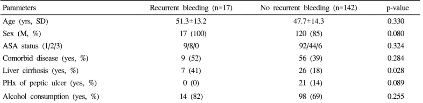
Table 1. Patients Characteristics according to the Presence of Recurrent Bleeding
Parameters Recurrent bleeding (n=17) No recurrent bleeding (n=142) p-value
Age (yrs, SD) 51.3±13.2 47.7±14.3 0.330
Sex (M, %) 17 (100) 120 (85) 0.080
ASA status (1/2/3) 9/8/0 92/44/6 0.324
Comorbid disease (yes, %) 9 (52) 56 (39) 0.284
Liver cirrhosis (yes, %) 7 (41) 26 (18) 0.028
PHx of peptic ulcer (yes, %) 0 (0) 21 (14) 0.089
Alcohol consumption (yes, %) 14 (82) 98 (69) 0.255
ASA, American society of anesthesiologists; SD, standard deviation; PHx, past history.
Table 2. Initial Clinical and Laboratory Findings according to the Presence of Recurrent Bleeding
Parameters Recurrent bleeding (n=17) No recurrent bleeding (n=142) p-value
Hematemesis or melena (yes, %) 17 (100) 129 (90) 0.193
Shock status (yes, %) 10 (58) 26 (18) <0.001
0.006
Systolic BP (mmHg, SD) 94.0±18.3 108.0±19.6
<0.001
Diastolic BP (mmHg, SD) 55.4±15.4 68.5±13.6
0.026
Pulse rate (SD) 118.5±28.8 105.2±22.1
0.043
Hemoglobin (g/dL, SD) 9.8±3.2 11.2±2.6
0.030
Platelet count (103/mm3, SD) 159.2±70.2 215.2±102.2
0.392
PT (INR, SD) 1.3±0.6 1.1±0.8
0.343
PTT (sec, SD) 31.3±12.1 29.0±8.9
BP, blood pressure; SD, standard deviation; PT, prothrombin time; INR, international normalized ratio; PTT, partial thromboplastin time.
김재우 외 8인. Mallory-Weiss 증후군 환자에서 재출혈의 예측인자 449
liver cirrhosis, diabetes mellitus, and cardiovascular disease, and a prior history of alcohol consumption or peptic ulcer was recorded. The clinical parameters included general physical status, presenting symptoms of melena or hematemesis as well as the pulse rate and blood pressure at initial manifestation.
General physical status was assessed according to the ASA five-categorial classification system.16 The following laboratory data from hospital database were also examined: hematocrit, platelet counts, and clotting parameters (prothrombin time, partial thromboplastin time, international normalized ratio [INR]). Shock was defined as a systolic pressure of <100 mmHg and a pulse rate >100 beat per minute accompanied by pallor or cold sweating. These variables were evaluated as being potential risk factors for recurrent bleeding. Comparison of the potential risk factors were made between the patients with and without recurrent bleeding.
2. Methods
Endoscopy was performed by one of 3 endoscopists using a standard upper endoscope (GIF-XQ 230, 240, or 260, Olympus Optical Co., Ltd., Tokyo, Japan). According to the endoscopic findings, the number and location of MW tears, the length of the longest tear, the presence of a hiatal hernia, and the type of stigmata were recorded. The type of stigmata was classified as active or non-active bleeding based on initial endoscopic finding. Active bleeding during endoscopy was defined as arterial spurting or oozing, whereas non-active bleeding was defined as non-bleeding focal endoscopic stigmata (visible vessel, fresh adherent clot, and black clot) or clean-based tears.
In addition, primary hemostasis was defined as an endos- copically verified cessation of bleeding after the first endos- copic session of the hemostatic procedure. Recurrent bleeding was defined as a recurrence of hematemesis, a bloody nasogas- tric aspirate, or a decrease in the hemoglobin concentration of
>1.5 g/dL within 24 hours after successful primary hemostasis.
Follow-up endoscopy was performed within 12 hours if recurrent bleeding was suspected. Permanent hemostasis was defined as an absence of recurrent bleeding during a 30-day period after the primary hemostasis.
3. Statistical analysis
Study database management and all statistical analyses were performed using the SPSS software, version 12.0 for Windows (SPSS Inc, Chicago, IL). The student t-test was used to compare the mean values of the continuous variables, and
chi-square test was used to analyze the categorical data. All results are expressed as a mean±SD unless otherwise stated. In addition, multiple logistic regression analysis for the risk factors of recurrent bleeding was carried out to determine the relative risks as odds ratio (OR) with 95% confidence intervals (CIs).
For multivariate analysis of the continuous variables, the patients were categorized after normalizing them to the mean values. p value <0.05 was considered significant. The study was carried out in accordance with the Helsinki Declaration as revised in 1989.
RESULTS
1. Characteristics of the patients
Of 159 patients with MWS, 17 patients (10.7%) presented with recurrent bleeding. These patients were compared with 142 patients who did not reveal recurrent bleeding. The demogra- phics of the patient and initial general physical status were not associated with the risk of recurrent bleeding. In addition, there was no significant differences in the risk of recurrent bleeding for those patients with comorbid diseases including diabetes or cardiovascular disease. A prior history of peptic ulcer or alcohol abuse had no influence on the risk of recurrent bleeding. However, the proportion of patients with comorbid liver cirrhosis was higher (41% vs. 18%, p=0.028) in those patients with recurrent bleeding (Table 1).
2. Clinical and laboratory findings
No difference was found between the patients with or without recurrent bleeding with respect to the presenting symptoms of hematemesis or melena (Table 2). However, the initial manifestation of shock was more frequently observed (58% vs. 18%, p<0.001) in those patients with recurrent bleeding. In addition, the systolic and diastolic blood pressures at initial presentation were lower in the patients with recurrent bleeding (p=0.006 and p<0.001, respectively), and the initial pulse rate was higher (p=0.026). The laboratory findings showed that the hematocrit level and platelet counts were significantly lower (p=0.043 and p=0.03, respectively) in those patients with recurrent bleeding. However, the clotting parameters in both groups were similar.
3. Endoscopic findings
We compared the initial endoscopic finding of the patients
450 The Korean Journal of Gastroenterology: Vol. 46, No. 6, 2005
with or without recurrent bleeding. The status of stigmata, and active bleeding such as spurting or active oozing from the MW tear were associated with recurrent bleeding. Active bleeding was endoscopically documented in 88% (15/17) of patients with recurrent bleeding as opposed to 38% (55/142) of patients without (p<0.001). On the other hand, the number and location of the tear, the length of the tear, the presence of hiatal hernia, and waiting time for endoscopy were similar in both groups (Table 3).
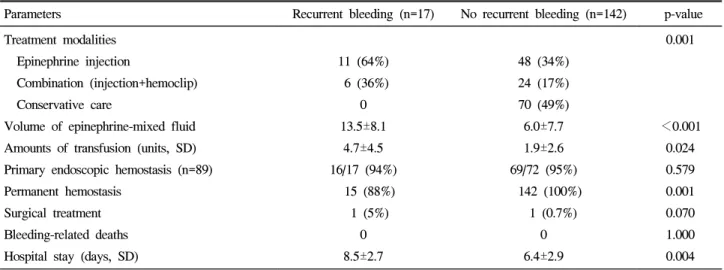
4. Clinical outcomes
We analyzed the clinical outcomes according to the presence of recurrent bleeding. Endoscopic hemostasis due to active bleeding and visible vessel on initial endoscopy was performed in 89 of the 159 patients. Of these, primary hemostasis was successfully achieved in 85/89 (95.5%) patients and was comparable in patients with or without recurrent bleeding (94%
vs. 95%; p=0.579). The treatment modalities were significantly
different between two groups (p=0.001). Endoscopic therapeutic intervention was required in all patients with recurrent bleeding, whereas half of the patients without recurrent bleeding did not require endoscopic hemostasis. In particular, 36% of patients with recurrent bleeding required more intensive endoscopic treatments requiring epinephrine injections and hemoclip to achieve the primary hemostasis. However, the rate of primary hemostasis was not significantly different between two groups treated by epinephrine injection alone and treated by both injection and hemoclip (p=0.878). Among patients treated with endoscopic injection therapy, alone larger volume of epinephrine injection was needed to control the bleeding (13.5 mL vs. 6.0 mL; p<0.001) in the patients with recurrent bleeding (13.5 mL vs. 6.0 mL; p<0.001). In addition, larger amounts of transfusion was required (4.7 vs. 1.9; p= 0.024) in the patients with recurrent bleeding. Among 17 patients with recurrent bleeding, 16 (94%) patients achieved successful primary hemostasis while one patient underwent surgery as a Table 3. Endoscopic Findings according to the Presence of Recurrent Bleeding
Parameters Recurrent bleeding (n=17) No recurrent bleeding (n=142) p-value
Number of tearing ≤1
≥2
13 (76%) 4 (24%)
117 (82%) 25 (18%)
0.550
Location of tearing Distal esophagus Gastroesophageal junction Cardia
6 (35%) 11 (65%)
0
71 (50%) 58 (40%) 13 (10%)
0.121
Length of tearing (cm) ≤1.5
>1.5
8 (47%) 9 (53%)
91 (64%) 51 (36%)
0.171
Hiatal hernia No Yes
17 (100%) 0
134 (94%) 8 (6%)
0.315
Waiting time for EGD (hours, SD) 5.1±3.8 14.2±25.5 0.147
EGD finding Active bleeding Spurting Oozing
Non-active bleeding Visible vessel Adherent clot Black base Clean base
3 (18%) 12 (70%)
2 (12%) 0 0 0
7 (5%) 48 (34%)
23 (16%) 20 (14%) 13 (9%) 31 (22%)
<0.001
EGD, esophagogastroduodenoscopy; SD, standard deviation.
Kim JW, et al. Predictive Factors of Recurrent Bleeding in Mallory-Weiss Syndrome 451
result of unsuccessful endoscopic hemostasis. Despite permanent hemostasis, one patient in the recurrent bleeding group died from hepatorenal syndrome. In patients without recurrent bleeding, primary hemostasis was achieved in 69 out of 72 (95%) patients which is similar to the recurrent bleeding group (p=0.579). Of these, one patient underwent surgery because of failed primary hemostasis and discharged with uneventful hospital course. Three patients died without further bleeding as a result of complications associated with liver cirrhosis;
alcoholic ketoacidosis or hepatic failure. Therefore, permanent hemostasis was successfully achieved in 15 (88%) patients with recurrent bleeding. In contrast, permanent hemostasis was achieved in all patients without recurrent bleeding (p=0.001).
The duration of the hospital stay was shorter (6.4 days vs. 8.5 days; p=0.004) in patients without recurrent bleeding (Table 4).
5. Multivariate analysis of risk factors for recurrent
bleeding
The results of multivariate analysis which includes the potential risk variables predicting recurrent bleeding, are sum- marized (Table 5). Multivariate analysis identified the initial manifestation of shock (odds ratio [OR] 3.71; 95% confidence interval [CI] 1.07-14.90) and the initial endoscopic finding of active bleeding (OR 9.89; 95% CI 1.88-51.98) as factors associated with increased risk of recurrent bleeding. Comorbid liver cirrhosis was associated with marginal but non-significant increase in risk. Other potential risk variables including age (≥
50 years), alcohol consumption, hemoglobin level or platelet count, and the number or the location of MW tear were not significant in predicting the recurrent bleeding of MWS.
Table 4. Clinical Outcomes according to the Presence of Recurrent Bleeding
Parameters Recurrent bleeding (n=17) No recurrent bleeding (n=142) p-value
Treatment modalities Epinephrine injection
Combination (injection+hemoclip) Conservative care
Volume of epinephrine-mixed fluid Amounts of transfusion (units, SD) Primary endoscopic hemostasis (n=89) Permanent hemostasis
Surgical treatment Bleeding-related deaths Hospital stay (days, SD)
11 (64%) 6 (36%)
0 13.5±8.1
4.7±4.5 16/17 (94%)
15 (88%) 1 (5%)
0 8.5±2.7
48 (34%) 24 (17%) 70 (49%)
6.0±7.7 1.9±2.6 69/72 (95%) 142 (100%)
1 (0.7%) 0 6.4±2.9
0.001
<0.001 0.024 0.579 0.001 0.070 1.000 0.004 SD, standard deviation.
Table 5. Multivariate Analysis of Risk Factors for Recurrent Bleeding in Mallory-Weiss Syndrome
Parameters Odds ratio 95% CI p-value
Age (<50 vs. ≥50 years) 1.85 0.57-6.04 0.310
Liver cirrhosis (yes vs. no) 2.74 0.75-10.04 0.129
Alcohol drinking (yes vs. no) 1.43 0.32-6.43 0.639
Shock (yes vs. no) 3.71 1.07-14.90 0.039
Hemoglobin (<11 g/dL vs. ≥11 g/dL ) 1.66 0.47-5.81 0.430
Platelet counts (<105/mm3 vs. ≥105/mm3) 0.47 0.11-2.04 0.312
EGD finding (active vs. non-active bleeding) 9.89 1.88-51.98 0.007
Number of tearing (>1 vs. ≤1) 1.22 0.30-4.90 0.785
Length of tearing (>1.5 cm vs. ≤1.5 cm) 1.03 0.30-3.56 0.964
CI, confidence interval; EGD, esophagogastroduodenoscopy.
452 대한소화기학회지: 제46권 제6호, 2005
DISCUSSION
MW tears are common source of non-variceal GI bleeding and generally have a benign course in more than 90% of cases.2,17 Although the recurrent bleeding rate in MWS is lower than that of other non-variceal hemorrhage, the prognosis of uncontrolled recurrent bleeding are worse than that of con- trolled bleeding.6,18 In particular, recurrent bleeding in MWS might require additional blood transfusions, repeated hemostatic procedures, and surgery in cases of inadequate treatment.18,19 Therefore, identifying the risk factors associated with recurrent bleeding will provide useful informations on risk stratification and eventually improve the patients' clinical outcomes through intensive care with close monitoring.
With respect to the conditions associated with MWS, several studies have suggested that chronic alcohol abuse,20,21 the presence of hiatal hernia,20,22 and portal hypertension22 are asso- ciated with MWS. However, the relationship between patient- associated variables and the risk of recurrent bleeding of MWS are unclear. In this study, the proportion of those with liver cirrhosis was significantly higher in the recurrent bleeding group. However, multivariate analysis revealed that this variable is only marginally associated with the risk of recurrent bleeding (OR 2.74, p=0.129). Moreover, the distribution of age, gender, the general physical status, the proportion of those with comorbid disease, and prior alcohol abuse were not significant risk factors. Therefore, the patients characteristics including comorbid diseases were not major determinants in recurrent bleeding of MWS.
In the aspect of clinical and laboratory parameters, initial manifestation of shock with lower blood pressure and higher pulse rate was more frequently observed in patients with recurrent bleeding. In addition, hemoglobin levels and platelet counts were significantly lower in these patients. Multivariate analysis revealed that the presence of shock at initial mani- festation is a predictive risk factor of recurrent bleeding.
Therefore, it is believed that initial clinical and laboratory findings are significantly related with the risk of recurrent bleeding, and special emphasis on these parameters is needed.
In the present study, the parameters of coagulopathy were not found to be different. Discrepancy was noted between the frequency of liver cirrhosis in the univariate comparison and the predictors for recurrent bleeding in multivariate analysis.
Univariate analysis showed higher frequency of combined liver cirrhosis in patients with recurrent bleeding. However, this
relationship was broken after the adjustment by multivariate analysis. This suggests that severe or massive bleeding at initial manifestation is a significant predictor of recurrent bleeding in MWS rather than the presence of coagulopathy or liver cirrhosis. However, further study is needed to determine the relative contributions of these variables to clinical outcomes.
The frequency and prognostic significance of active bleeding or nonbleeding focal stigmata at initial endoscopy vary in MWS on account of the differences in the time interval between the onset of bleeding, symptoms and initial endos- copy.6,15 In this study, most patients (88%) with recurrent bleeding showed active (spurting or oozing) bleeding at initial endoscopy, whereas only 38% of patients without recurrent bleeding showed active (spurting or oozing) bleeding (p<
0.001). This relationship is supported by multivariate analysis, which found a risk of recurrent bleeding in MWS patients with an odds ratio of 9.89. However, the number, length and location of MW tearing, and the presence of a hiatal hernia did not have any significant effect. These results emphasize the need for endoscopists to take every effort to prevent recurrent bleeding in patients who presented with active bleeding at initial endoscopy.
Mallory-Weiss patients with hemostatic treatment showed more severe hemorrhagic episodes and had higher risk of upper GI bleeding (higher heart rate, lower systolic pressure, lower hemoglobin values, and higher transfusion requirements) than those without.7 It also showed that endoscopic hemostatic procedures including combination treatments are performed more frequently in patients with recurrent bleeding. In addition, it has been reported that a low initial hemoglobin level is associated with recurrent bleeding.15 In addition, the volume of injected epinephrine-mixed fluid was larger in those patients with recurrent bleeding. Therefore, there appears to be a relationship between the need for endoscopic intervention upon admission and the increased risk of recurrent bleeding, an index of poor outcome. In this study, the requirement of transfusion and the length of hospital stay were higher and longer in patients with recurrent bleeding which reflects poor clinical outcomes.
In summary, these results show that several risk factors are positively associated with the risk of recurrent bleeding. Uni- variate analysis revealed following significant risk factors: the presence of liver cirrhosis or shock, low hemoglobin levels and platelet counts, low blood pressure and a high pulse rate at admission as well as an endoscopic finding of active bleeding.
김재우 외 8인. Mallory-Weiss 증후군 환자에서 재출혈의 예측인자 453
Multivariate analysis identified two independent factors which could predict the recurrent bleeding in MWS: shock at an initial presentation and active bleeding at an initial endoscopy.
Therefore, careful consideration of these variables can provide optimal therapeutic strategy which would improve the clinical outcome in these patients.
요 약
목적: Mallory-Weiss 증후군 환자의 대다수는 양호한 임 상경과를 보이나 출혈이 재발하면 집중 치료가 필요할 뿐만 아니라 예후가 나쁘다. 이번 연구는 재출혈 여부에 따른 환 자군의 특성을 비교하고 이에 관여하는 위험 인자들을 알아 보고자 하였다. 대상 및 방법: 1999년 1월부터 2003년 12월 사이에 본원에서 Mallory-Weiss 증후군으로 진단 받은 환자 들을 후향 분석하였다. 재출혈 유무에 따른 환자 특성, 내원 당시 임상 및 혈액학 변수, 그리고 내시경 소견을 비교하였 고 재출혈에 대한 관련 인자를 평가하였다. 재출혈은 토혈 재발, 위관을 통한 출혈성 흡인물의 확인 또는 초기 지혈술 후 24시간 내에 혈색소가 1.5 g/dL 이상 감소한 경우로 정의 하였다. 결과: 대상 환자는 총 159명(여자 22명, 남자 137명) 이었으며 평균 나이는 48세였다. 재출혈은 17명(10.7%)에서 발생하였다. 재발군은 비재발군에 비하여 내원 당시 쇼크와 간경변증 동반율이 높았으며 내시경 소견에서 활동성 출혈 빈도가 높았고 내시경 지혈 시 에피네프린 함유 용액의 요 구량이 많았다. 또한 내원 당시 혈색소와 혈소판수가 낮았 으며 수혈 요구량과 입원 기간이 비재발군에 비해 증가하였 다. 다변량 분석 결과 Mallory-Weiss 병변의 재출혈을 예측 할 수 있는 위험 인자로는 초기 내원 시 쇼크 유무(비교위 험도 3.71, 95% CI 1.07-14.90)와 내시경으로 확인된 활동성 출혈(비교위험도 9.89, 95% CI 1.88-51.98)이 의미 있는 인자 로 나타났다. 결론: 초기 내원 시 쇼크의 존재와 내시경에 서의 활동성 출혈은 Mallory-Weiss 환자에서 재출혈과 관련 된 독립적인 위험 요소가 되므로 이러한 소견이 있는 경우 적극적인 감시와 집중 치료가 필요하리라 생각한다.
ꠏꠏꠏꠏꠏꠏꠏꠏꠏꠏꠏꠏꠏꠏꠏꠏꠏꠏꠏꠏꠏꠏꠏꠏꠏꠏꠏꠏꠏꠏꠏꠏꠏꠏꠏꠏꠏꠏꠏꠏꠏꠏꠏꠏꠏꠏꠏꠏꠏꠏꠏꠏꠏꠏ 색인단어: Mallory-Weiss 증후군, 일차 지혈, 재출혈
REFERENCES
1. Church NI, Palmer KR. Ulcers and nonvariceal bleeding.
Endoscopy 2003;35:22-26.
2. Sugawa C, Benishek D, Walt AJ. Mallory-Weiss syndrome. A study of 224 patients. Am J Surg 1983;145:30-33.
3. Huang SP, Wang HP, Lee YC, et al. Endoscopic hemoclip
placement and epinephrine injection for Mallory-Weiss syn- drome with active bleeding. Gastrointest Endosc 2002;55:842- 846.
4. Peng YC, Tung CF, Chow WK, et al. Efficacy of endoscopic isotonic saline-epinephrine injection for the management of active Mallory-Weiss tears. J Clin Gastroenterol 2001;32:119- 122.
5. Chung IK, Kim EJ, Hwang KY, et al. Evaluation of endos- copic hemostasis in upper gastrointestinal bleeding related to Mallory-Weiss syndrome. Endoscopy 2002;34:474-479.
6. Bharucha AE, Gostout CJ, Balm RK. Clinical and endoscopic risk factors in the Mallory-Weiss syndrome. Am J Gastroen- terol 1997;92:805-808.
7. Bataller R, Llach J, Salmeron JM, et al. Endoscopic sclerother- apy in upper gastrointestinal bleeding due to the Mallory- Weiss syndrome. Am J Gastroenterol 1994;89:2147-2150.
8. Olmos JA, Marcolongo M, Pogorelsky V, Varela E, Davolos JR. Argon plasma coagulation for prevention of recurrent bleed- ing from GI angiodysplasias. Gastrointest Endosc 2004;60:
881-886.
9. Park CH, Lee SJ, Park JH, et al. Optimal injection volume of epinephrine for endoscopic prevention of recurrent peptic ulcer bleeding. Gastrointest Endosc 2004;60:875-880.
10. Bianco MA, Rotondano G, Marmo R, Piscopo R, Orsini L, Cipolletta L. Combined epinephrine and bipolar probe coagul- ation vs. bipolar probe coagulation alone for bleeding peptic ulcer: a randomized, controlled trial. Gastrointest Endosc 2004;
60:910-915.
11. Jensen DM. Management of severe ulcer bleeding. N Engl J Med 1999;340:799-801.
12. Wong SK, Yu LM, Lau JY, et al. Prediction of therapeutic failure after adrenaline injection plus heater probe treatment in patients with bleeding peptic ulcer. Gut 2002;50:322-325.
13. Adler DG, Leighton JA, Davila RE, et al. ASGE guideline:
The role of endoscopy in acute non-variceal upper-GI hemor- rhage. Gastrointest Endosc 2004;60:497-504.
14. Lau JY, Sung JJ, Lam Yh, et al. Endoscopic retreatment compared with surgery in patients with recurrent bleeding after initial endoscopic control of bleeding ulcers. N Engl J Med 1999;340:751-756.
15. Kortas DY, Haas LS, Simpson WG, Nickl NJ, Gates LK.
Mallory-Weiss tear: predisposing factors and predictors of a complicated course. Am J Gastroenterol 2001;96:2863-2865.
16. Owens WD, Felts JA, Spitznagel EL Jr. ASA physical status classification: a study of consistency ratings. Anesthesiology 1978;49:239-243.
17. Boonpongmanee S, Fleischer DE, Pezzullo JC, et al. The
454 The Korean Journal of Gastroenterology: Vol. 46, No. 6, 2005
frequency of peptic ulcer as a cause of upper-GI bleeding is exaggerated. Gastrointest Endosc 2004;59:788-794.
18. Skok P. Fatal hemorrhage from a giant Mallory-Weiss tear.
Endoscopy 2003;35:635.
19. Harris JM, DiPalma JA. Clinical significance of Mallory-Weiss tears. Am J Gastroenterol 1993;88:2056-2058.
20. Paquet KJ, Mercado-Diaz M, Kalk JF. Frequency, significance and therapy of the Mallory-Weiss syndrome in patients with
portal hypertension. Hepatology 1990;11:879-883.
21. Lee HL, Han DS, Kim JP, et al. A study on clinical charac- teristics of Mallory-Weiss syndrome with complicated course.
Korean J Gastrointest Endosc 2003;26:405-409.
22. Knauer CM. Mallory-Weiss syndrome. Characterization of 75 Mallory-weiss lacerations in 528 patients with upper gastro- intestinal hemorrhage. Gastroenterology 1976;71:5-8.