there is an increased need for alternative sources of easily accessible and rapidly expandable cells. Mesenchymal stem cells (MSCs) are an attractive cell source for the regeneration of nerve tissue as they are able to self-renew with a high growth rate and possess multipotent differentiation [3].
In fact, according to Gimble and Guilak [4], the ideal transplantable cell source should be easily accessible, proliferate rapidly in culture and successfully integrate into host tissue without any immunological problem [3]. In recent years, adipose tissue has been known to contain a population of multipotent stem cells called adipose-derived stem cells (ASCs) [4, 5] and that the ASCs can also transdifferentiate into Schwann-like (SC-like) cells under specific conditions [1, 3]. Furthermore, it has been elucidated that ASCs can promote the axonal regeneration, enhance myelination, and functional recovery after peripheral nerve injury [6, 7]. However, SC
Introduction
Axons in peripheral nervous system (PNS) have a capacity to regenerate after injury and application of Schwann cells (SCs) is one of the promising methods for stimulating injured axons in PNS or even central nervous system to grow.
However, autologous SCs have limited clinical application because of lack of donor site in the body and its difficult and time consuming isolation and expansion [1, 2]. Therefore,
Corresponding author:
Vahid Bayati
Cellular and Molecular Research Center, Faculty of Medicine, Ahvaz Jundishapur University of Medical Sciences, Ahvaz 61357-15794, Iran Tel: +98-613-3336380, Fax: +98-613-3336380,
E-mail: [email protected]
Differentiation of adipose-derived stem cells into Schwann-like cells: fetal bovine serum or human serum?
Elham Younesi
1,2, Vahid Bayati
1,2, Mahmoud Hashemitabar
1,2, Seyyed Saeed Azandeh
1,2, Dariush Bijannejad
1,2, Amin Bahreini
31Cellular and Molecular Research Center and 2Department of Anatomical Sciences, Faculty of Medicine, Ahvaz Jundishapur University of Medical Sciences, Ahvaz, 3Transplant Ward, Ahvaz Golestan Hospital, Ahvaz Jundishapur University of Medical Sciences, Ahvaz, Iran
Abstract: Access to autologous Schwann cells is limited due to lack of donor site and its difficult isolation and culture.
Therefore, one of the possible ways to obtain to Schwann cells is to differentiate mesenchymal stem cells into glial pathway using various materials and protocols. The aim of this study was to compare the effects of fetal bovine serum and human serum on Schwann cell differentiation of adipose-derived stem cells to choose the best serum for use in future research. For this purpose, after isolation of human adipose-derived stem cells, it was characterized and differentiated into Schwann cell lineage using two protocols which one of them contained fetal bovine serum and the other human serum. At the end, morphological evaluation declared an increased detachment of cells in response to human serum. On the other side, immunocytochemistry showed that there was a significant increase in the number of cells expressing glial fibrillary acidic proteins and S100 in fetal bovine serum-treated group when compared to human serum-treated one (P<0.05). It was concluded that fetal bovine serum was more effective than allogeneic human serum in Schwann cell differentiation of adipose-derived stem cells.
Key words: Adipose-derived stem cells, Schwann cell, Differentiation, Human serum Received May 4, 2015; Revised June 15, 2015; Accepted September 2, 2015
differentiation of ASCs is a very important matter of clinical researches. To utilize stem cells extensively in clinical settings, such as cell therapy and tissue engineering, a large number of stem cells are needed, so fetal bovine serum (FBS) is added as an effective supplement to the medium in most protocols of MSCs isolation and expansion [8]. It is noteworthy that in all studies, 10% FBS plays a central role in differentiation of stem cells into SC-like cells [1, 3, 6, 7]. Although FBS is widely used in cell culture laboratories, zoonotic infection and allergic side effects pose serious obstacles in the path of its clinical application [9, 10]. Moreover, administration of FBS in human studies has created problems with immune system, particularly in patients that repeated administrations are required [11-13].
Hence, it seems necessary to look for a material that serves as an acceptable substitute for FBS in all protocols for human cell therapy. Up to now, various materials has been investigated as substitute for FBS such as egg yolk [14], serum from outdated human platelet concentrates [15], human serum (HS) [16], human plasma [17], bovine ocular fluid [18], mouse serum [19], rat serum [20], and horse serum [21]. However, HS has been shown to be more effective than FBS in in-vitro expansion of human bone marrow-derived MSCs (BMSCs) in some studies [22, 23]. Furthermore, it was indicated that the effectiveness of HS as the culture medium for MSCs might depend on the origin of the MSCs, and that there has been little research using human ASCs (hASCs) [24].
Since no study has been conducted to examine the effect of HS on differentiation of ASCs into the SC-like cells, this study was aimed to evaluate the effect of HS on this differentiation process and to compare its effectiveness with FBS.
Materials and Methods
Isolation and expansion of ASCshASCs were isolated as previously described [4]. Briefly, samples of human subcutaneous adipose tissue were taken from healthy donors during reconstructive surgery after obtaining their informed consent. Then, the fat was enzymatically dissociated for 1 hour at 37oC using 0.1%
collagenase type I (Gibco, Grand Island, NY, USA). The partially digested tissue was neutralized by adding growth medium composed of Dulbecco’s modified Eagle’s media (DMEM; Gibco) containing 20% FBS (cat. No. 16000-044, Gibco) and centrifuged at 2,000 rpm for 5 minutes. The stromal cell pellet was re-suspended in growth medium and
re-centrifuged to better isolate floating mature adipocytes from the pelleted cells of stromal vascular fraction (SVF).
Finally, SVF was suspended in growth medium and cultured in 25-cm2 flask. Cultures were maintained at subconfluent levels in a 37oC incubator with 5% CO2. By reaching confluent, cells were passaged using 0.25% trypsin (Sigma, New York, NY, USA) containing 0.1% ethylenediaminetetraacetic acid (Sigma) solution. Cells at fourth passage were characterized by evaluating their surface markers and their differentiation potential, and used for differentiation into SC-like cells.
Evaluation of surface markers
Surface antigens of the undifferentiated hASCs were identified by flow cytometry using anti-human CD29 (eBiosciences, San Diego, CA, USA), CD90 (eBiosciences), CD44 (eBiosciences), and CD45 (eBiosciences) antibodies.
After being detached subconfluent hASCs, the cells were re-suspended in phosphate-buffered saline (PBS; Gibco) plus 1% bovine serum albumin (BSA; Gibco) to achieve a concentration of 1×10⁷ cells/ml, and incubated with antibodies for 30 minutes on ice. Flow cytometry analysis was performed on the FACS Vantage SE (BD Biosciences, San Jose, CA, USA) and the data was analyzed using Flow Jo Cytometry Analysis Software (version 7.6.4).
Differentiation potential assay
ASCs were plated at a concentration of 7×10³ cells per well. Adipogenic differentiation was started using DMEM containing 0.5 mM isobutylmethylxanthine (Sigma), 0.1 mM dexamethasone (Sigma), 0.1% insulin transferrin selenium (Sigma), and 0.1 mM indomethacin (BD Bio- sciences, Bedford, MA, USA) and followed for 14 days.
The differentiation was assessed using Oil Red O stain (Sigma) as an indicator of intracellular lipid accumulation after fixation in 4% formalin for 10 minutes. Osteogenesis was induced using culture medium supplemented with 1 nM dexamethasone (Sigma), 2 mM β-glycerolphosphate (Sigma), and 50 mM ascorbate-2-phosphate (Sigma) and last for 3 weeks. Mineralization was assessed by staining the cells with 40 mM Alizarin Red (pH 4.1, Sigma) after fixation in 4%
formalin.
Differentiation of hASCs into SC-like cells
The cells were counted and seeded in 4-well plates at a concentration 1.4×10⁴ cells per well. hASCs were differentiated into SC-like cells as previously described [3].
Briefly, subconfluent ASCs were cultured in a growth medium containing 1 mM mercaptoethanol (Sigma) for 24 hours. The cells were then washed and growth medium supplemented with 35 ng/ml all-trans-retinoic acid (Sigma) was replaced.
After 72 hours, the cells were washed and the medium was replaced with differentiation medium as follows.
In FBS-treated group, DMEM with 10% FBS supplemented with 5 ng/ml platelet-derived growth factor (PeproTech Ltd., Rocky Hill, NJ, USA), 10 ng/ml basic fibroblast growth factor (PeproTech Ltd.), 5.7 μg/ml forskolin (Sigma), and 200 ng/ml recombinant human heregulin-b1 (PeproTech Ltd.). In HS- treated group, DMEM with 10% HS [24, 25] (cat. No. H4522, Sigma) was replaced and the other materials were the same as FBS-treated groups. In both groups, the cells were incubated for 10 days to achieve full differentiation while the medium was changed every 3 days. At the end of differentiation period, cell number was determined by counting cells with hemocytometer and compared between HS- and FBS-treated groups.
Immunocytochemistry
For immunocytochemistry, the cells were fixed by 4%
paraformaldehyde (Sigma) for 20 minutes, followed by permeabilization using 0.2% Triton X (Merck, Kenilworth, NJ, USA) for 10 minutes. To prevent background staining, cells were treated 3% BSA for 3 hours at room temperature.
In order to continue blocking, all washing steps and antibody dilution were done using PBS containing 0.1% BSA. Finally, the cells were then incubated at 4oC with specific primary antibodies against SCs, i.e., mouse monoclonal anti-S100 and anti-glial fibrillary acidic protein (anti-GFAP) overnight. In the following days, the cells were washed three times, and treated with goat anti-mouse FITC-conjugated secondary antibody (1:100) for 2 hours at room temperature. Then, DAPI as a nuclear marker was added to the cells for 10
minutes at room temperature. At the end, cells were examined using an inverted fluorescence microscope and the number of immunopositive cells counted in a minimum total of 100 cells per experiment. It should be mentioned that the appropriate positive and negative controls were set using new born rat SCs and 3T3 fibroblasts, respectively.
Statistical analysis
Data are presented as mean±SEM from 5 independent cell cultures. The t tests was employed to test the statistical significance of group differences. A 0.05 significance level was used for statistical tests.
Results
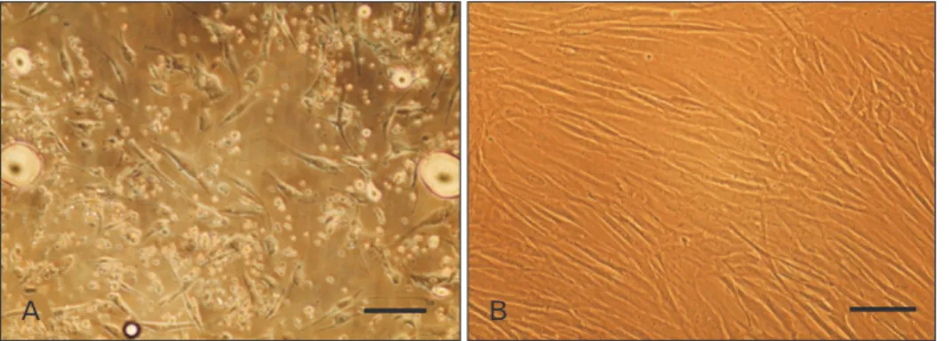
After the cells adhered to the tissue culture flask, non- adherent cells, such as red blood cells, were removed by changing the culture medium after 2 days. The initially adherent cells grew into spindle or stellate-shaped cells, which then developed into visible colonies 3 days after the initial plating (Fig. 1A). The cells began to proliferate rapidly and were passaged by trypsination every 3–4 days until they were completely confluent. After the second passage, hASCs appeared to adopt a more uniform fibroblast-like shape with regular directions (Fig. 1B).
Most of the hASCs expressed high levels of CD44 (99.97%), CD29–integrin b1 (98.97%), and CD90 Thy-1 (99.73%) but they were almost negative for CD45 (0.57%). The plasticity of hASCs was also assessed by induction of adipogenic and osteogenic differentiations and was demonstrated by lipid vacuoles and calcium deposits, respectively (Fig. 2). These results confirmed the mesenchymal nature of the isolated cells as well as their multipotentiality.
hASCs at passage 4 were exposed to the glial growth factors for 2 weeks. At the end, morphology of SC-like cells and the
Fig. 1. Adipose-derived stem cells (ASCs) isolation and culture. (A) Cultured cells of stromal vascular fraction of fat tissue on day 3 of primary culture. (B) ASCs at passage 2 at confluence. Scale bars=30 mm (A, B).
A B
Fig. 2. Differentiation potential assay of adipose-derived stem cells (ASCs) at fourth passage. (A) Lipid vacuoles inside the cytoplasm of adipoblasts appear as red circular spots after staining with oil Red O which stains triglyceride and neutral lipid. (B) Calcium deposition was shown by alizarin red S staining as dark red spots in the plate. Scale bars=30 mm (A, B).
A B
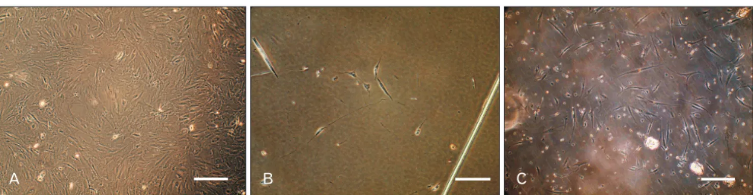
Fig. 3. Morphological assessment of Schwann-like (SC-like) cells after 14 days of induction. (A) Adipose-derived stem cells at fourth passage on day 0 of induction by b-mercaptoethanol. (B) SC-like cells with deformed morphologies in human serum (HS)-treated groups. Most of cells detached in response to the differentiation medium containing allogeneic HS. (C) SC-like cells appeared mostly bipolar or tripolar, typical morphology of Schwann cells when ex posed to the differentiation medium containing fetal bovine serum. Scale bars=40 mm (A–C).
A B C
Fig. 4. Immunofluorescence staining in- dicated that the differentiated Schwann- like cells expressed S100 (A, C) and glial fibrillary acidic protein (GFAP) (B, D) in both human serum (HS)- and fetal bovine serum (FBS)-treated groups.
Fibroblasts 3T3 and undifferentiated adipose-derived stem cells were taken as negative control and new born rat Schwann cells was considered as positive control. Scale bars=30 mm (A–D).
HS-treated/S100
FBS-treated/S100
HS-treated/GFAP
FBS-treated/GFAP
A B
C D
expression of two SCs-related proteins, GFAP and S100, were evaluated. After beginning the differentiation, some cells started to change their morphology and adopted a bipolar or tripolar morphologies which were similar to SCs in both groups. Interestingly, as it was assessed qualitatively, many cells seemed to be detached in HS-treated groups after adding differentiation medium while the attached differentiated cells displayed a deformed shape, indicative of being under stress.
In contrast, these events were less evident in FBS-treated groups (Fig. 3). Cell counting at the end of differentiation period also confirmed this matter and indicated that there are more cells left in wells containing FBS (9×10³ cells/well) as statistically compared to wells containing HS (3×10³ cells/
well) (P<0.001).
None of SCs-related proteins was detected in undiffer- entiated ASCs (data not shown) but both were expressed in differentiated ASCs treated by both FBS and HS (Fig. 4).
To compare the effects of two different serums on the differentiation capacity of ASCs into SC-like cells, the number of SC-like cells was counted using two markers in both groups. Quantitative analysis indicated that 2.3±0.0%
of the cells expressed GFAP in HS-treated groups while 11.6±1.0% expressed the same marker in FBS-treated groups (Fig. 5). Furthermore, S100-positive cells were 2.6±1.0%
and 14.0±2.08% in HS-treated and FBS-treated groups, respectively.
Discussion
Up to now, many studies has been designed to look for possible alternatives for FBS and it was shown that human
blood supplements, as being free from ethical concerns and infection, could act as promising substitutes for cell culture [23, 24, 26, 27]. Human blood supplements include autologous and allogeneic serum, and platelet derivatives. It has been shown that autologous serum is similar or better than FBS with respect to the isolation, expansion, and differentiation of stem cells [24]. Up to now, no study had not been conducted to investigate the effect of HS on SC differentiation of ASCs compared with FBS. Therefore, we compared the effects of 10% HS with 10% FBS on differentiation of ASCs into SC like-cells to test the efficiency of HS as a suitable substitute in this process for basic and clinical researches. Collectively, present study had one major conclusion and it was that the FBS was more efficient than allogeneic HS in this process based on immunostaining.
A major controversy exists concerning use of autologous and allogeneic HS in MSCs isolation and differentiation.
Many studies had been demonstrated that autologous HS could modulate osteogenesis and adipogenesis of BMSCs [28], had more favorable effects on osteoblastic differentiation of BMSCs [29], be good as 10% FBS with respect to both isolation, expansion, and differentiation of hMSCs [27, 30, 31]. In addition, it was indicated that autologous HS is a valid substitute for FBS in culture and in vitro differentiation of human pulp stem cells to obtain a successful bone regeneration in vivo [32]. On the other side, because the amount of autologous serum is restricted, allogeneic HS has been replaced and used in further researches [26]. Of course, the effectiveness of autologous HS as a supplement for culture of MSCs may also depend on the origin of MSCs, and there has been little research involving ASCs [24]. However, it was Fig. 5. Quantitative analysis of cells expressing glial fibrillary acidic protein (GFAP) (A) and S100 (B) in both fetal bovine serum (FBS)- and human serum (HS)-treated groups (data are mean cells±SEM, %). *P<0.05.
HS treated 12.50
GFAPpositivecells(%)
.00
A
FBS treated Experimental groups 10.00
7.50 5.00 2.50
*
HS treated 15.00
S100positivecells(%)
.00
B
FBS treated Experimental groups 10.00
5.00 20.00 *
shown previously that pooled allogeneic HS could maintains the characteristics of ASCs even after long-term expansion and, therefore, could play a role as an alternative to FBS [25]. In other study, effectiveness of autologous HS has been reported as a suitable alternative for FBS in ASCs engineering and it has indicated that 10% HS appeared to be at least as good as 10% FBS with respect to expansion of ASCs, while the lower concentrations of HS were not efficient. These studies apparently contrast with our study that shows that FBS is a better option for glial differentiation than HS. On the other side, it was previously declared that 10% FBS was more effective than autologous HS regardless of the concentration for adipogenic differentiation [24] and this might be because 10% FBS had a higher degree of adipogenic factors and lipid content [27]. However, there is enough evidence that using HS contains two major faults: (1) in large-scale expansion of cells to be used in human cellular therapy, large volumes of peripheral blood is forbiddingly needed to obtain autologous HS [33]; (2) allogeneic serum has been shown to lead to MSCs growth arrest and death, so pooled HS may not meet the above mentioned problem [22]. Besides, nowadays in most clinical trials using MSCs for the treatment of various disorders, FBS is used as good supplement for MSCs culture and differentiation in spite of reported complications and side-effects [34-36]. However, there has been few clinical trials involving ASCs expanded in human blood supplements.
These are consistent with our study’s results which show FBS can support ASCs differentiation better than HS and HS put great stress on differentiating ASCs as it is evident by enormous cells detachment and deformation.
In conclusion, although the use of FBS may contain potential hazards (although reliable tests, such as for the detection of prion proteins, are now available), this remains as a more effective supplement for SC differentiation as compared to allogeneic HS in order to continue basic researches and clinical trials for treating the variety of irreversible pathological disorders that do not respond to conventional therapies until a suitable alternative arises.
Acknowledgements
This paper was issue from thesis of Elham Younesi and was supported by a Grant (CMRC-121) from the research council of the Ahvaz Jundishapur University of Medical Sciences in 2014.
References
1. Tomita K, Madura T, Sakai Y, Yano K, Terenghi G, Hosokawa K. Glial differentiation of human adipose-derived stem cells:
implications for cell-based transplantation therapy. Neuroscience 2013;236:55-65.
2. Jung J, Cai W, Jang SY, Shin YK, Suh DJ, Kim JK, Park HT.
Transient lysosomal activation is essential for p75 nerve growth factor receptor expression in myelinated Schwann cells during Wallerian degeneration. Anat Cell Biol 2011;44:41-9.
3. Kingham PJ, Kalbermatten DF, Mahay D, Armstrong SJ, Wiberg M, Terenghi G. Adipose-derived stem cells differentiate into a Schwann cell phenotype and promote neurite outgrowth in vitro.
Exp Neurol 2007;207:267-74.
4. Gimble J, Guilak F. Adipose-derived adult stem cells: isolation, characterization, and differentiation potential. Cytotherapy 2003;
5:362-9.
5. Taha MF, Hedayati V. Isolation, identification and multipotential differentiation of mouse adipose tissue-derived stem cells. Tissue Cell 2010;42:211-6.
6. di Summa PG, Kingham PJ, Raffoul W, Wiberg M, Terenghi G, Kalbermatten DF. Adipose-derived stem cells enhance peripheral nerve regeneration. J Plast Reconstr Aesthet Surg 2010;63:1544-52.
7. Tomita K, Madura T, Mantovani C, Terenghi G. Differentiated adipose-derived stem cells promote myelination and enhance functional recovery in a rat model of chronic denervation. J Neurosci Res 2012;90:1392-402.
8. Baksh D, Song L, Tuan RS. Adult mesenchymal stem cells:
characterization, differentiation, and application in cell and gene therapy. J Cell Mol Med 2004;8:301-16.
9. Erfanmanesh A, Gholampuor H, Gharibi S. The effect of different doses of gamma irradiation on the sterilization of fetal bovine serum. J Microb Biotechnol 2011;3:1-6.
10. Russell KA, Koch TG. Equine platelet lysate as an alternative to fetal bovine serum in equine mesenchymal stromal cell culture:
too much of a good thing? Equine Vet J 2015 Mar 16 [Epub].
http://dx.doi.org/10.1111/evj.12440.
11. Mackensen A, Drager R, Schlesier M, Mertelsmann R, Lindemann A. Presence of IgE antibodies to bovine serum albumin in a patient developing anaphylaxis after vaccination with human peptide-pulsed dendritic cells. Cancer Immunol Immunother 2000;49:152-6.
12. Selvaggi TA, Walker RE, Fleisher TA. Development of antibodies to fetal calf serum with arthus-like reactions in human immunodeficiency virus-infected patients given syngeneic lymphocyte infusions. Blood 1997;89:776-9.
13. Tuschong L, Soenen SL, Blaese RM, Candotti F, Muul LM.
Immune response to fetal calf serum by two adenosine deaminase-deficient patients after T cell gene therapy. Hum Gene Ther 2002;13:1605-10.
14. Sasse M, Lengwinat T, Henklein P, Hlinak A, Schade R.
Replacement of fetal calf serum in cell cultures by an egg yolk factor with cholecystokinin/gastrin-like immunoreactivity.
Altern Lab Anim 2000;28:815-31.
15. Schwartz KA, Lu G, Trosko JE, Chang CC. Serum from outdated human platelet concentrates: an alternative supplement for tissue (fibroblast) culture media. Am J Hematol 1984;17:23-7.
16. Clemmons DR, Isley WL, Brown MT. Dialyzable factor in human serum of platelet origin stimulates endothelial cell replication and growth. Proc Natl Acad Sci U S A 1983;80:1641-5.
17. Pietschmann P, Stöckl J, Draxler S, Majdic O, Knapp W.
Functional and phenotypic characteristics of dendritic cells generated in human plasma supplemented medium. Scand J Immunol 2000;51:377-83.
18. Filipic B, Shehata M, Toth S, Schwarzmeier J, Koren S. Novel serum replacement based on bovine ocular fluid: a useful tool for cultivation of different animal cells in vitro. ALTEX 2002;19:15-20.
19. Grace S, Guthrie LA, Johnston RB Jr. The use of mouse serum and the presence of non-adherent cells for the culture of mouse macrophages. J Immunol Methods 1988;114:21-6.
20. Reisser D, Fady C, Pelletier H, Lagadec P, Jeannin JF, Olsson NO.
Comparative effect of rat and fetal calf serum on measurement of the natural tumoricidal activity of rat lymphocytes, macrophages and polymorphonuclear cells. Cancer Immunol Immunother 1989;28:34-6.
21. Mangalo R, Marcovich H. Mitogenic factors for BALB/c 3T3 cells isolated from the serum of horses by affinity chromatography on a column using fetal calf serum as the ligand. C R Acad Sci III 1984;299:445-50.
22. Shahdadfar A, Frønsdal K, Haug T, Reinholt FP, Brinchmann JE.
In vitro expansion of human mesenchymal stem cells: choice of serum is a determinant of cell proliferation, differentiation, gene expression, and transcriptome stability. Stem Cells 2005;23:1357-66.
23. Kobayashi T, Watanabe H, Yanagawa T, Tsutsumi S, Kayakabe M, Shinozaki T, Higuchi H, Takagishi K. Motility and growth of human bone-marrow mesenchymal stem cells during ex vivo expansion in autologous serum. J Bone Joint Surg Br 2005;
87:1426-33.
24. Choi J, Chung JH, Kwon GY, Kim KW, Kim S, Chang H.
Effectiveness of autologous serum as an alternative to fetal bovine serum in adipose-derived stem cell engineering. Cell Tissue Bank 2013;14:413-22.
25. Bieback K, Hecker A, Schlechter T, Hofmann I, Brousos N, Redmer T, Besser D, Klüter H, Müller AM, Becker M. Replicative aging and differentiation potential of human adipose tissue- derived mesenchymal stromal cells expanded in pooled human or fetal bovine serum. Cytotherapy 2012;14:570-83.
26. Kocaoemer A, Kern S, Klüter H, Bieback K. Human AB serum
and thrombin-activated platelet-rich plasma are suitable alternatives to fetal calf serum for the expansion of mesenchymal stem cells from adipose tissue. Stem Cells 2007;25:1270-8.
27. Stute N, Holtz K, Bubenheim M, Lange C, Blake F, Zander AR. Autologous serum for isolation and expansion of human mesenchymal stem cells for clinical use. Exp Hematol 2004;
32:1212-25.
28. Oreffo RO, Virdi AS, Triffitt JT. Modulation of osteogenesis and adipogenesis by human serum in human bone marrow cultures.
Eur J Cell Biol 1997;74:251-61.
29. Yamamoto N, Isobe M, Negishi A, Yoshimasu H, Shimokawa H, Ohya K, Amagasa T, Kasugai S. Effects of autologous serum on osteoblastic differentiation in human bone marrow cells. J Med Dent Sci 2003;50:63-9.
30. Nimura A, Muneta T, Koga H, Mochizuki T, Suzuki K, Makino H, Umezawa A, Sekiya I. Increased proliferation of human synovial mesenchymal stem cells with autologous human serum:
comparisons with bone marrow mesenchymal stem cells and with fetal bovine serum. Arthritis Rheum 2008;58:501-10.
31. Tateishi K, Ando W, Higuchi C, Hart DA, Hashimoto J, Nakata K, Yoshikawa H, Nakamura N. Comparison of human serum with fetal bovine serum for expansion and differentiation of human synovial MSC: potential feasibility for clinical applications. Cell Transplant 2008;17:549-57.
32. Pisciotta A, Riccio M, Carnevale G, Beretti F, Gibellini L, Maraldi T, Cavallini GM, Ferrari A, Bruzzesi G, De Pol A. Human serum promotes osteogenic differentiation of human dental pulp stem cells in vitro and in vivo. PLoS One 2012;7:e50542.
33. Sotiropoulou PA, Perez SA, Salagianni M, Baxevanis CN, Papamichail M. Cell culture medium composition and translational adult bone marrow-derived stem cell research.
Stem Cells 2006;24:1409-10.
34. Smith SR, Munavalli G, Weiss R, Maslowski JM, Hennegan KP, Novak JM. A multicenter, double-blind, placebo-controlled trial of autologous fibroblast therapy for the treatment of nasolabial fold wrinkles. Dermatol Surg 2012;38(7 Pt 2):1234-43.
35. Kølle SF, Fischer-Nielsen A, Mathiasen AB, Elberg JJ, Oliveri RS, Glovinski PV, Kastrup J, Kirchhoff M, Rasmussen BS, Talman ML, Thomsen C, Dickmeiss E, Drzewiecki KT. Enrichment of autologous fat grafts with ex-vivo expanded adipose tissue- derived stem cells for graft survival: a randomised placebo- controlled trial. Lancet 2013;382:1113-20.
36. Chhetri DK, Berke GS. Injection of cultured autologous fibroblasts for human vocal fold scars. Laryngoscope 2011;121:785-92.