Prevalence of Low Bone Mineral Density and Associated Risk Factors in Korean Puerperal Women
Although pregnancy is a medical condition that contributes to bone loss, little information is available regarding bone mineral density (BMD) in puerperal women. This cross sectional study aimed to evaluate the prevalence of low BMD in puerperal women and to identify associated risk factors. We surveyed all puerperal women who had BMD measurements taken 4-6 weeks after delivery in a tertiary university hospital, and did not have any bone loss-related comorbidities. Among the 1,561 Korean puerperal women, 566 (36.3%) had low BMD at the lumbar spine, total hip, femoral neck, and/or trochanter. Multivariate analysis revealed that underweight women had a significantly higher risk of low BMD compared with obese women at pre-pregnancy (adjusted odds ratio [aOR], 3.21; 95%
confidence interval [CI], 1.83-5.63). Also, women with inadequate gestational weight gain (GWG) were 1.4 times more likely to have low BMD than women with excessive GWG (aOR, 1.42; 95% CI, 1.04-1.94). One-way ANOVA showed that BMDs at the lumbar spine and total hip were significantly different between the 4 BMI groups (both P < 0.001) and also between the 3 GWG groups (both P < 0.001). In conclusion, this study identifies a high prevalence of low BMD in puerperal women and thus suggests the need for further evaluation about the change of BMD in pregnancy and postpartum period.
Keywords: Bone Density; Postpartum Period; Osteoporosis; Body Mass Index; Weight Gain; Korean
Dong Gyu Jang,* Ji Young Kwon,*
Sae Kyung Choi, Hyun Sun Ko, Jong Chul Shin, and In Yang Park Department of Obstetrics and Gynecology, The Catholic University of Korea, Seoul, Korea
* Dong Gyu Jang and Ji Young Kwon contributed equally to this work.
Received: 24 December 2015 Accepted: 5 August 2016 Address for Correspondence:
In Yang Park, MD
Department of Obstetrics and Gynecology, The Catholic University of Korea, Seoul St. Mary’s Hospital, 222 Banpodaero, Seocho-gu, Seoul 06591, Korea
E-mail: [email protected]
http://dx.doi.org/10.3346/jkms.2016.31.11.1790 • J Korean Med Sci 2016; 31: 1790-1796
INTRODUCTION
Osteoporosis is common health problem in postmenopausal women that can have serious adverse impacts on life quality (1).
Despite the importance of osteoporosis, little information is available regarding osteoporosis in premenopausal women, probably due to its low incidence in women of younger ages.
However, since having a low peak bone mass early in life is an important contributing factor to developing osteoporosis later in life, bone health should be carefully monitored from an early age (2). Among several medical conditions that may decrease bone density, pregnancy is an important physiologic event ex- perienced by most women. A pregnant woman’s body is an im- portant source of calcium that is transferred to the fetus. The in- creased calcium requirement during pregnancy is usually met by increased intestinal absorption, decreased renal excretion, and the mobilization of minerals from the skeleton (3,4). Thus, during pregnancy the rate of bone resorption often exceeds the rate of bone formation, leading to bone loss (5).
Despite these important physiological changes during preg- nancy, the precise effects of pregnancy on bone mineral density (BMD) have not yet been established. Small-scale prospective studies using dual energy X-ray absorptiometry (DXA) to mea- sure BMD at pre-pregnancy and during pregnancy have report-
ed that BMD decreases during pregnancy (6-8). In addition, a number of studies using ultrasound measurements of BMD have demonstrated significant decreases of BMD during preg- nancy (9-11). However, these studies had small sample sizes and diverse research methods; thus, no established consensus has yet been reached regarding bone density in pregnant and puerperal women.
To address these issues, this study analyzed BMD in Korean puerperal women. The goals of this study were to determine the prevalence of puerperal low BMD and to identify the risk factors associated with puerperal low BMD. This is the first large- scale study of BMD in puerperal women and the first to define the risk factors in women for low BMD. The results of this study are expected to contribute to our understanding of bone densi- ty in puerperal women and to help establish guidelines for pre- natal and postnatal care of women to ensure optimal bone health.
MATERIALS AND METHODS
This cross-sectional study surveyed all puerperal women who had given birth to a child at a university hospital in Korea from 2007 to 2009 and whose BMD was measured 4-6 weeks after childbirth. During the study period, 2,397 childbirths occurred.
Postpartum BMD measurement was recommended to all wom- Obstetrics & Gynecology
Jang DG, et al. • Low Bone Mineral Density in Puerperal Women
en; 1,672 women consented to and underwent this measure- ment. Of these women, 110 had comorbid conditions known to be associated with secondary bone loss and were excluded. These conditions included hyperthyroidism, hypothyroidism, system- ic lupus erythematous, rheumatoid arthritis, insulin-dependent diabetes mellitus, kidney disease, epilepsy, depression, schizo- phrenia, hematologic disease, and treatment for infertility. In addition, 55 women who delivered before 32 weeks of pregnan- cy were excluded. Finally, a total of 1,561 puerperal women were included in this study.
Baseline data included medical conditions, smoking status, parity, newborn birth weight, gestational age at delivery, deliv- ery method, experience of hospitalized bed rest, treatment with dexamethasone for fetal lung maturation, calcium intake dur- ing the prenatal and postnatal periods, and breastfeeding be- havior. Calcium intake was assessed by self-reported medical records. Only supplemental calcium intake was investigated;
dietary calcium intake was not considered. Most calcium sup- plements consisted of daily multivitamins containing 125 mg calcium each. Subjects were divided into 2 groups according to the duration of calcium intake. Breastfeeding behavior during the first 6 weeks after delivery was evaluated by self-reporting and was classified as either exclusive breastfeeding or formula feeding (with or without supplemental breastfeeding).
Additional anthropometric data, including pre-pregnancy weight, pre-pregnancy height, and weight at delivery date were collected. Height and weight were used to calculate body mass index (BMI; kg/m2) at pre-pregnancy. All enrolled women were categorized according to the World Health Organization (WHO) BMI classification system for Asians as follows: underweight, BMI of 18.6 kg/m2 or less; normal weight, BMI of 18.5 to 22.9 kg/m2; overweight, BMI of 23 to 24.9 kg/m2; obese, BMI of 25 kg/m2 or higher (12). Gestational weight gain (GWG) was cal- culated as the difference between the maternal pre-pregnancy weight and the last weight before delivery and then classified as inadequate, adequate, or excessive according to the 2009 Insti- tute of Medicine (IOM) recommendation (13). Weekly weight gains according to the 2009 IOM recommendations were used to adjust the recommended total GWG ranges to the gestational weeks at delivery (13).
The BMDs (g/cm2) of the lumbar spine (composite L1-L4) and the hip (total femur, femoral neck, and trochanter) were as- sessed 4-6 weeks postpartum by DXA using a Lunar Prodigy scanner equipped with enCORE software (GE, Madison, WI, USA). Quality assurance of the densitometric technique was checked according to the manufacturer’s protocol. The long- term percent coefficients of variance (%CV) for the different sites were 1.2% for the lumbar spine, 1.2% for the femoral neck, and 1.4% for the total hip region. The Z-scores were calculated from manufacturer-provided Korean normative data matched for sex and age. Low BMD was defined according to the Nation-
al Osteoporosis Foundation guidelines as a Z-score of -2.0 or lower for the appropriate age group at any site among the lum- bar spine, total hip, femoral neck, and trochanter (14).
Statistics
All maternal and newborn characteristics were summarized using descriptive statistics. The characteristics of women in the low BMD group were compared to those of women in the nor- mal BMD group using the independent sample t-test for con- tinuous variables and the χ2 test for categorical variables. Vari- ables found to be significantly associated with low BMD (P < 0.10) were then entered into multivariate logistic regression analysis to identify risk factors for low BMD. The associations between the variables are expressed as odds ratios (ORs), 95% confidence intervals (95% CIs), and P values. Statistical significance was set at P < 0.05. One-way ANOVA tests were also performed to com- pare mean BMDs at the lumbar and hip regions for each BMI group and each gestational weight gain group. Statistical analy- ses were performed using SPSS 17.0 (SPSS Inc., Chicago, IL, USA).
Ethics statement
The study was approved by the institutional review board of the College of Medicine of the Catholic University of Korea (KC150 IS10051). The board waived informed consent from the subject patients. It was conducted in accordance with the Declaration of Helsinki.
RESULTS
The general characteristics of the study population are present- ed in Table 1. The pre-pregnancy BMI was classified as normal in 1,033 (66.2%) women, underweight in 265 (17.0%) women, overweight in 147 (9.4%) women, and obese in 116 (7.4%) wom- en. Preterm delivery occurred for 134 (8.6%) women (median, 35 weeks). Information about puerperal breastfeeding was only available for 1,453 subjects, 407 of whom self-reported exclusive breastfeeding. No current smokers were in either of the groups.
The mean lumbar spine BMD was 1.170 g/cm2 and the mean total hip BMD was 0.941 g/cm2. Out of all the women analyzed, 13.8%, 16.4%, 24.8%, and 24.1% had low BMD in the lumbar spine, total hip, femoral neck, and trochanter, respectively (Ta- ble 2). Moreover, 566 (36.3%) women had low BMD in at least one of the four measured sites, 139 (8.9%) had low BMD in two of the sites, 135 (8.6%) in three of the sites, and 87 (5.6%) in all four sites. In contrast, low-trauma fracture occurred in only 3 of the women during pregnancy (1.9 per 1,000 pregnant women).
All of these women had severely low BMDs, with Z-scores less than -2.5 in each of the four sites.
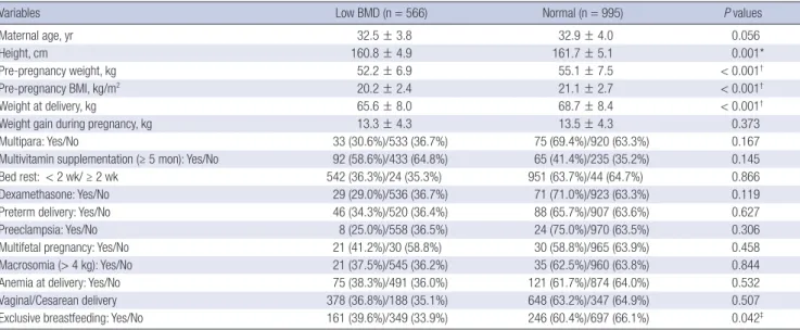
Women with low BMD were significantly different from wom- en with normal BMD with respect to pre-pregnancy height, pre- pregnancy weight, pre-pregnancy BMI, weight at delivery, and
Table 1. General and pregnancy-related characteristics of the study populations
Variables n = 1,561
Maternal age, yr 32.8 ± 3.9
Height, cm 161.4 ± 5.0
Pre-pregnancy weight, kg 54.0 ± 7.4
Pre-pregnancy BMI, kg/m2 20.8 ± 2.6
Weight at delivery 67.5 ± 8.4
Weight gain during pregnancy 13.5 ± 4.3
Previous deliveries (≥ 2) 196 (12.6%)
Multivitamin supplementation (> 5 mon) during pregnancy 157 (19.0%)
Bed rest (> 2 wk) 68 (4.4%)
Treatment of dexamethasone 100 (6.4%)
Gestational age at delivery, wk 38.7 ± 1.7
Preterm delivery 134 (8.6%)
Preeclampsia 32 (2.0%)
Multifetal pregnancy 51 (3.3%)
Newborn weight, g 3,199.5 ± 489.8
Anemia at delivery 196 (12.6%)
Mode of delivery (Cesarean delivery) 535 (34.3%)
Exclusive breastfeeding* 407 (26.1%)
All values are expressed as mean (± standard deviation) or number (%).
*Data about puerperal breastfeeding were available for 1,453 cases.
Table 2. Bone mineral density (BMD) measures and proportion of low BMD Skeletal sites BMD, g/cm2 BMD (Z-score) Low BMD (Z-score ≤ -2) Lumbar spine
L1 L2 L3 L4
1.170 ± 0.136*
-0.59 ± 1.04 -0.61 ± 1.12 -0.04 ± 1.12 -0.27 ± 1.12
215 (13.8%)† 117 (7.5%) 160 (10.2%)
55 (3.5%) 95 (6.1%) Total hip 0.941 ± 0.115 -0.59 ± 1.04 256 (16.4%)
Femoral neck -1.08 ± 0.93 387 (24.8%)
Trochanter -1.33 ± 0.88 376 (24.1%)
All values are expressed as mean (± standard deviation) or number (%).
*Total BMD for the lumbar spine (L1-L4); †No. of subjects with low BMD at any lum- bar spine (L1-L4).
Table 3. Comparison between puerperal women with low bone mineral density (BMD) versus women with normal BMD
Variables Low BMD (n = 566) Normal (n = 995) P values
Maternal age, yr 32.5 ± 3.8 32.9 ± 4.0 0.056
Height, cm 160.8 ± 4.9 161.7 ± 5.1 0.001*
Pre-pregnancy weight, kg 52.2 ± 6.9 55.1 ± 7.5 < 0.001†
Pre-pregnancy BMI, kg/m2 20.2 ± 2.4 21.1 ± 2.7 < 0.001†
Weight at delivery, kg 65.6 ± 8.0 68.7 ± 8.4 < 0.001†
Weight gain during pregnancy, kg 13.3 ± 4.3 13.5 ± 4.3 0.373
Multipara: Yes/No 33 (30.6%)/533 (36.7%) 75 (69.4%)/920 (63.3%) 0.167
Multivitamin supplementation (≥ 5 mon): Yes/No 92 (58.6%)/433 (64.8%) 65 (41.4%)/235 (35.2%) 0.145
Bed rest: < 2 wk/ ≥ 2 wk 542 (36.3%)/24 (35.3%) 951 (63.7%)/44 (64.7%) 0.866
Dexamethasone: Yes/No 29 (29.0%)/536 (36.7%) 71 (71.0%)/923 (63.3%) 0.119
Preterm delivery: Yes/No 46 (34.3%)/520 (36.4%) 88 (65.7%)/907 (63.6%) 0.627
Preeclampsia: Yes/No 8 (25.0%)/558 (36.5%) 24 (75.0%)/970 (63.5%) 0.306
Multifetal pregnancy: Yes/No 21 (41.2%)/30 (58.8%) 30 (58.8%)/965 (63.9%) 0.458
Macrosomia (> 4 kg): Yes/No 21 (37.5%)/545 (36.2%) 35 (62.5%)/960 (63.8%) 0.844
Anemia at delivery: Yes/No 75 (38.3%)/491 (36.0%) 121 (61.7%)/874 (64.0%) 0.532
Vaginal/Cesarean delivery 378 (36.8%)/188 (35.1%) 648 (63.2%)/347 (64.9%) 0.507
Exclusive breastfeeding: Yes/No 161 (39.6%)/349 (33.9%) 246 (60.4%)/697 (66.1%) 0.042‡
*P < 0.01; †P < 0.001; ‡P < 0.05.
breastfeeding habits according to univariate analysis (all P <
0.001 except for height [P = 0.001] and breastfeeding [P = 0.042]).
No significant associations were observed between age, parity, or multivitamin supplementation status during pregnancy and low BMD (P = 0.056, 0.167, and 0.145, respectively) (Table 3).
Since height, weight, and BMI at pre-pregnancy and weight at delivery were significantly correlated with each other (all P <
0.001), only the BMI at pre-pregnancy was used in the following analysis.
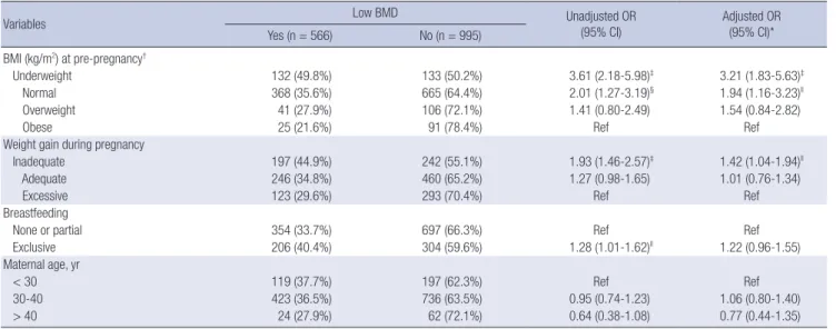
To attempt to adjust for any potential bias, adjusted odds ra- tios (aORs) for low BMD were calculated with a logistic regres- sion model including BMI at pre-pregnancy, weight gain dur-
ing pregnancy, breastfeeding status, and maternal age. The re- sults were comparable to those obtained by univariate analysis (crude ORs) (Table 4). Women who were underweight at pre- pregnancy had an elevated risk for low BMD compared with women who were obese at pre-pregnancy. This elevated risk persisted even after adjusting for the aforementioned variables (aOR, 3.21; 95% CI, 1.83-5.63; P < 0.001). With respect to GWG, women with inadequate weight gain were 1.4 times more likely to have low BMD than women with excessive weight gain (aOR, 1.42; 95% CI, 1.04-1.94; P = 0.030). The unadjusted OR of the re- lationship of exclusive breastfeeding with low BMD was 1.28 (95% CI, 1.01-1.62; P = 0.042). However, after adjusting for the aforementioned variables, the risk of low BMD was no longer elevated (P = 0.116).
One-way ANOVA showed that the BMDs at the lumbar spine and total hip were significantly different between the 4 BMI groups (P < 0.001). Also, regarding GWG, BMDs at the lumbar spine and total hip were significantly different between the 3 GWG groups (both P < 0.001) (Fig. 1 and 2).
Jang DG, et al. • Low Bone Mineral Density in Puerperal Women
DISCUSSION
This study found that the prevalence of low BMD (i.e. “below the expected range for the age”) among Korean puerperal wom- en was quite high (36.3%). Considering the relatively young age of the subjects, the prevalence of low BMD was higher than ex- pected. Few studies have examined the prevalence of low BMD among puerperal women, not just for Asians but also for other ethnicities. To the best of our knowledge, this is the first report to evaluate the prevalence of low BMD in puerperal women.
Low-trauma fracture occurred in three women during preg- nancy. Each of these women had severely low BMD. Premeno-
pausal women do not have a high risk of fracture, even with low BMD (15). This finding can be explained by their greater mus- cle mass, thicker cortices, normal trabecular connectivity, and fewer falls compared with postmenopausal women (16). How- ever, since the high rate of bone turnover observed in pregnant women is an important risk factor for fracture fragility, pregnant women are likely to have a relatively higher risk of fracture than nonpregnant premenopausal women (3). Accordingly, preg- nant and puerperal women who are at risk of severely low BMD should be strongly encouraged to strive to prevent fractures, even though the incidence of fracture is relatively low.
Low peak bone mass is known to be a critical determinant of Table 4. Risk factors and odds ratios (ORs) for low bone mineral density (BMD)
Variables Low BMD Unadjusted OR
(95% CI) Adjusted OR
(95% CI)*
Yes (n = 566) No (n = 995) BMI (kg/m2) at pre-pregnancy†
Underweight 132 (49.8%) 133 (50.2%) 3.61 (2.18-5.98)‡ 3.21 (1.83-5.63)‡
Normal 368 (35.6%) 665 (64.4%) 2.01 (1.27-3.19)§ 1.94 (1.16-3.23)ll
Overweight 41 (27.9%) 106 (72.1%) 1.41 (0.80-2.49) 1.54 (0.84-2.82)
Obese 25 (21.6%) 91 (78.4%) Ref Ref
Weight gain during pregnancy
Inadequate 197 (44.9%) 242 (55.1%) 1.93 (1.46-2.57)‡ 1.42 (1.04-1.94)ll
Adequate 246 (34.8%) 460 (65.2%) 1.27 (0.98-1.65) 1.01 (0.76-1.34)
Excessive 123 (29.6%) 293 (70.4%) Ref Ref
Breastfeeding
None or partial 354 (33.7%) 697 (66.3%) Ref Ref
Exclusive 206 (40.4%) 304 (59.6%) 1.28 (1.01-1.62)ll 1.22 (0.96-1.55)
Maternal age, yr
< 30 119 (37.7%) 197 (62.3%) Ref Ref
30-40 423 (36.5%) 736 (63.5%) 0.95 (0.74-1.23) 1.06 (0.80-1.40)
> 40 24 (27.9%) 62 (72.1%) 0.64 (0.38-1.08) 0.77 (0.44-1.35)
*Adjusted ORs obtained with a logistic regression model including pre-pregnancy BMI, weight gain during pregnancy, breastfeeding status and maternal age; †Underweight, overweight, and obesity are defined as BMIs less than 18.5 kg/m2, between 23 and 24.9 kg/m2, or 25 kg/m2 or more, respectively; ‡P < 0.001; §P < 0.01; llP < 0.05.
Fig. 1. Mean bone mineral densities (BMDs) at the lumbar spine and total hip in the 4 body mass index (BMI) groups. Data are presented as means; vertical bars show the 95% confidence intervals. At both sites, the BMD tended to increase as the BMI increased (both P < 0.001, ANOVA test). Post hoc analysis revealed significant differences in BMD at the lumbar spine and total hip between the different BMI groups (all P < 0.05), with the exception of BMD at the lumbar spine in the overweight versus obese groups (P
= 0.216).
Figure1
Lumbar spine BMI (g/cm2)
underweight normal overweight obese underweight normal overweight obese
P < 0.001 P = 0.025 P= 0.216 P < 0.001
P< 0.001 P< 0.001 P = 0.012 P< 0.001
BMI at pre-pregnancy BMI at pre-pregnancy
Lumbar spine BMD (g/cm2) Total hip BMD (g/cm2)
Lumbar spine BMD (g/cm2)
BMI at pre-pregnancy Underweight Normal Overweight Obese 1.30
1.25 1.20 1.15 1.10 1.05
P < 0.001 P < 0.001 P = 0.025 P = 0.216
Figure1
Lumbar spine BMI (g/cm2)
underweight normal overweight obese underweight normal overweight obese
P < 0.001 P = 0.025 P= 0.216 P < 0.001
P< 0.001 P< 0.001 P = 0.012 P< 0.001
BMI at pre-pregnancy BMI at pre-pregnancy
Lumbar spine BMD (g/cm2) Total hip BMD (g/cm2)Total hip BMD (g/cm2 )
BMI at pre-pregnancy Underweight Normal Overweight Obese 1.10
1.05 1.00 0.95 0.90 0.85
P < 0.001 P < 0.001 P < 0.001 P = 0.012
Jang DG, et al. • Low Bone Mineral Density in Puerperal Women
postmenopausal osteoporosis (2). Since pregnancy is an im- portant condition that may lower bone density in young wom- en, it is necessary to identify pregnant or puerperal women at risk for low BMD not only for preventing fracture during the puerperal period, but also for suppressing the subsequent de- velopment of osteoporosis later in life. In this study, low pre- pregnancy BMI and inadequate GWG were found to be impor- tant risk factors for low BMD. A number of studies have report- ed that low BMI is significantly associated with low BMD (17,18).
The present study extends these findings and suggests that BMI impacts bone density even in pregnant women. As women who have a low BMI before they become pregnant are likely to have low BMD during pregnancy, they should be more concerned about achieving optimal GWG during pregnancy. Also regular exercise and healthy eating may be helpful for bone health.
Obesity appeared to be protective against low BMD in this study. Pregnant women with normal BMI had an increased risk for low BMD compared with obese pregnant women at an odds ratio of 1.94. However, the relationship between obesity and low BMD has been quite controversial so far (19,20). Body weight is made up of lean mass and fat mass and fat mass per whole body weight is suggested to be negatively associated with BMD (21).
This retrospective study did not analyze the effect of lean mass and fat mass on the BMD. Further study about the influence of body composition on the BMD during pregnancy will be nec- essary.
This is also the first study to indicate that achieving the opti- mal weight gain during pregnancy is important for maintaining a healthy BMD in pregnant women. For pregnant women, in- adequate GWG indicates that the calorie intake is lower than required, which may be associated with low dietary calcium in- take if calcium supplements were not taken. For the develop-
ment of the fetal skeleton, 30 g of calcium is required over the course of pregnancy (3). This requirement is mainly met through an increased calcium intake, the doubling of maternal intesti- nal calcium absorption, increased skeletal resorption, and in- creased renal mineral conservation (3,4). Accordingly, low di- etary calcium intake can induce excessive resorption of calcium from the skeleton, and may cause severe loss of maternal bone mineral content during pregnancy.
An increased calcium intake is expected to have a positive ef- fect on maternal bone mineral content. Previous studies have suggested that daily intake of a calcium supplement can decre- ase the levels of bone resorption markers and increase postpar- tum lumbar BMD (22,23). However, the effect of calcium intake on BMD was difficult to analyze in the present study because daily calcium intake was not recorded. Breastfeeding is also known to be a risk factor for low BMD (3,4). The univariate anal- ysis performed in the present study revealed that the risk of low BMD was significantly higher in the exclusively breastfeeding group. However, after adjusting the analysis according to pre- pregnancy BMI and GWG, breastfeeding did not have a signifi- cant effect on BMD. Considering the present study analyzed women within six weeks of delivery, any conclusions that can be drawn regarding the effect of breastfeeding on BMD are some- what limited.
Although the present study did not identify the precise cause underlying the high prevalence of low BMD in puerperal wom- en, the possibility of ethnic effects should be considered. Obe- sity rates of Korea are among the lowest in the OECD (24). Al- though relatively strict obesity criteria for Asia were applied in the present study, a relatively high percent (17.0%) of women were underweight at pre-pregnancy, whereas the rate of obesity was relatively low (7.4%). Korean women generally prefer to Fig. 2. Mean bone mineral densities (BMDs) at the lumbar spine and hip in the 3 gestational weight gain (GWG) groups. Data are presented as means; vertical bars show the 95% confidence intervals. At both sites, the BMD tended to increase as the GWG increased (both P < 0.001, ANOVA test). Post hoc analysis revealed significant differences in BMD at both sites between the different GWG groups (all P < 0.05).
inadequate adequate excessive inadequate adequate
Gestational weight gain excessive Gestational weight gain
Lumbar spine BMD (g/cm2) Total hip BMD (g/cm2)
P = 0.015 P< 0.001 P < 0.001
P = 0.006 P = 0.003 P< 0.001
Lumbar spine BMD (g/cm2)
Gestational weight gain
Inadequate Adequate Excessive 1.25
1.20
1.15
1.10
P < 0.001 P < 0.001 P = 0.015
inadequate adequate excessive inadequate adequate
Gestational weight gain excessive Gestational weight gain
Lumbar spine BMD (g/cm2) Total hip BMD (g/cm2)
P = 0.015 P< 0.001 P < 0.001
P = 0.006 P = 0.003 P< 0.001
Total hip BMD (g/cm2)
Gestational weight gain
Inadequate Adequate Excessive 1.00
0.98 0.96 0.94 0.92 0.90
P < 0.001 P = 0.003 P = 0.006
Jang DG, et al. • Low Bone Mineral Density in Puerperal Women
manage weight through diet control, even during pregnancy. In addition, Koreans get less dietary calcium from meals than West- erners do (25). These ethnic characteristics of Koreans may con- tribute to the high prevalence of low BMI in this study. To com- prehensively understand the BMD characteristics of puerperal women, further studies of different ethnicities are required.
Also, our data do not necessarily imply that pregnancy itself is a significant risk factor for low BMD in puerperal women. Since pre-pregnancy BMD measurements were not performed for the subjects in our study group, we cannot exclude the possibil- ity that they had already had low BMD at pre-pregnancy. More- over, it is difficult to tell whether low BMD in the puerperal pe- riod is reversible and whether it affects peak bone mass. How- ever, this study did identify a high prevalence of low BMD in puerperal women and thus highlights the need for interest in bone health when caring for pregnant or puerperal women, re- gardless of whether low BMD is caused by pregnancy.
The major finding of the present study was that approximate- ly 36% of all puerperal women had low BMD and the risk of low BMD was higher when pre-pregnancy BMI was low and when the recommended GWG was not attained. Our study implies that women with an elevated risk for low BMD should be en- couraged to achieve optimal weight gain and carefully guard against falls and fractures during pregnancy. Further studies of women of different ethnicities during the prenatal and postna- tal periods are necessary to determine the extent to which our findings are broadly applicable.
DISCLOSURE
The authors have no potential conflicts of interest to disclose.
AUTHOR CONTRIBUTION
Study design: Jang DG, Park IY. Literature review: Choi SK, Ko HS, Shin JC. Data management: Choi SK, Ko HS, Park IY. Statis- tical analysis: Kwon JY. Interpretation of the findings: Jang DG, Kwon JY, Park IY. Drafting of the manuscript: Jang DG, Kwon JY.
Revision of manuscript: Kwon JY, Park IY. Final approval: all au- thors.
ORCID
Dong Gyu Jang http://orcid.org/0000-0003-2375-3526 Ji Young Kwon http://orcid.org/0000-0002-7788-7303 Sae Kyung Choi http://orcid.org/0000-0001-6264-4256 Hyun Sun Ko http://orcid.org/0000-0001-6310-6206 Jong Chul Shin http://orcid.org/0000-0002-3059-3842 In Yang Park http://orcid.org/0000-0002-9458-2886
REFERENCES
1. Sambrook P, Cooper C. Osteoporosis. Lancet 2006; 367: 2010-8.
2. Berger C, Goltzman D, Langsetmo L, Joseph L, Jackson S, Kreiger N, Te- nenhouse A, Davison KS, Josse RG, Prior JC, et al. Peak bone mass from longitudinal data: implications for the prevalence, pathophysiology, and diagnosis of osteoporosis. J Bone Miner Res 2010; 25: 1948-57.
3. Kovacs CS. Calcium and bone metabolism during pregnancy and lacta- tion. J Mammary Gland Biol Neoplasia 2005; 10: 105-18.
4. Ward KA, Adams JE, Mughal MZ. Bone status during adolescence, preg- nancy and lactation. Curr Opin Obstet Gynecol 2005; 17: 435-9.
5. Hellmeyer L, Ziller V, Anderer G, Ossendorf A, Schmidt S, Hadji P. Bio- chemical markers of bone turnover during pregnancy: a longitudinal study. Exp Clin Endocrinol Diabetes 2006; 114: 506-10.
6. More C, Bettembuk P, Bhattoa HP, Balogh A. The effects of pregnancy and lactation on bone mineral density. Osteoporos Int 2001; 12: 732-7.
7. Black AJ, Topping J, Durham B, Farquharson RG, Fraser WD. A detailed assessment of alterations in bone turnover, calcium homeostasis, and bone density in normal pregnancy. J Bone Miner Res 2000; 15: 557-63.
8. Kaur M, Pearson D, Godber I, Lawson N, Baker P, Hosking D. Longitudi- nal changes in bone mineral density during normal pregnancy. Bone 2003; 32: 449-54.
9. Yamaga A, Taga M, Minaguchi H, Sato K. Changes in bone mass as deter- mined by ultrasound and biochemical markers of bone turnover during pregnancy and puerperium: a longitudinal study. J Clin Endocrinol Metab 1996; 81: 752-6.
10. To WW, Wong MW, Leung TW. Relationship between bone mineral den- sity changes in pregnancy and maternal and pregnancy characteristics: a longitudinal study. Acta Obstet Gynecol Scand 2003; 82: 820-7.
11. Hellmeyer L, Hahn B, Fischer C, Hars O, Boekhoff J, Maier J, Hadji P. Quan- titative ultrasonometry during pregnancy and lactation: a longitudinal study. Osteoporos Int 2015; 26: 1147-54.
12. World Health Organization Western Pacific Region; International Associ- ation for the Study of Obesity; International Obesity Task Force. The Asia- Pacific Perspective: Redefining Obesity and Its Treatment. Sydney: Health Communications Australia Pty Limited, 2000.
13. Institute of Medicine, National Research Council, Committee to Reexam- ine IOM Pregnancy Weight Guidelines (US). Weight Gain during Preg- nancy: Reexamining the Guidelines. Washington, D.C.: National Acade- mies Press, 2009.
14. National Osteoporosis Foundation (US). Clinician’s Guide to Prevention and Treatment of Osteoporosis. Washington, D.C.: National Osteoporosis Foundation, 2008.
15. Martínez-Morillo M, Grados D, Holgado S. Premenopausal osteoporosis:
how to treat? Reumatol Clin 2012; 8: 93-7.
16. Bhalla AK. Management of osteoporosis in a pre-menopausal woman.
Best Pract Res Clin Rheumatol 2010; 24: 313-27.
17. Coin A, Sergi G, Benincà P, Lupoli L, Cinti G, Ferrara L, Benedetti G, To- masi G, Pisent C, Enzi G. Bone mineral density and body composition in underweight and normal elderly subjects. Osteoporos Int 2000; 11: 1043- 50.
18. Tanaka S, Kuroda T, Saito M, Shiraki M. Overweight/obesity and under- weight are both risk factors for osteoporotic fractures at different sites in Japanese postmenopausal women. Osteoporos Int 2013; 24: 69-76.
19. Zhao LJ, Jiang H, Papasian CJ, Maulik D, Drees B, Hamilton J, Deng HW.
Correlation of obesity and osteoporosis: effect of fat mass on the deter- mination of osteoporosis. J Bone Miner Res 2008; 23: 17-29.
20. Cao JJ. Effects of obesity on bone metabolism. J Orthop Surg Res 2011; 6:
30.
21. Zhu K, Hunter M, James A, Lim EM, Cooke BR, Walsh JP. Discordance between fat mass index and body mass index is associated with reduced bone mineral density in women but not in men: the Busselton healthy ageing study. Osteoporos Int. Forthcoming 2016.
22. Ettinger AS, Lamadrid-Figueroa H, Mercado-García A, Kordas K, Wood RJ, Peterson KE, Hu H, Hernández-Avila M, Téllez-Rojo MM. Effect of cal- cium supplementation on bone resorption in pregnancy and the early
postpartum: a randomized controlled trial in Mexican women. Nutr J 2014; 13: 116.
23. Janakiraman V, Ettinger A, Mercado-Garcia A, Hu H, Hernandez-Avila M.
Calcium supplements and bone resorption in pregnancy: a randomized crossover trial. Am J Prev Med 2003; 24: 260-4.
24. Organisation for Economic Co-operation and Development. Obesity and the economics of prevention: fit not fat (key facts - Korea, update 2012) [Internet]. Availabe at http://www.oecd.org/els/health-systems/49712651.
pdf [accessed on 21 February 2012].
25. Park SY, Paik HY, Skinner JD, Spindler AA, Park HR. Nutrient intake of Ko- rean-American, Korean, and American adolescents. J Am Diet Assoc 2004;
104: 242-5.