• 검색 결과가 없습니다.

Assessment of β-Lactamase Inhibitor Potential of Medicinal Plant Extracts against Antibiotic-resistant Staphylococcus aureus

N/A
N/A
Protected

Academic year: 2021

Share "Assessment of β-Lactamase Inhibitor Potential of Medicinal Plant Extracts against Antibiotic-resistant Staphylococcus aureus"

Copied!
8
0
0

로드 중.... (전체 텍스트 보기)

전체 글

(1)

Introduction

Over the last few decades, the misuse and overuse of antibiotics has contributed to the development and rapid spread of multidrug-resistant (MDR) bacteria, leading to global threat due to the increased therapeutic failures (Lamers et al., 2013). MDR bacteria exposed to antibiotics have developed their own survival strategies underlying several mechanisms such as enzyme production and efflux pump activity (Rumbo et al., 2013). The development of resistance to β-lactam antibiotics in Staphylococcus aureus is mainly attributed to the production of β-lactamases (Sibanda and Okoh, 2007). S. aureus produces β-lactamases that hydrolyze β-lactam antibiotics, responsible for the variants of methicillin- resistant S. aureus (MRSA) (Fuda et al., 2005). In addition, the mutational alterations in and overproduction of penicillin-

binding proteins (PBPs) result in the increased resistance to β- lactam antibiotics (Sun et al., 2014). The discovery of new classes of antibiotic has become a global priority. However, the development of novel antibiotics has been faced with difficulty because it lags far behind the rapid evolution of antibiotic resistance in bacteria. Therefore, novel strategy for tackling antibiotic resistance problem is urgently needed to protect global public health.

Recently, potentiators have gained growing attention towards potential adjuvants used in combination with anti- biotics (Zabawa et al., 2016). The potentiators act as inhibitors of β-lactamases, known as β-lactamase inhibitors (BLIs) such as clavulanate, sulbactam, and tazobactam (Bush and Bradford, 2016). The combined treatments of β-lactams and BLIs can extend the spectrum of antibiotics against not only Gram-positive bacteria but also Gram-negative bacteria (Sood, 2013). BLIs are commercially used for enhancing broad-spectrum activity, including ampicillin/sulbactam,

Assessment of β-Lactamase Inhibitor Potential of Medicinal Plant Extracts against Antibiotic-resistant Staphylococcus aureus

Jirapat Dawan 1 and Juhee Ahn 2 *

1

Graduate Student, Department of Biomedical Science, Kangwon National University, Chuncheon 24341, Korea

2

Professor, Department of Biomedical Science and Institute of Bioscience and Biotechnology, Kangwon National University, Chuncheon 24341, Korea

Abstract - This study was designed to assess the possibility of using medicinal plant extracts as β-lactamase inhibitors to control antibiotic-resistant Staphylococcus aureus. The susceptibilities of S. aureus ATCC 15564 (SA

WT

), ciprofloxacin- induced S. aureus ATCC 15564 (SA

CIP

), oxacillin-induced S. aureus ATCC 15564 (SA

OXA

), and clinically-isolated S.

aureus CCARM 3008 (SA

CLI

) to ampicillin were determined in the absence and presence of medicinal plant extracts, including Cleyera japonica (CJ), Carpinus laxiflora (CL), Euphorbia helioscopia (EH), Euscaphis japonica (EJ), Oenothera erythrosepala (OE), and Rosa multiflora (RM). The phenotypic change in the clear inhibition zones around ampicillin disc was observed for SA

WT

, SA

CIP

, and SA

OXA

, indicating the production of ampicillinase. Compared to the controls, the MICs of ampicillin against SA

WT

, SA

CIP

, and SA

OXA

were decreased from 4 to 0.5 ㎍/mL in the presence of CL, 16 to 4 ㎍/mL in the presence of RM, and 32 to 2 ㎍/mL in the presence of CL, EH, and RM, respectively. The medicinal plant extracts, OE, EJ, and CL, effectively inhibited the β-lactamase activities of SA

WT

(78%), SA

CIP

(57%), and SA

OXA

(76%) when compared to the control. This results suggest that the medicinal plant extracts can be used as BLIs to control the antibiotic-resistant S. aureus.

Key words – Ampicillin, Antibiotic resistance, β-Lactamase inhibitor, β-Lactamase activity, Medicinal plant extract, Staphylococcus aureus

*Corresponding author. E-mail : [email protected] Tel. +82-33-250-6564

Original Research Article

(2)

cefoperazone/sulbactam, piperacillin/tazobactam, ticarcillin/

clavulanate, cefepime/clavulanate, and carbapenem/clavulanate (Akova, 2008; Cheng et al., 2019). In addition to the enhanced antibiotic activity, BLIs can extend the use of conventional antibiotics by re-considering sub-potent and outdated antibiotics (Zabawa, et al., 2016). Medicinal plants, which show antimicrobial properties, are well recognized for antibiotic development with less or no side effects (Lee et al., 2019; Rubens et al., 2015). The screening of plant extracts have gained more attention for the discovery of novel drugs in the treatment of life-threatening bacterial infections.

Medicinal plants are desirable to consider possible sources of BLIs due to the increasing demand for naturally occurring antibiotics (Aparna et al., 2014). Therefore, the objective of this study was to assess the possibility of using medicinal plants as BLIs in combination with antibiotics that can fight antibiotic-resistant S. aureus.

Materials and Methods

Bacterial strains and culture conditions

Strains of Staphylococcus aureus ATCC 15564 (SA

WT

) and clinically-isolated S. aureus CCARM 3008 (SA

CLI

) were obtained from American Type Culture Collection (ATCC, Manassas, VA, USA) and Culture Collection of Antibiotic Resistant Microbes (CCARM, Seoul, Korea), respectively.

SA

WT

was serially exposed to stepwise increasing concen- trations of ciprofloxacin and oxacillin to induce antibiotic- resistant strains (Michéa-Hamzehpour et al., 1994), which were assigned as ciprofloxacin-induced S. aureus ATCC 15564 (SA

CIP

) and oxacillin-induced S. aureus ATCC 15564 (SA

OXA

), respectively. All strains were cultured in typical soy broth (TSB; BD, Becton, Dickinson and Co., Sparks, MD) at 37℃ for 20 h. The cultured cells were centrifuged with phosphate-buffered saline (PBS, pH 7.2) at 6,000 × g for 10 min at 4℃ to remove cell debris and then suspended in PBS to a concentration of 10

8

cfu/mL. No noticeable changes in the antibiotic resistance profiles of SA

CIP

and SA

OXA

were observed through 10 serial passages in antibiotic-free TSB.

Plant materials

The extracts of medicinal plants, including Cleyera japonica

(CJ), Carpinus laxiflora (CL), Euphorbia helioscopia (EH), Euscaphis japonica (EJ), Oenothera erythrosepala (OE), and Rosa multiflora (RM) were kindly provided by Dr. Kil-Nam Kim from the Korea Basic Science Institute (KBSI, Chun- cheon, Korea). All medicinal plants were extracted with water at 60℃ for 24 h. The extracts were lyophilized using a freeze-dryer and stored at -20℃ prior to use. The extracts were dissolved in water to obtain 30 ㎎/mL of stock solutions.

Hodge test

Modified Hodge test was used to evaluate the β-lactamase- producing ability of SA

WT

, SA

CIP

, SA

OXA

, and SA

CLI

in the presence of cefoxitin, ceftriaxone, imipenem, meropenem, ampicillin, and piperacillin (Amjad et al., 2011; Anderson et al., 2007). In brief, Escherichia coli as a control strain was evenly spread on the surface of Mueller-Hinton (MH) agar.

The spread-plated MH agar was cut straightly from the center to the edge using a blade smeared with SA

WT

, SA

CIP

, SA

OXA

, and SA

CLI

. Antibiotic discs, including cefoxitin (30 ㎍), ceftriaxone (30 ㎍), imipenem (10 ㎍), meropenem (10 ㎍), ampicillin (10 ㎍), and piperacillin (100 ㎍), were placed on the surface of MH agar and then incubated at 37℃ for 20 h.

Distortion in the clear zone of inhibition was observed to determine the production of β-lactamases.

Antimicrobial susceptibility assay

The susceptibilities of SA

WT

, SA

CIP

, SA

OXA

, and SA

CLI

to ampicillin in the absence and presence of medicinal plant extracts, including CJ, CL, EH, EJ, OE, and RM, were determined using a broth microdilution assay according to the Clinical Laboratory Standards Institute (CLSI) procedure (CLSI, 2015). The ampicillin (100 µL) was serially (1:2) diluted with TSB in 96-well microtiter plates. Approximately 10

5

cfu/mL of each strain were inoculated, and the prede- termined quarter MIC of each extract (MICs = 0.4-15 ㎎/mL) was added in the 96-well microtiter plates. The prepared microtiter plates were incubated at 37℃ for 20 h. MICs were determined at the lowest concentration where no growths were visually observed.

β-Lactamase activity assay

The β-lactamase activity of SA

WT

, SA

CIP

, SA

OXA

, and SA

CLI

(3)

was evaluated by using a nitrocefin-hydrolyzing assay (Matsumoto et al., 2011). Each strain was cultured in the absence and the presence of a quarter MIC of medicinal plant extracts (CJ, CL, EH, EJ, OE, and RM) at 37℃ for 20 h.

BLI-489 was used as a positive β-lactamase inhibitor. The cultures were centrifuged at 6000 × g for 10 min at 4℃. The cell-free supernatants were mixed with 10 μL of 1.5 mM nitrocefin and incubated at 37℃ for 30 min. The absorbance was measured every 5 min at 515 ㎚ using a microplate reader.

Statistical analysis

All experiments were conducted in three replicates. The collected data were analyzed by the Statistical Analysis System (SAS). The General Linear Model (GLM) and least significant difference (LSD) procedures were used to compare treatments at p < 0.05.

Results

Antibiotic susceptibilities of SA

WT

, SA

CIP

, SA

OXA

, and SA

CLI

in the presence of medicinal plant extracts

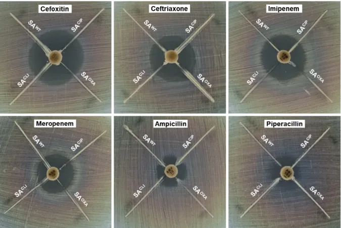
The β-lactamase-producing strains were screened by the modified Hodge assay (Fig. 1). Antibiotics are hydrolyzed by β-lactamases produced by the test strains (SA

WT

, SA

CIP

, SA

OXA

, and SA

CLI

), resulting in the growth of sensitive control strain (E. coli). No noticeable phenotypic change in the clear inhibition zones around cefoxitin, ceftriaxone, imipenem, meropenem, and piperacillin discs was observed at all test strains. In contrast, the distortion in the clear zone of inhibition around ampicillin disc was observed at SA

WT

, SA

CIP

, and SA

OXA

, except SA

CLI

. The ampicillin suscep- tibilities of SA

WT

, SA

CIP

, SA

OXA

, and SA

CLI

were evaluated in the absence and presence of medicinal plant extracts, including Cleyera japonica (CJ), Carpinus laxiflora (CL), Euscaphis japonica (EJ), Euphorbia helioscopia (EH),

Fig. 1. Modified Hodge test for β-lactamase-producing Staphylococcus aureus ATCC 15564 (SA

WT

), ciprofloxacin-induced S.

aureus ATCC 15564 (SA

CIP

), oxacillin-induced S. aureus ATCC 15564 (SA

OXA

), and clinically-isolated S. aureus CCARM 3008

(SA

CLI

) exposed to different antibiotics, including cefoxitin, ceftriaxone, imipenem, meropenem, ampicillin, and piperacillin.

(4)

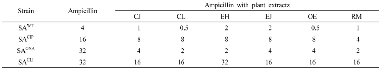
Oenothera erythrosepala (OE), and Rosa multiflora (RM) (Table 1). SA

OXA

and SA

CLI

(MIC=32 ㎍/mL) were highly resistance to ampicillin in the absence of medicinal plant extracts, followed by SA

CIP

(MIC=16 ㎍/mL) and SA

WT

(MIC=4 ㎍/mL). SA

WT

, SA

CIP

, SA

OXA

, and SA

CLI

were susceptible to all medicinal plant extracts with the exception of EH against SA

CLI

, showing no change in the MIC (32 ㎍ /mL). The MICs of ampicillin against SA

WT

, SA

CIP

, SA

OXA

, and SA

CLI

were decreased from 2 to 8 folds, 2 to 4 folds, 8 to16 folds, and 1 to 2 folds, respectively, in the presence of CJ, CL, EH, EJ, OE, and RM.

Inhibitory effect of medicinal plant extracts on the produc- tion of β-lactamases by SA

WT

, SA

CIP

, SA

OXA

, and SA

CLI

The extracellular β-lactamase activities of SA

WT

, SA

CIP

, SA

OXA

, and SA

CLI

were estimated in the presence of CJ, CL, EH, EJ, OE, and RM, compared to the control (Fig. 2). The highest β-lactamase activities were observed in SA

WT

, SA

CIP

, and SA

OXA

in the absence of medicinal plant extracts, showing 39, 32, and 43 µmol/min/mL, respectively. SA

CLI

exhibited the lowest β-lactamase activity (3 µmol/min/mL), which was not significantly changed in the presence of CJ, CL, EH, EJ, OE, and RM (p > 0.05). The β-lactamase inhibitor, Table 1. MICs of ampicillin against Staphylococcus aureus ATCC 15564 (SA

WT

), ciprofloxacin-induced S. aureus ATCC 15564 (SA

CIP

), oxacillin-induced S. aureus ATCC 15564 (SA

OXA

), and clinically-isolated S. aureus CCARM 3008 (SA

CLI

) treated without and with medicinal plant extracts

z

Strain Ampicillin Ampicillin with plant extractz

CJ CL EH EJ OE RM

SA

WT

4 1 0.5 2 2 0.5 1

SA

CIP

16 8 8 8 8 8 4

SA

OXA

32 4 2 2 4 4 2

SA

CLI

32 16 16 32 16 16 16

z

Medicinal plant extracts include Cleyera japonica (CJ), Carpinus laxiflora (CL), Euscaphis japonica (EJ), Euphorbia helioscopia (EH), Oenothera erythrosepala (OE), and Rosa multiflora (R M).

Fig. 2. Hydrolyzing activity of β-lactamases produced by Staphylococcus aureus ATCC 15564 (SA

WT

), ciprofloxacin-induced S.

aureus ATCC 15564 (SA

CIP

), oxacillin-induced S. aureus ATCC 15564 (SA

OXA

), and clinically-isolated S. aureus CCARM 3008

(SA

CLI

) treated with BLI-489 and medicinal plant extracts (Cleyera japonica, CJ; Carpinus laxiflora, CL; Euphorbia helioscopia,

EH; Euscaphis japonica, EJ; Oenothera erythrosepala, OE; Rosa multiflora, RM). Letters (a-c) on the bars are significantly different

among treatments within each strain at p < 0.05.

(5)

BLI-489, used as a positive control effectively reduced the β -lactamase activities of SA

WT

, SA

CIP

and SA

OXA

up to less than 7 µmol/min/mL (> 80% reduction).

Discussion

Since some bacteria can evolve antibiotic resistance mediated by the production of β-lactamases, the discovery of natural β -lactamase inhibitors can provide a clue for the control of antibiotic-resistant pathogens. Novel β-lactamase inhibitors can help to make the best reuse of unnoticed and outdated antibiotics.

The distortion in the clear zone of inhibition around ampicillin disc (Fig. 1) suggests that the reduced susceptibility of E. coli to ampicillin was mediated by ampicillin-hydrolyzing enzymes produced by SA

WT

, SA

CIP

, and SA

OXA

, whereas SA

CLI

did not produce β-lactamases. As a result, ampicillin was selected for further β-lactamase inhibition study. The specific types of β-lactamases, such as penicillinases and cephalosporinases, have their own specificity for β-lactam antibiotics based on the substrate specificity (Bauernfeind, 1986). The β-lactamases are divided into four classes, A (Bla-A, SHV, and TEM), B (IMP, SPM, and VIM), C (AmpC, Bla-B, and FOX), and D (OXA), and also classified into class I through VI (Barry and Barlow, 2005; Bauern- feind, 1986). The class A β-lactamases (class II, III, and V) are resistant to ESBLs, associated with the overexpression of AmpC and TEM (Féria et al., 2002). The class B β -lactamases are resistant to most β-lactam antibiotics (carbapenem) and β-lactamase inhibitors (clavulanic acid) (Hanson and Sanders, 1999; Payne et al., 1994). The class C β-lactamases (class I) include an inducible cephalosporinase (Hanson and Sanders, 1999; Manchanda and Singh, 2003).

The class D β-lactamases (class IV) include OXA β -lactamase (Bauernfeind, 1986).

Previous studies reported that the medicinal plant extracts, CJ, CL, EH, EJ, OE, and RM, inhibited the growth of Helicobacter pylori, Listeria monocytogenes, S. aureus, S.

epidermidis, Salmonella enterica, and Escherichia coli (Cendrowski et al., 2020; Elizaquível et al., 2013; Hou et al., 2003). The antimicrobial activity of CJ, CL, EH, EJ, OE, and RM was not considered in this study because the ampicillin

treatments were combined with a quarter MIC of each extract that might not affect the viability of SA

WT

, SA

CIP

, SA

OXA

, and SA

CLI

. The decrease in the MICs of ampicillin combined with CJ, CL, EH, EJ, OE, and RM might be due to the inhibition of β-lactamase activity and/or PBP2a (Catteau et al., 2017). The medicinal plant extracts containing flavonoids, tannins, flavan-3-oleflavonoid, and epicatechin gallate can inhibit β -lactamase activity, leading to the inhibition of hydrolysis of β-lactam antibiotics (Eumkeb et al., 2012; Stapleton et al., 2004; Zhao et al., 2002). Therefore, the increased suscepti- bilities of SA

WT

, SA

CIP

, and SA

OXA

to ampicillin in the presence of medicinal plant extracts imply that the medicinal plant extracts used in this study acted as β-lactamase inhibitor.

However, the ampicillin-resistant SA

CLI

(32 ㎍/mL) might be more associated with modified PBPs (PBP2a) that contributes to the resistance to β-lactam antibiotics, rather than the production of β-lactamases (Llarrull et al., 2009).

The β-lactamase inhibitor, BLI-489, effectively reduced the β-lactamase activities of SA

WT

, SA

CIP

and SA

OXA

(Fig. 2).

BLI-489 is a broad-spectrum β-lactamase inhibitor, which is effective against class A, C, and D β-lactamases when combined with piperacillin (Venkatesan et al., 2004). The common β-lactamase inhibitors, sulbactam and clavulanic acid, effectively inhibit the activity of extended-spectrum β -lactamases (ESBLs), but less effective against AmpC (Liu et al., 2016). The ability of CJ, CL, EH, EJ, OE, and RM to inhibit β-lactamases varied with strains used in this study (Fig. 2). The intrinsic β-lactamase activities of SA

WT

, SA

CIP

, and SA

OXA

were decreased by 55-78%, 27-57%, and 65-76%, respectively, in the presence of medicinal plant extracts (Fig.

2). The highest reductions in β-lactamase activities of SA

WT

, SA

CIP

, and SA

OXA

were 78%, 57%, and 76%, respectively, for OE, EJ, and CL. These results are in a good agreement with previous report that medicinal plant extracts containing quercetin, naringenin, and catechin could act as β-lactamase inhibitors for enhancing antimicrobial activity (Boussoualim and Abderrahmane, 2011; Eumkeb et al., 2010). These β -lactamase inhibitors can directly interfere with β-lactamases or compete for the binding sites of β-lactamases (Catteau, et al., 2017).

In conclusion, the most significant finding was that

medicinal plant extracts, including CJ, CL, EH, EJ, OE, and

(6)

RM, potentiated the ampicillin activity against SA

WT

, SA

CIP

, and SA

OXA

. The β-lactamase activities of SA

WT

, SA

CIP

, and SA

OXA

were significantly reduced by 55-78%, 27-57%, and 65-76%, respectively, in the presence of CJ, CL, EH, EJ, OE, and RM, which were corresponded to the increased susceptibilities of SA

WT

, SA

CIP

, and SA

OXA

treated with CJ, CL, EH, EJ, OE, and RM. Therefore, the medicinal plant extracts used in this study could be used as BLIs to enhance the ampicillin activity against SA

WT

, SA

CIP

, and SA

OXA

. The antibiotic resistance mechanism-targeted approach can be a promising alternative for the control of MRSA. However, further study is needed to identify bioactive compounds in the medicinal plant extracts and elucidate their mechanisms for therapeutic use in combination with other antibiotics.

Abbreviations

Staphylococcus aureus ATCC 15564, SA

WT

; Clinically- isolated Staphylococcus aureus CCARM 3008, SA

CLI

; Cipro- floxacin-induced Staphylococcus aureus ATCC 15564, SA

CIP

; Oxacillin-induced Staphylococcus aureus ATCC 15564, SA

OXA

; Cleyera japonica, CJ; Carpinus laxiflora, CL;

Euphorbia helioscopia, EH; Euscaphis japonica, EJ; Oeno- thera erythrosepala, OE; Rosa multiflora, RM.

Acknowledgements

This research was supported by Basic Science Research Program through the National Research Foundation of Korea (NRF) funded by the Ministry of Education (NRF-2016R1D1 A3B01008304).

Conflict of Interest

There are no conflicts of interest to declare.

References

Akova, M. 2008. Sulbactam-containing b-lactamase inhibitor combinations. Clin. Microbiol. Infect. 14:185-88.

Amjad, A., I. Mirza, S. Abbasi, U. Farwa, N. Malik and F. Zia.

2011. Modified Hodge test: A simple and effective test for detection of carbapenemase production. Iran J Microbiol

3:189-93.

Anderson, K. F. , D. R. Lonsway, J. K. Rasheed, J. Biddle, B.

Jensen, L.K. McDougal, R.B. Carey, A. Thompson, S.

Stocker, B. Limbago and J.B. Patel. 2007. Evaluation of methods to identify the Klebsiella pneumoniae carbapenemase in Enterobacteriaceae. J. Clin. Microbiol. 45:2723-25.

Aparna, V., K. Dineshkumar, N. Mohanalakshmi, D. Velmuru- gan and W. Hopper. 2014. Identification of natural compound inhibitors for multidrug efflux pumps of Escherichia coli and Pseudomonas aeruginosa using In silico high-throughput virtual screening and in vitro validation. PLoS One 9:

e101840.

Barry, G.H. and M. Barlow. 2005. Revised Ambler classification of b-lactamases. J. Antimicrob. Chemother. 55:1050-51.

Bauernfeind, A. 1986. Classification of β-lactamases. Rev.

Infect. Dis. 8:S470-S81.

Boussoualim, N. and B. Abderrahmane. 2011. Kinetic study of different flavonoids as inhibitors of beta-lactamase enzyme.

Afr. J. Biochem. Res. 5:321-27.

Bush, K. and P.A. Bradford. 2016. β-Lactams and β-lactamase inhibitors: An overview. Cold Spring Harbor Perspect. Med.

6:a025247.

Catteau, L., N.T. Reichmann, J. Olson, M.G. Pinho, V. Nizet, F.

Van Bambeke and J. Quetin-Leclercq. 2017. Synergy between ursolic and oleanolic acids from Vitellaria paradoxa leaf extract and β-lactams against methicillin-resistant Staphy- lococcus aureus: In vitro and in vivo activity and underlying mechanisms. Molecules 22:2245.

Cendrowski, A., K. Kraśniewska, J. Przybył, A. Zielińska and S. Kalisz. 2020. Antibacterial and antioxidant activity of extracts from rose fruits (Rosa rugosa). Molecules 25:E1365.

Cheng, M.P., R.S. Lee, A.P. Cheng, S. De L’étoile-Morel, K.

Demir, C.P. Yansouni, P. Harris, E.G. McDonald and T.C.

Lee. 2019. Beta-lactam/beta-lactamase inhibitor therapy for potential AmpC-producing organisms: A systematic review and meta-analysis. Open Forum Infect. Dis. 6:ofz248.

CLSI. 2015. Methods for dilution antimicrobial susceptibility tests for bacteria that grow aerobically. Approved Standard M07-A10.

Elizaquível, P., M. Azizkhani, G. Sánchez and R. Aznar. 2013.

Evaluation of Zataria multiflora Boiss. essential oil activity against Escherichia coli O157:H7, Salmonella enterica and Listeria monocytogenes by propidium monoazide quantitative PCR in vegetables. Food Cont. 34:770-776.

Eumkeb, G., S. Sakdarat and S. Siriwong. 2010. Reversing β

(7)

-lactam antibiotic resistance of Staphylococcus aureus with galangin from Alpinia officinarum Hance and synergism with ceftazidime. Phytomedicine 18:40-45.

Eumkeb, G., S. Siriwong, S. Phitaktim, N. Rojtinnakorn and S.

Sakdarat. 2012. Synergistic activity and mode of action of flavonoids isolated from smaller galangal and amoxicillin combinations against amoxicillin‐resistant Escherichia coli.

J. Appl. Microbiol. 112:55-64.

Féria, C., E. Ferreira, J.D. Correia, J. Gonçalves and M. Caniça.

2002. Patterns and mechanisms of resistance to b-lactams and b-lactamase inhibitors in uropathogenic Escherichia coli isolated from dogs in Portugal. J. Antimicrob. Chemother.

49:77-85.

Fuda, C.C.S., J.F. Fisher and S. Mobashery. 2005. β-Lactam resistance in Staphylococcus aureus: The adaptive resistance of a plastic genome. Cell. Mol. Life Sci. 62:2617.

Hanson, N.D. and C.C. Sanders. 1999. Regulation of inducible AmpC beta-lactamase expression among Enterobacteriaceae.

Curr. Pharmaceut. Design. 5:881-894.

Hou, W.C., R.D. Lin, K.T. Cheng, Y.T. Hung, C.H. Cho, C.H.

Chen, S.Y. Hwang and M.H. Lee. 2003. Free radical- scavenging activity of Taiwanese native plants. Phytomedicine 10:170-175.

Lamers, R.P., J.F. Cavallari and L.L. Burrows. 2013. The efflux inhibitor phenylalanine-arginine beta-naphthylamide (PAβ N) permeabilizes the outer membrane of gram-negative bacteria. PLoS One 8:e60666.

Lee, S.-H., T.-W. Jang, J.-S. Choi, J.-Y. Mun and J.-H. Park.

2019. Inhibitory effects of pine cone Pinus densiflora on melanogenesis in B16F10 melanoma cells. Korean J. Plant Res. 32:275-81.

Liu, X., K. Thungrat and D.M. Boothe. 2016. Occurrence of OXA-48 carbapenemase and other b-lactamase genes in ESBL-producing multidrug resistant Escherichia coli from dogs and cats in the United States, 2009-2013. Front.

Microbiol. 7:1057.

Llarrull, L.I., J.F. Fisher and S. Mobashery. 2009. Molecular basis and phenotype of methicillin resistance in Staphy- lococcus aureus and insights into new β-lactams that meet the challenge. Antimicrob. Agent Chemother. 53:4051-63.

Manchanda, V. and N.P. Singh. 2003. Occurrence and detection of AmpC b-lactamases among Gram-negative clinical isolates using a modified three-dimensional test at Guru Tegh Bahadur Hospital, Delhi, India. J. Antimicrob. Chemother.

51:415-18.

Matsumoto, Y., K. Hayama, S. Sakakihara, K. Nishino, H. Noji, R. Iino and A. Yamaguchi. 2011. Evaluation of multidrug efflux pump inhibitors by a new method using microfluidic channels. PLoS One 6:e18547.

Michéa-Hamzehpour, M., A. Kahr and J.C. Pechère. 1994. In vitro stepwise selection of resistance to quinolones, β-lactams and amikacin in nosocomial gram-negative bacilli. Infection 22:S105-S110.

Payne, D.J., R. Cramp, J.H. Bateson, J. Neale and D. Knowles.

1994. Rapid identification of metallo- and serine b-lactamases.

Antimicrob. Agent. Chemother. 38:991-96.

Rubens, D.M., O.O. Constantin, A.-A. Moevi, G.K. Nathalie, T. Daouda, N.J. David, D. Mireille and D.A. Joseph. 2015.

Anti Staphylococcus aureus activity of the aqueous extract and hexanic fraction of Thonningia sanguinea (Cote ivoire).

Int. J. Pharmaco. Phytochem. Res. 7:301-06.

Rumbo, C., E. Gato, M. López, C. Ruiz de Alegría, F. Fernández- Cuenca, L. Martínez-Martínez, J. Vila, J. Pachón, J.M.

Cisneros, J. Rodríguez-Baño, A. Pascual, G. Bou and M.

Tomás. 2013. Contribution of efflux pumps, porins, and b-lactamases to multidrug resistance in clinical isolates of Acinetobacter baumannii. Antimicrob. Agent. Chemother.

57:5247-5257.

Sibanda, T. and A. Okoh. 2007. The challenges of overcoming antibiotic resistance: Plant extracts as potential sources of antimicrobial and resistance modifying agents. Afr. J.

Biotechnol. 6:2886-2896.

Sood, S. 2013. Comparative evaluation of the in-vitro activity of six β-lactam/β-lactamase inhibitor combinations against Gram negative Bacilli. J. Clin. Diagn. Res. 7:224-28.

Stapleton, P., S. Shah, J. Anderson, Y. Hara, J. Hamilton-Miller and P. Taylor. 2004. Modulation of b-lactam resistance in Staphylococcus aureus by catechins and gallates. Int. J.

Antimicrob. Agents 23:462-67.

Sun, S., M. Selmer and D.I. Andersson. 2014. Resistance to β- lactam antibiotics conferred by point mutations in penicillin- binding proteins PBP3, PBP4 and PBP6 in Salmonella enterica. PLoS One 9:e97202.

Venkatesan, A.M., A. Agarwal, T. Abe, H. Ushirogochi, I.

Yamamura, T. Kumagai, P.J. Petersen, W.J. Weiss, E. Lenoy, Y. Yang, D.M. Shlaes, J.L. Ryan and T.S. Mansour. 2004.

Novel imidazole substituted 6-methylidene-penems as broad-spectrum β-lactamase inhibitors. Bioorg. Med.

Chem. 12:5807-5817.

Zabawa, T.P., M.J. Pucci, T.R. Parr and T. Lister. 2016.

(8)

Treatment of Gram-negative bacterial infections by potenti- ation of antibiotics. Curr. Opin. Microbiol. 33:7-12.

Zhao, W.-H., Z.-Q. Hu, Y. Hara and T. Shimamura. 2002.

Inhibition of penicillinase by epigallocatechin gallate

resulting in restoration of antibacterial activity of penicillin against penicillinase-producing Staphylococcus aureus.

Antimicrob. Agent Chemother. 46:2266-68.

(Received 20 May 2020 ; Revised 14 September 2020 ; Accepted 14 September 2020)

수치

Fig. 1. Modified Hodge test for β-lactamase-producing Staphylococcus aureus ATCC 15564 (SA WT ), ciprofloxacin-induced S
Fig. 2. Hydrolyzing activity of β-lactamases produced by Staphylococcus aureus ATCC 15564 (SA WT ), ciprofloxacin-induced S

참조

관련 문서