Purification of an Antibacterial Peptide from the Gills of the Pufferfish Takifugu pardalis
Tae Young Kim, Hye-Jin Go and Nam Gyu Park*
Department of Biotechnology, College of Fisheries Sciences, Pukyong National University, Busan 48513, Korea Received September 28, 2016 /Revised November 7, 2016 /Accepted November 25, 2016
An antibacterial peptide was purified from an acidified gill extract of the pufferfish Takifugu pardalis.
The acidified gill extract was put through a Sep-Pak C18 solid phase extraction cartridge using a step- wise gradient and divided into a flow-through (F.T.) and 60% methanol fraction (RM 60). Among the eluents, RM 60 had potent antibacterial activity against Bacillus subtilis KCTC 1021. RM 60 was parti- ally purified on a cationic-exchange column (SP-5PW) by a linear gradient, and the antibacterial pep- tide was then further purified, using a series of cationic-exchange and C
18reversed-phase HPLC columns. For characterization of the purified peptide, its molecular weight and amino acid sequence were analyzed by MALDI-TOF MS and Edman degradation. The molecular weight of the peptide was about 1171.6 Da. The amino acid sequence of the peptide was partially determined as: STKEKAPRKQ.
A comparison of the N-terminal amino acid sequence of the purified peptide with that of other known polypeptides revealed high homology with the N-terminus of the histone H3 protein, which belongs to the histone H3 family. Thus, this peptide was designated as a puffer fish gill (PFG)-related antimicrobial peptide. This is the report to describe an antimicrobial function for the N-terminus of histone H3 of an animal species. The findings suggest that this peptide plays a significant role in the innate defense system of the pufferfish.
Key words : Antibacterial peptide, gill, pufferfish, purification
*Corresponding author
*Tel : +82-51-629-5867, Fax : +82-51-629-5863
*E-mail : [email protected]
This is an Open-Access article distributed under the terms of the Creative Commons Attribution Non-Commercial License (http://creativecommons.org/licenses/by-nc/3.0) which permits unrestricted non-commercial use, distribution, and reproduction in any medium, provided the original work is properly cited.
Journal of Life Science 2017 Vol. 27. No. 1. 50~56 DOI : https://doi.org/10.5352/JLS.2017.27.1.50
서 론
나방(Hyatophora cecropia)의 hemolymph로부터 항균성 펩 타이드인 cecropin이 발견된[13] 이후로, 비교적 저분자량을 지닌 항균성 펩타이드의 정제 및 특성에 관한 많은 연구가 활발히 진행되어 오고 있다. 지금까지 항균성 펩타이드는 척 추동물, 무척추동물, 곤충, 식물 및 미생물에 이르기까지 다양 한 생물로부터 발견되었다[8, 10, 37]. 이들 물질들은 그램 양성 및 음성균, 효모, 곰팡이, 바이러스 및 기생충 등을 포함한 다 양한 병원체들에 대해 광범위한 항균효과를 나타낸다[4, 11, 26]. 어류는 병원균이 많이 존재하는 수용성 환경에서 생활을 하고 있기 때문에, 수많은 병원균에 항상 노출되어 있다. 이러 한 이물질의 침입에 방어하기 위해서 어류들은 효과적인 일차 적인 방어 기능인 선천성 면역 반응이 필요하다[17]. 어류들은 세포-매개체를 통한 반응과 항체를 포함한 특이적인 면역반응 체계를 지니고 있지만, 육상과는 달리 고압, 저온, 고염을 포함 한 극한 환경하에서 생활하고 있어서 어류의 면역체계가 정상 적으로 활성화되기까지는 비교적 오랜 시간이 필요하다[7,
35]. 그러나 어류의 비특이적 면역체계는 침입한 병원체에 대 해 신속하게 대응하여 비특이적으로 병원체에 대항하는 물질 을 발현하고, 생성시켜 자신을 보호한다[2, 12, 15]. 그러므로 어류는 그들의 환경에 광범위하게 존재하는 병원균 및 환경의 변화에 대해 생체방어 또는 항상성을 유지하기 위해서 일차적 으로 그들의 비특이적인 면역반응에 의존하게 된다[35]. 비특 이적인 면역반응의 요소들 중에서 특히 항균활성 펩타이드가 어류의 면역에 관해 중요한 역할을 지닌다는 보고가 발표되고 있다[1, 24, 32]. 최근 어류의 껍질, 점액질, 및 아가미와 같은 일차적인 방어 기관으로부터 piscidin 및 pleurocidin을 포함 한 다양한 종류의 항균활성 펩타이드가 정제되었다[6, 21, 22, 28, 31, 32, 34]. 이들 항균활성 펩타이드들은 곰팡이 뿐만 아니 라 다양한 세균들에 대해 항균작용을 나타낸다[5, 14, 33, 36].
어류로부터 이들 항균활성 펩타이드들의 발견과 항균활성의 발현은 비특이적인 면역체계가 일차적인 방어기관에서 매우 중요한 역할을 하고 있다는 것을 제시하고 있다. 최근 생물 자원의 제한성 및 환경적인 문제로 인해 어류의 가공으로부터 폐기되는 부산물의 이용과 재활용에 관한 관심이 점차적으로 증대되고 있다[18]. 한편, 어류의 미이용자원인 아가미를 포함 한 두부, 내장 및 껍질은 다양한 종류의 생리활성 물질을 다량 포함하고 있다고 알려져 있다[9]. 이러한 비가식부인 폐기물들 을 이용하여 어류 질병의 항생제로 사용될 수 있는 새로운 항균활성 펩타이드를 개발한다면, 미이용자원으로부터 고부 가가치를 지닌 새로운 물질을 발견할 수 있으리라 생각된다.
어류로부터 다양한 종류의 항균활성 펩타이드가 발견되었지
만 아직까지 졸복의 부산물인 아가미로부터 새로운 기능을 지니는 생리활성 물질 개발에 관한 연구는 그다지 많지 않다.
따라서, 본 연구에서는 졸복의 아가미 추출물을 사용하여 항 균활성을 나타내는 물질을 정제하고자 하였다.
재료 및 방법
실험동물
실험에 사용한 졸복(Takifugu pardalis, 30마리, 체장 5~7 cm) 는 2015년 2월에 부산시 남구 섬마을수산에서 구입하였다. 졸 복으로부터 아가미를 분리한 후, 즉시 액체질소에 동결시켜 실험 전까지 -75℃에 보관하였다.
시약 및 재료
Sep-Pak Vac C18 cartridge는 Waters사 (Waters associates, Miliford, MA, USA)에서 구입하였다. Tryptic soy broth (TSB) 와 Lactose-Boullion (LB)는 Merck사 (Merck, Darmstadt, Germany)에서, Agarose (Low EEO Agar)는 Sigma사 (St.
Louis, MO, USA)에서 각각 구입하였다. HPLC-grade의 water (H
2O) 및 acetonitrile (CH
3CN)은 J.T. Baker사 (NJ, USA)에서 구입하였고, 그 이외의 모든 시약은 특급을 사용하였다.
졸복 아가미 조직으로부터 추출물의 조제
동결 보관된 졸복의 아가미 조직(50 g)에 200 ml의 1% 초산 을 첨가한 뒤 100℃에서 5분간 끓인 후 냉각시켰다. 조직을 homogenizer (IKAⓇ basic ULTRA-TURRAX, USA)로 파쇄 하여 시료를 4℃에서 30분 동안 13,000 x g로 원심분리를 하여 상층액을 농축하였다(65 ml). 그 후, Sep-Pak C18 cartridge (20 ml, Waters)에 농축액을 주입하여 0.1% trifluoroacetic acid (TFA), 0.1% TFA가 포함된 60% 메탄올(RM60)로 물질을 용출시켰다(50 ml). 항균활성을 측정하기 위해 RM60의 1/
10,000, 1/5,000 및 1/2,500에 해당하는 양을 사용하였으며, 사 용 균주로는 Bacillus subtilis KCTC 1021을 사용하였다.
항균활성의 측정
정제과정 및 정제한 물질에 대한 항균활성은 이전의 방법과 동일한 방법으로 측정하였다[27]. 우선 배지 및 배양온도에서 18시간 동안 전 배양한 다음, BioMerieux Vitek, Inc. Colorim- eter (Product No. 52-1210, BioMerieux, Inc., USA)를 사용하 여 농도를 84%T (≒ 1×10
8CFU/ml)가 되게 맞추었다. 그 후, 9.5 ml의 0.03% TSB, 1% Type I agarose 및 10 mM phosphate buffer (pH 6.5)를 포함하는 underlay gel에 84%T로 희석된 균 0.5 ml을 넣고 잘 섞은 후에 plate에 편평하게 부어 굳히고, 굳은 plate에 punch를 사용하여 직경 2.5 mm의 well을 뚫은 후에, 추출물 총 부피의 1/100을 도입시켰다. 모든 sample은 0.01% acetic acid (pH 4.0)에 녹여 활성을 측정하였으며, neg-
ative control로 0.01% acetic acid (pH 4.0) 5 μl를 사용하여 용매에 의한 영향이 없음을 확인하였다. 추출물이 배지에 스 며들면 3시간 동안 1차 배양한 후, 그 위에 10 ml의 6% TSB, 1% Type I agarose 및 10 mM phosphate buffer (pH 6.5)를 포함하는 overlay gel을 붓고 굳힌 후에 동일한 온도에서 18시 간 동안 2차 배양하였다. 다음 날, well 주위의 clear zone의 유무 및 clear zone diameter (mm)를 확인하여 활성의 세기를 측정하였다. 한편, 정제한 물질의 pH, 온도, 및 trypsin 효과에 대한 실험은 이전의 방법과 동일한 방법으로 측정하였다[28].
실험에 사용한 물질은 20 μg/ml을 사용하였으며, 사용 균주는
B. subtilis KCTC 1021을 사용하였다. 우선, 온도에 대한 효과를 알아보기 위해서 물질은 100℃에서 10분간 처리하여 항균 효과를 측정하였다. 물질의 pH 변화, 산성 및 염기성에 대한 항균효과는 0.01% HCl 및 0.01% NaOH를 각각 처리하여 37℃
에서 10분간 방치한 후 측정하였다. Trypsin에 대한 물질의 항균효과는 250 mg/ml의 crystalline trypsin (Fisher Scientific, Fairlawn, NJ, USA)을 첨가하고 37 ℃에서 60분간 방치한 후 측정하였다.
항균활성 펩타이드의 정제
강한 항균활성을 나타내는 RM60 (50 ml) 용액을 농축시킨 뒤, 농축시킨 용액(2 ml)을 High Performance Liquid Chro- matography (HPLC)로 정제하였다. 첫 번째 단계로 cation-ex- change column인 TSK-gel SP-5PW (7.5 mm X 75 mm, Tosoh, Japan)를 사용하여 다음과 같은 분리조건으로 정제하였다: A 용매; 20 mM citric buffer (pH 6.0), B용매; 1.0 M NaCl을 포함 하는 20 mM citric buffer (pH 6.0), 분리조건; 0 M NaCl (45 min), 0 M → 1 M NaCl (50 min), 유속; 1.0 ml/min. 분리된 각각의 분획들은 1/200에 해당하는 양을 사용하여 URDA로 활성을 측정하였다. 두 번째 단계로, 첫 번째 단계에서 활성을 나타낸 분획들을 CapcellPak C
18(4.6 mm × 250 mm, Shisei- do, Japan) column에 주입하여 정제하였으며 분리조건은 다 음과 같다: A 용매; 0.1% TFA를 포함하는 H
2O (pH2.2), B용매;
0.1% TFA를 포함하는 CH
3CN (pH 2.2), 분리조건; 5 → 65%
(60 min), 파장; 220 nm, 유속; 1.0 ml/min. 분획은 fraction collector를 이용하여 1 분당 1 ml씩 받았으며, 분리된 각각의 분획들은 1/50에 해당하는 양을 사용하여 URDA로 활성을 측정하였다. 세 번째 단계로, 활성을 지닌 분획들을 cation-ex- change column인 Mono-S
ⓇHR5/5 (Pharmacia Biotech, Sweden)를 사용하여 다음과 같은 분리조건으로 정제하였다.
A 용매; 20 mM citric buffer (pH 6.0), B 용매; 1.0 M NaCl을 포함하는 20 mM citric buffer (pH 6.0), 분리조건; 0 M→ 1 M NaCl (50 min), 파장; 220 nm, 유속; 0.5 ml/min. 네 번째 단계로 활성분획을 CapcellPak C
18(4.6×250 mm) column에 주입하여 정제하였으며 분리조건은 다음과 같다: A 용매; 0.1%
TFA를 포함하는 H
2O (pH2.2), B용매; 0.1% TFA를 포함하는
Fig. 1. Procedures for the HPLC purification of antibacterial peptide from the gill extract of pufferfish Takifugu pardalis.
Fig. 2. Antimicrobial activity of the pufferfish’ gill RM60 eluted by Sep-Pak C18 cartridge. A; 1/2,500 of RM 60, B;
1/5,000 of RM 60, C; 1/10,000 of RM 60, D; positive con- trol (Piscidin 3 μl of 1 mg/ml), E; negative control (sterilized water).
Table 1. Effect of pH, temperature, and protease treatment on the stability of PFG against Bacillus. subtilis KCTC1021.
A; PFG, B; trypsin treatment, C; heating, D; 0.01% HCl, E; 0.01% NaOH
A B C D E CH
3CN (pH 2.2), 분리조건; 12 → 17% (10 min), 파장; 220
nm, 유속; 1.0 ml/min. 분리된 각각의 분획들은 1/5에 해당하 는 양을 사용하여 URDA로 활성을 측정하였다. 마지막 정제 단계로 활성을 지닌 분획은 동일한 CapcellPak C
18column을 사용하여 isocratic 조건으로 진행하였으며, 분리조건은 다음 과 같다: A 용매; 0.1% TFA를 포함하는 H
2O (pH2.2), B용매;
0.1% TFA를 포함하는 CH
3CN (pH 2.2), 분리조건; 14%의 iso- cratic (30 min), 파장; 220 nm, 유속; 1.0 ml/min (Fig. 1). 모든 실험은 실온에서 진행되었으며, 각각의 과정에서 분리된 분획 들은 B. subtilis KCTC 1021을 사용하여 항균활성을 측정하였 다.
분자량 및 서열분석
최종 정제한 항균활성물질의 분자량을 측정하기 위해서 Matrix-Assisted Laser Desorption Ionization (MALDI) Mass spectrometer (Voyager-DETM STR spectrometer, perseptive Biosystem, USA)를 사용하였으며, Matrix는 α-cyano-4-hy- droxycinamic acid를 사용하였다. 또한 정제한 천연물의 아미 노산 서열을 분석하기 위해서 Edman 분해법을 이용한 Shi- madzu (PPSQ-21A, Japan) 아미노산 분석기(부경대학교 공동 실험실습관)를 사용하였다.
결과 및 고찰
졸복의 아가미로부터 항균물질 추출 및 정제
졸복의 아가미 추출물을 사용하여 Sep-Pak C18 cartridge로 부분 정제한 분획인 RM 60의 1/10,000, 1/5,000, 1/2,500에 해당하는 양의 농도로 B. subtilis KCTC 1021에 대한 항균활성
을 측정하였다. 그 결과, RM 60은 각각 해당하는 농도에서 항균 반응을 나타내었다(Fig. 2). 그 결과 RM 60은 11.6~14.1 mm 크기의 항균활성을 나타내었다. Positive control로 항균 성 펩타이드인 piscidin (1 mg/ml의 3 μl)을 사용하였으며, 이 물질의 항균반응의 크기는 12.8 mm를 나타내었다.
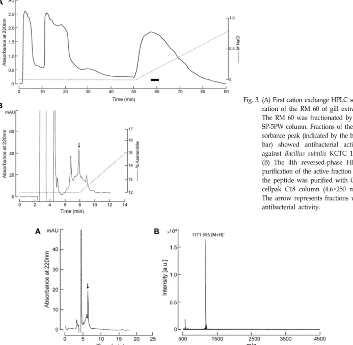
아가미 추출물(RM60)로부터 항균활성 펩타이드의 정제 첫 번째 정제단계로 RM 60을 cation-exchange column인 TSK-Gel SP-5PW를 사용하였으며, 얻어진 분획들은 각 분획 의 1/200에 해당하는 양을 사용하여 B. subtilis KCTC 1021에 대해 항균활성을 측정하였다. 그 결과 약 0.13~0.17 M에서 강 한 활성을 나타내었다(Fig. 3A의 black bar). 반응을 나타낸 활성 분획은 Fig. 1에 나타낸 것처럼 역상 및 양이온 교환 HPLC를 교차 반복적으로 사용하여 정제하였다. 마지막 단계 의 역상 HPLC (Capcellpak C
18, 4.6×250 mm)에서 14%의 iso- cratic 조건으로 최종정제 하였다(Fig. 4A). 이 물질은 Puffer- fish gill에서 정제되었기 때문에 편의상 PFG로 명명하였다.
PFG의 pH, 온도 및 trypsin 처리에 대한 항균 효과
정제한 물질의 화학적 특성인 안정성 효과를 알아보기 위해
서 pH, 온도 및 효소처리를 하여 항균활성을 측정하였다
(Table 1). PFG는 단백질 분해효소인 trypsin을 처리하기 전에
A
B
Fig. 3. (A) First cation exchange HPLC sepa-ration of the RM 60 of gill extracts.
The RM 60 was fractionated by the SP-5PW column. Fractions of the ab- sorbance peak (indicated by the black bar) showed antibacterial activity against Bacillus subtilis KCTC 1021.
(B) The 4th reversed-phase HPLC purification of the active fraction and the peptide was purified with Cap- cellpak C18 column (4.6×250 mm).
The arrow represents fractions with antibacterial activity.
A B
Fig. 4. (A) Final purification of the active fraction. The peptide was purified with Capcellpak C18 column (4.6×250 mm) column and an isocratic gradient of 14% acetonitrile in 0.1% TFA at a flow rate of 1.0 ml/min. The eluate was monitored at 220 nm. (B) The molecular weight of the purified peptide was determined using an MALDI-TOF mass spectrometer.
는 항균활성을 나타내었지만(Table 1A), trypsin을 처리한 후 에는 대부분 활성이 소실되었다(Table 1B). 그러나 열(100℃) 을 가하거나 산성 및 염기성 조건하에서는 활성이 그대로 유 지되었다(Table 1C - Table 1E). 이러한 결과로부터 PFG의 활 성은 온도 및 pH 변화에 대해 안정하지만, trypsin 처리 후에 는 반응에 민감하다는 것을 알 수 있다.
PFG의 분자량 및 아미노산 서열
정제한 펩타이드의 분자량 및 아미노산 서열을 알아보기 위해 MALDI-TOF Mass와 아미노산서열 분석기로 측정하였
다. PFG의 분자량은 1171.595 Da이었고(Fig. 4B), 이 물질의
일차구조는 10개의 아미노산 잔기로 구성된 STKEKAPRKQ-
OH로 판명되었다. 아미노산서열 분석으로부터 얻어진 PFG
의 분자량을 계산한 결과, 분자량은 1172.35 Da으로 이론치와
잘 일치하였다. 또한 이 물질의 pI값은 10.29로 매우 높은 염기
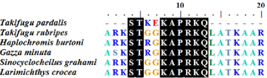
성을 지니고 있다. PFG의 아미노산 서열과 기존에 밝혀진 서
열과의 동일성(homology)을 알아보기 위해 NCBI blast를 통
해 조사한 결과, 다른 종의 졸복(Takifugu rubripes), 담수어 시
클리드(Haplochromis burtoni), 농어목 어류(Gazza minuta), 담
수어 cyprinid 어류(Sinocyclocheilus grahami), 부세(large yel-
Fig. 5. Alignment of the purified peptide amino acid sequence with the N-terminus of other histone H3: Takifugu rubripes (XP_
011607083.1), Haplochromis burtoni (XP_005947953.1), Gazza minuta |gb (AEI90816.1), Sinocyclocheilus grahami (XP_016114114.1), Larimichthys crocea (XP_010749697.1). Conserved amino acids are indicated by black boxes.
low croaker, Larimichthys crocea)에 존재하는 histone H3의 N- 말단영역과 부분적으로 유사한 것을 알 수 있었다(Fig. 5).
Histone은 염색질을 구성하는 중심 단백질이며, H1, H2, H2A, H2B, H3 및 H4와 같이 6종류의 histone이 알려져 있다.
이들은 DNA의 응축을 도우며, 유전자 발현조절에 중요한 역 할을 한다. 또한 histone은 염기성 아미노산이 많이 있기 때문 에 전체적으로 음전하를 띠는 DNA의 인산골격과의 반응에 유리할 뿐만 아니라 histone의 수용성에도 기여한다. 이러한 생화학적 특성 이외에 histone 유래의 물질들이 항균활성을 나타낸다고 보고되었다. 항균성 펩타이드는 일반적으로 산성 보다는 염기성 아미노산을 많이 포함하고 있는 물질로 알려 져 있기 때문에, 염기성 아미노산을 많이 포함하고 있는 his- tone 유래 물질도 항균성 펩타이드와 유사하게 항균효과를 나타내었으리라 생각된다. 지금까지 메기, 넙치, 연어 및 굴로 부터 histone H1, H2A 및 H2B 유래의 물질들이 항균성 펩타 이드로 발견되었다[3, 19, 20, 23, 25, 29, 30]. 그러나 histone H3과 H4에서 유래된 항균활성 펩타이드에 대한 보고는 아직 까지 알려져 있지 않다[16]. 본 연구에서 졸복의 아가미로부 터 정제된 PFG는 histone H3의 N-말단영역과 부분적으로 동 일한 서열을 나타내고 있기 때문에, PFG는 처음으로 정제된 histone H3 유래의 항균활성 펩타이드라 생각된다. 이러한 결 과는 본 연구에서 정제된 항균활성 물질이 기존에 밝혀진 histone H1, H2A 및 H2B 유래의 물질과 다른 종류의 물질이 라는 것을 의미한다. 따라서 새로이 정제된 물질은 졸복의 아 가미에 존재하고 있는 histone H3관련 항균성 물질이라 생각 되며, 이 물질은 졸복의 아가미에서 선천성 방어 시스템에서 중요한 역할을 하고 있다고 여겨진다. 현재 이 결과를 토대로 정확한 서열을 얻기 위해 추후 cDNA 클로닝을 진행할 예정 이다.
감사의 글
이 논문은 부경대학교 자율창의학술연구비(2015년)에 의하 여 연구되었음(과제번호; CD20150958).
References
1. Acosta, J., Montero, V., Carpio, Y., Velázquez, J., Garay, H.
E., Reyes, O., Cabrales, A., Masforrol, Y., Morales, A. and Estrada, M. P. 2013. Cloning and functional characterization of three novel antimicrobial peptides from tilapia (Oreochro- mis niloticus). Aquaculture 372-375, 9-18.
2. Bartlett, T. C., Cuthbertson, B. J., Shepard, E. F., Chapman, R. W., Gross, P. S. and Warr, G. W. 2002. Crustins, homo- logues of an 11.5-kDa antibacterial peptide, from two spe- cies of penaeid shrimp, Litopenaeus vannamei and Litopenaeus setiferus. Mar. Biotechnol. 4, 278-293.
3. Birkemo, G. A., Luders, T., Andersen, O., Nes, I. F. and Nissen-Meyer, J. 2003. Hipposin, a histone-derived anti- microbial peptide in Atlantic halibut (Hippoglossus hippo- glossus L.), Biochim. Biophys. Acta. 1646, 207-215.
4. Boulanger, N., Munks, R. J. L., Hamilton, J. V., Vovelle, F., Brun, R., Lehane, M. J. and Bulet, P. 2002. Epithelial innate immunity. A novel antimicrobial peptide with antiparasitic activity in the blood-sucking insect Stomoxys calcitrans. J.
Biol. Chem. 277, 49921-49926.
5. Bulet, P., Stöcklin, R. and Menin, L. 2004. Anti-microbial peptides: from invertebrates to vertebrates. Immunol. Rev.
198, 169-184.
6. Cole, A. M., Weis, P. and Diamond, G. 1997. Isolation and characterization of pleurocidin, an antimicrobial peptide in the skin secretions of winter flounder. J. Biol. Chem. 272, 12008-12013.
7. Ellis, A. E. 2001. Innate host defense mechanisms of fish against viruses and bacteria. Dev. Comp. Immunol. 25, 827- 839.
8. Fernandes, J. M. O., Molle, M. G., Kemp, G. D. and Smith, V. J. 2004. Isolation and characterisation of oncorhyncin II, a histone H1-derived antimicrobial peptide from skin secre- tions of rainbow trout, Oncorhynchus mykiss. Dev. Comp.
Immunol. 28, 127-138.
9. Ferraro, V., Cruz, I. B., Jorge, R. F., Malcata, F. X., Pintado, M. E. and Castro, P. M. L. 2010. Valorization of natural ex- tracts from marine source focused on marine byproducts:
a review. Food Res. Int. 43, 2221-2233.
10. Fu, P., Wu, J. W. and Guo, G. 2009. Purification and molec- ular identification of an antifungal peptide from the hemo-
lymph of Musca domestica (housefly). Cell. Mol. Immunol. 6, 245-251.
11. Hancock, R. E. W. 2001. Cationic peptides: effectors in innate immunity and novel antimicrobials. Lancet Infect Dis. 1, 156- 164.
12. House, R. V. and Hastings, K. L. 2004. Opinion in im- munotoxicology: multidimensional immunomodulation. J.
Immunotoxicol. 1, 123-129.
13. Hultmark, D., Steiner, H., Rasmuson, T. and Boman, H. G.
1980. Insect immunity. Purification and properties of three inducible bactericidal proteins from hemolymph of immu- nized pupae of Hyalophora cecropia. Eur. J. Biochem. 106, 7-16.
14. Izadpanah, A. and Gallo, R. L. 2005. Antimicrobial peptides.
J. Am. Acad. Dermatol. 52, 381-390.
15. Jenssen, H., Hamill, P. and Hancock, R. E. 2006. Peptide antimicrobial agents. Clin. Microbiol. Rev. 19, 491-511.
16. Kawasaki, H. and Iwamuro, S. 2008. Potential roles of histo- nes in host defense as antimicrobial agents, Infect. Disord.
Drug Targets 8, 195-205.
17. Kimbrell, D. A. and Beutler, B. 2001. The evolution and ge- netics of innate immunity. Nat. Rev. Genet. 2, 256-267.
18. Klomklao, S., Kishimura, H., Nonami, Y. and Benjakul, S.
2009. Biochemical properties of two isoforms of trypsin pu- rified from the Intestine of skipjack tuna (Katsuwonus pela- mis). Food Chem. 115, 155-162.
19. Luders, T., Birkemo, G. A., Nissen-Meyer, J., Andersen, O.
and Nes, I. F. 2005. Proline conformation-dependent anti- microbial activity of a proline-rich histone h1 N-terminal Peptide fragment isolated from the skin mucus of Atlantic salmon, Antimicrob. Agents Chemother. 49, 2399-2406.
20. Noga, E. J., Fan, Z. and Silphaduang, U. 2001. Histone-like proteins from fish are lethal to the parasitic dinoflagellate Amyloodinium ocellatum, Parasitology 123, 57-65.
21. Oren, Z. and Shai, Y. 1996. A class of highly potent anti- bacterial peptides related from pardaxin, a pore-forming peptide from the Moses sole fish Pardachirus marmoratus.
Eur. J. Biochem. 237, 304-310.
22. Park, C. B., Lee, J. H., Park, I. Y., Kim, M. S. and Kim, S.
C. 1997. A novel antimicrobial peptide from the loach, Misgurnus anguillicaudatus. FEBS. Lett. 411, 173-178.
23. Park, I. Y., Park, C. B., Kim, M. S. and Kim, S. C. 1998.
Parasin I, an antimicrobial peptide derived from histone H2A in the catfish, Parasilurus asotus, FEBS. Lett. 437, 258- 262.
24. Pasupuleti, M., Schmidtchen, A. and Malmsten, M. 2012.
Antimicrobial peptides: key components of the innate im- mune system. Crit. Rev. Biotechnol. 32, 143-171.
25. Richards, R. C., O'Neil, D. B., Thibault, P. and Ewart, K.
V. 2001. Histone H1: an antimicrobial protein of Atlantic salmon (Salmo salar), Biochem. Biophys. Res. Commun. 284, 549-555.
26. Robinson, W. E. J., McDougall, B., Tran, D. and Selsted, M.
E. 1998. Anti-HIV-1 activity of indolicidin, an antimicrobial peptide from neutrophils. J. Leukoc. Biol. 63, 94-100.
27. Seo, J. K., Crawford, J. M., Stone, K. L. and Noga, E. J. 2005.
Purification of a novel arthropod defensin from the American oyster, Crassostrea virginica. Biochem. Biophys. Res.
Commun. 338, 1998-2004.
28. Seo, J. K., Lee, M. J., Go, H. J., Park, T. H. and Park, N.
G. 2012. Purification and characterization of YFGAP, a GAPDH- related novel antimicrobial peptide, from the skin of yellowfin tuna, Thunnus albacares. Fish Shellfish Immunol.
33, 743- 752.
29. Seo, J. K., Stephenson, J., Crawford, J. M., Stone, K. L. and Noga, E. J. 2010. American oyster, Crassostrea virginica, ex- presses a potent antibacterial histone H2B protein. Mar.
Biotechnol. (NY) 12, 543-551.
30. Seo, J. K., Stephenson, J. and Noga, E. J. 2011. Multiple anti- bacterial histone H2B proteins are expressed in tissues of American oyster, Comp. Biochem. Physiol. B. Biochem. Mol.
Biol. 158, 223-229.
31. Shike, H., Lauth, X., Westerman, M. E., Ostland, V. E., Carlberg, J. M., Van Olst, J. C., Shimizu, C., Bulet, P. and Burns, J. C. 2002. Bass hepcidin is a novel antimicrobial pep- tide induced by bacterial challenge. Eur. J. Biochem. 269, 2232-2237.
32. Silphaduang, U. and Noga, E. J. 2001. Peptide antibiotics in mast cells of fish. Nature 414, 268-269.
33. Stark, M., Liu, L. P. and Deber, C. M. 2002. Cationic Hydrophobic Peptides with Antimicrobial Activity. Antimicrob.
Agents Chemother. 46, 3585-3590.
34. Subramanian, S., Ross, N. W. and MacKinnon, S. L. 2009.
Myxinidin, a novel antimicrobial peptide from the epi- dermal mucus of hagfish, Myxine glutinosa L. Mar. Biotechnol.
11, 748-757.
35. Ullal, A. J., Litaker, R. W. and Noga, E. J. 2008.
Antimicrobial peptides related from hemoglobin are ex- pressed in epithelium of channel catfish (Ictalurus punctatus, Rafinesque). Dev. Comp. Immunol. 32, 1301-1312.
36. van't Hof, W., Veerman, E. C., Helmerhorst, E. J. and Amerongen, A. V. 2001. Antimicrobial peptides: properties and applicability. Biol. Chem. 382, 597-619.
37. Zasloff, M. 2002. Antimicrobial peptides of multicellular organisms. Nature 415, 389-396.
초록:졸복의 아가미로부터 항균성 펩타이드의 정제
김태영․고혜진․박남규*
(부경대학교 수산과학대학 생물공학과)