Phenylarsine Oxide and Adenosine-sensitive Trans-golgi Complex Targeting of GFP Fused to Modified Sulfatide-binding Peptide
Yong-Woo Jun1, Jin-A Lee2* and Deok-Jin Jang1*
1Department of Ecological Science, College of Ecology and Environment, Kyungpook National University, 2559 Gyeongsang-daero, Sangju-si, Gyeongsangbuk-do 37224, Korea
2Department of Biotechnology and Biological Science, College of Life Science and Nanotechnology, Hannam University, 1646 Yuseong-daero, Yuseong-gu, Daejeon 34054, Korea
Received September 28, 2017 /Revised December 5, 2017 /Accepted December 7, 2017
Many cytoplasmic proteins are targeted to the cytoplasmic membrane of the trans-Golgi network (TGN) via an N-terminal short helix. We previously showed that the 20 N-terminal amino acids of Aplysia phosphodiesterase 4 (ApPDE4) long form are sufficient for its targeting to the plasma mem- brane and the TGN. The N-terminus of the ApPDE4 long form binds to PI4P and sulfatide in vitro.
Therefore, in order to decipher the roles of sulfatide in Golgi complex targeting, we examined the cel- lular localization of sulfatide-binding peptides. In this study, we found that enhanced green fluo- rescent protein (EGFP) fused to the C-terminus of modified sulfatide- and heparin-binding peptides (mHSBP-EGFP) was localized to the TGN. On the other hand, its mutant, in which tryptophan was replaced with an alanine, leading to the impairment of heparin and sulfatide binding, was localized to cytosol. We also found that the TGN targeting of mHSBP-EGFP is impaired by the treatment of antimycin A, phenylarsine oxide (PAO), and adenosine but not a high concentration of wortmannin.
These results suggest that PAO and adenosine-sensitive kinases, including phosphatidylinositol 4-kin- ase II, may play key roles in the recruitment of mHSBP-EGFP.
Key words : PI4KII, PI4P, sulfatide, Trans-golgi network
*Corresponding authors
*Tel : +82-42-629-8785, Fax : +82-42-629-8789
*E-mail : [email protected] (Jin-A Lee)
*Tel: +82-54-530-1213, Fax: +82-54-530-1218
*E-mail address: [email protected] (Deok-Jin Jang)
This is an Open-Access article distributed under the terms of the Creative Commons Attribution Non-Commercial License (http://creativecommons.org/licenses/by-nc/3.0) which permits unrestricted non-commercial use, distribution, and reproduction in any medium, provided the original work is properly cited.
Journal of Life Science 2018 Vol. 28. No. 2. 162~169 DOI : https://doi.org/10.5352/JLS.2018.28.2.162
Introduction
Phospholipids are the major constituents of the cellular membrane. In mammals, many proteins have structured phospholipid-binding domains, which include PROPPIN, ENTH, ANTH, the FYVE, Phox homology (PX), and pleck- strin homology (PH) domains [17]. In addition, phosphoino- sitides, which are derivatives of phosphatidylinositol (PI), are minor phospholipid components in eukaryotic mem- branes that have crucial roles in many cellular functions [4, 17]. Phosphoinositides are not randomly distributed within intracellular membranes, but rather, each species has a unique cellular localization [4, 11]. For example, phosphati- dylinositol 4-phosphate (PI4P) is enriched in the trans-Golgi
network (TGN) of cells. Therefore, many PI4P binding pro- teins are localized to the TGN. Moreover, the PH domain of oxysterol-binding protein (OSBP) and PI4P adaptor pro- tein 1/2 (FAPP1/2) binds specifically to PI4P and is mainly concentrated in the TGN [6, 8, 18].
Many proteins have a short N-terminal amphipathic helix domain, which is required for selective intracellular mem- brane targeting. For example, the N-terminal helix region of phosphodiesterase 4 (PDE4) A1 is required for TGN tar- geting, and directly interacts with the membrane by binding to phosphatidic acid (PA) [1, 9, 10]. Moreover, an isoform of the Sec14 domain in kalirin (cKalSec14) is localized to the TGN via its N-terminal amphipathic helix, which binds to cholesterol and phophoinositide [19]. The 35 N-terminal amino acids of glutamic acid decarboxylase 65 (GAD65 (N50)-EGFP) also localized to the TGN in a palmitoyla- tion-dependent manner. Additionally, we showed that the N-terminus of Aplysia PDE4 short-form was localized to the plasma membrane via electrostatic interaction mainly gen- erated by PI4P and PI(4,5)P2 at the inner leaflet of the plasma membrane [16].
Previously, it was reported that the 20 N-terminal amino acids of ApPDE4 long-form are sufficient for its targeting
to the plasma membrane and the TGN [12, 16]. The N-termi- nus of the ApPDE4 long-form binds to PI4P and sulfatide in vitro [12], and a mutant of this protein, ApPDE4 L (N20/
C14,15S), is localized to the TGN in a phosphatidylinositol 4-kinase II (PI4KII)-dependent manner [13]. Therefore, in or- der to decipher the roles of sulfatide in Golgi complex target- ing, we examined the cellular localization of sulfatide-bind- ing peptides [12]. As a result, we found that EGFP-fused modified heparin- and sulfatide-binding peptides (mHSBP) were localized to the TGN of HEK293T cells. These local- izations were impaired by the treatment of a phenylarsine oxide (PAO) and intracellular adenosine. These results sug- gest that PAO and intracellular adenosine sensitive kinase such as PI4KII play a crucial role in recruiting many cyto- plasmic proteins through its association with a protein’s N-terminal helix.
Materials and Methods
Cloning of DNA constructs
The plasmids were constructed with pcDNA3.1-EGFP [16]. The HSBP motif of the type I repeats of the human endothelial cell thrombospondin (hTHR) gene, correspond- ing to residues 1,641-1,682 of hTHR (GenBank accession no.
NM_003247.3), was generated by primer extension with syn- thetic primers and subcloned into the pcDNA3.1-EGFP vec- tor at HindIII-XbaI sites. In order to generate EGFP-fused proteins for Growth-Associated Protein-43 (GAP-43; GAP43 (N40)-EGFP), postsynaptic density protein-95 (PSD95; PSD95 (N160)-EGFP), MLCCM-HSBP-EGFP, MDCLC-HSBP-EGFP, MGSNKS-HSBP-EGFP, and MLAAM-HSBP-EGFP, the cor- responding DNA sequences were amplified by primer ex- tension with synthetic primers and inserted at the HindIII- XbaI site of the pcDNA3.1-EGFP vector. In order to generate pcDNA3.1-MLAAM-HSBP (AA)-EGFP, we amplified the PCR primers with a recombinant PCR technique and per- formed subcloning at the HindIII-XbaI site in pcDNA3.1- EGFP.
We used previously described methods to produce EGFP- and monomeric red fluorescent protein (mRFP)-fused pro- teins, including pcDNA3.1-galactose-1-phosphate uridylyl- transferase (GalT)-mRFP [16]. EGFP-FAPP1 (PH) was a gift from Dr. T. Balla [2], and mouse PSD95 was a gift from Dr.
Eun-Jun Kim [15].
Cell culture
HEK293T tumor cells were grown in Dulbecco’s modified Eagle’s medium supplemented with 10%(v/v) fetal bovine serum and penicillin/streptomycin in a humidified atmos- phere of 5%(v/v) CO2 at 37℃. For transient transfection, HEK293T cells were plated at a density of 5–7´105 cells per well in 6-well plates and cultured for 24 hr. The cells were then transfected with DNA constructs with Lipofectamine 2000 (Thermo Fisher Scientific Inc., Waltham, MA, USA) and incubated for an additional 24 hr. Images of the cells were captured with a laser scanning confocal microscope (LSM 510; Carl Zeiss AG, Jena, Germany). The anti-GM130 (BD Biosciences, San Jose, CA, USA) antibody was used as a cis- Golgi marker.
Heparin-binding assays
In order to check for protein-heparin interactions, trans- fected HEK293T cells were washed twice with 1X phos- phate-buffered saline (PBS) and lysed with 500 μl of binding buffer containing 10 mM HEPES (pH 7.4), 150 mM NaCl, 0.25% NP-40, and a protease-inhibitor cocktail (#118735800001, Roche Diagnostics Corporation, Indianapolis, IN, USA) at 4°C for 20 min. Subsequently, the sample was centrifuged at 12,000× g for 10 min. Next, 20 μl of heparin-agarose (Sigma-Aldrich Co.) was added to the cell lysate, incubated at 4 °C for 1 hr, and centrifuged at 2,500× g for 2 min. For elution, the agarose pellet was resuspended in 1 mL of bind- ing buffer and centrifuged. This step was repeated three times, and the eluted samples were then dissolved in sodium dodecyl sulfate sample buffer. Protein expression levels were detected by western blotting with an anti-GFP antibody (#75-131, NeuroMab, Davis, CA, USA).
Drug treatment
To disrupt palmitoylation, HEK293T cells were treated with 100 μM 2-bromopalmitate (2-BrP) in culture media for 4 hr. The lipid derivatives generated by various lipid kin- ases, including phosphoinositides, were depleted by treating the cells with 200 nM Antimycin A in PBS for 1 hr. The cells were treated with 10 μM phenylarsine oxide (PAO) for 30 min to inhibit the activities of PI4KIIs and PI4KIIIs. To further inhibit the activity of PI4KIIIs in cells, the cells were incubated with 10 μM wortmannin in PBS for 30 min.
Adenosine, which is a PI4KII-specific inhibitor, was ad- ministered through a combined treatment of 0.01% digitonin and 500 μM adenosine in culture media for 30 min.
A
B
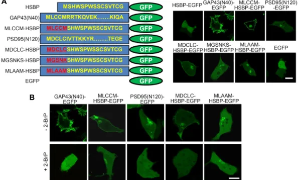
Fig. 1. Cellular expression of enhanced green fluorescent protein (EGFP)-fused heparin- and sulfatide-binding peptides (HSBP)- binding domains in HEK293T cells. (A) Schematic diagrams (left) and cellular expression of EGFP-fused modified HSBP-bind- ing domains in HEK293T cells. The EGFP-fused 40 N-terminal amino acids of human growth-associated protein-43 (GAP43;
GAP43 (N40)-EGFP) or the 120 N-terminal amino acids of human postsynaptic density protein-95 (PSD95; PSD95(N120)-EGFP) were used as controls. (B) The effects of 2-bromopalmitate (2-BrP) on the cellular localization of various EGFP-fused modified HSBP (mHSBP)-binding proteins in HEK293T cells. Scale bar, 20 μm.
Results and Discussion
Generation of EGFP-fused mHSBPs
We previously found that the N-terminus of the ApPDE4 long-form was sufficient for its targeting to the plasma mem- brane and the Golgi complex, and the N-terminus of the ApPDE4 long-form might bind to sulfatide or PI4P in vitro [12]. We also recently showed that one of the ApPDE4 long-form mutants, ApPDE4 L (N20/C14,15S), was localized to the TGN in PI4KII activity-dependent manner [13]. There- fore, we next evaluated whether sulfatide binding is in- volved in the Golgi complex targeting. To this end, we searched the literature on sulfatide-binding peptides and found the peptide SHWSPWSSCSVTCG, which belongs to an HSBP from the type I repeat of hTHR [7]. We then gen- erated an EGFP-fusion construct containing this HSBP (HSBP-EGFP) and expressed it in HEK293T cells. However, as shown in Fig. 1A, HSBP-EGFP was localized to the cytoplasm, which was similar to the control EGFP in HEK293T cells.
In order for a protein to be specifically targeted for a membrane, it usually requires a hydrophobic motif that can be stably associated with the membrane and an intracellular
targeting domain. For instance, the N-terminal amino acids 1–23 of GAD65 are involved in TGN targeting, whereas the N-terminal amino acids 31–42 of GAD65, which have dual palmitoylation sites, are involved in membrane association [14]. For H-Ras, which is localized to the plasma membrane, myristoylation is required to produce the hydrophobicity needed for membrane association, and a polybasic amino acid domain is required for plasma membrane targeting [3].
Therefore, in order to increase the membrane-binding affin- ity of HSBP-EGFP, we used two alternative strategies: (1) the addition of lipid-modifying sites to the N-terminal re- gion, as in GAD65 and H-Ras, and (2) the addition of a mod- erate hydrophobic motif within the N-terminus.
First, we added lipid-modification sites to the N-terminus of HSBP by adding palmitoylation sites either with the MLCCM sequence from the N-terminus of GAP43 (MLCCM- HSBP-EGFP) or the MDCLC sequence from the N-terminus of PSD95 (MDCLC-HSBP-EGFP), which contains dual-pal- mitoylation sequences. Alternatively, we added a myristoy- lation site, the MGSNKS sequence from the nonreceptor ty- rosine kinase, c-Src, to the N-terminus of HSBP-EGFP, which contains one myristoylation sequence (MGSNKS-HSBP-EGFP).
As a result, MLCCM-HSBP-EGFP and MDCLC- HSBP-EGFP were localized to the plasma membrane and intracellular or- ganelles (Fig. 1A). Conversely, MGSNK-HSBP- EGFP was weakly localized to intracellular organelles and not to the plasma membrane (Fig. 1A). The EGFP-fused 40 N-terminal amino acids of GAP43 (GAP43 (N40)-EGFP) or EGFP-fused 120 N-terminal amino acids of PSD95 (PSD95 (N120)-EGFP), which were controls, were localized only to the plasma membrane or intracellular organelles, respectively (Fig. 1A).
These results indicated that the addition of lipid-mod- ification sites to the N-terminus of HSBP-EGFP might induce stable intracellular membrane associations with both the plasma membrane and intracellular organelles.
In order to examine whether palmitoylation was involved in these localizations, 2-BrP (100 μM), a palmitoylation in- hibitor, was applied to cells expressing MLCCM-HSBP- EGFP or MDCLC-HSBP-EGFP. The plasma membrane local- ization of MLCCM-HSBP-EGFP was impaired after 2-BrP treatment, whereas the intracellular localization of MLCCM- HSBP-EGFP was retained (Fig. 1B). However, the membrane localization of MDCLC-HSBP-EGFP was completely dis- rupted by 2-BrP. The intracellular localization of the controls GAP43(N40)-EGFP and PSD95(N120)-EGFP was completely impaired by 2-BrP treatment (Fig. 1B). These results sug- gested that palmitoylation was involved in the plasma mem- brane localization of MLCCM-HSBP-EGFP but not in the in- tracellular localization of MLCCM-HSBP-EGFP. On the other hand, the palmitoylation of MDCLC-HSBP-EGFP was pri- marily involved in the association of MDCLC-HSBP-EGFP to both the plasma membrane and intracellular organelles.
Next, as an alternative approach, we generated a construct that would only increase the hydrophobicity of MLCCM sequences. Specifically, we replaced the third and fourth cys- teine residues with hydrophobic alanine residues (MLAAM- HSBP-EGFP). These modified domains have higher hydro- phobicity and do not have any palmitoylation sites. As shown in Fig. 1A, MLAAM-HSBP-EGFP was localized only to intracellular organelles, which was similar to the findings obtained with MGSNK-HSBP-EGFP and the 2-BrP-treated MLCCM-HSBP-EGFP (Fig. 1A, Fig. 1B). This Golgi targeting of MLAAM-HSBP-EGFP was not palmitoylation-dependent because the 2-BrP treatment had no effect on its intracellular localization (Fig. 1B).
TGN targeting of EGFP-fused modified HSBPs In order to identify the intracellular localization of
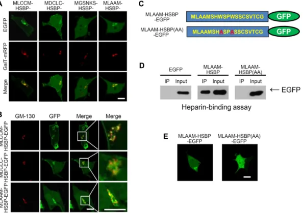
MLCCM-, MDCLC-, MGSNKN-, and MLAAM-HSBP-EGFP, each construct was co-expressed with a TGN marker, GalT- mRFP. Fig. 2A shows that all of the constructs were co-lo- calized with GalT-mRFP, thus indicating mainly TGN targeting. In addition, we examined the Golgi complex local- ization of MLCCM-HSBP-EGFP, MDCLC-HSBP-EGFP, and MLAAM-HSBP-EGFP with the cis-Golgi marker, GM130.
Fig. 2B shows that anti-GM130 signals were partially co-lo- calized with GFP signals and that the majority of the signals were localized next to GFP signals, thus indicating that modified HSBP-EGFPs (mHSBP-EGFP), including MLCCM- HSBP-EGFP, MDCLC-HSBP-EGFP, and MLAAM-HSBP-EGFP, were localized primarily to the TGN.
Next, we investigated whether peptide mutations, which resulted in a failure to bind heparin and sulfatide, would disrupt TGN targeting in HEK293T cells. A previous study has shown that the replacement of a tryptophan (W) with an alanine (A) within HSBP disrupts heparin and sulfatide binding [7]. Therefore, we constructed W-to-A-replacement- mutant peptides that were fused to EGFP (MLAAM-HSBP (AA)-EGFP) (Fig. 2C) and examined heparin binding. As shown in Fig. 2D, MLAAM-HSBP (AA)-EGFP did not bind to heparin-agarose. In addition, MLAAM-HSBP (AA) was not localized to the TGN (Fig. 2E). These results suggested that the tryptophan (W) in HSBP was critical for the specific targeting and intracellular localization of MLAAM-HSBP- EGFP. Thus, the lipid-binding properties of HSBP may be related to the TGN targeting of mHSBP-EGFP. Together, our results suggested that MLAAM-HSBP-EGFP was localized to the TGN and that the HSBP mutant, which impaired hep- arin binding disrupted its TGN localization.
Antimycin A, PAO, and intracellular adenosine- sensitive TGN targeting of MLAAM-HSBP-EGFP
Although we demonstrated that mHSBP-EGFPs were lo- calized to the Golgi complex in HEK293T cells, the molecular mechanisms underlying these localizations were not clear.
In order to investigate the molecular mechanisms of the TGN targeting of MLAAM-HSBP-EGFP in HEK293T cells, we examined the effects of pharmacological inhibitors on the cellular localization of MLAAM-HSBP-EGFP. Antimycin A, which is an ATP synthesis inhibitor, was used to deplete the lipid derivatives generated by various lipid kinases, in- cluding various PIs in cells [16]. The TGN targeting of MLAAM-HSBP-EGFP was disrupted by Antimycin A treat- ment, which was similar to the results obtained with EGFP-
A
B
C
D
E
Fig. 2. Trans-Golgi network (TGN) targeting of EGFP-fused modified HSBP (mHSBP)-binding domains. (A) The EGFP-fused mHSBP- binding domains were co-localized with galactose-1-phosphate uridylyltransferase-monomeric red fluorescent protein (GalT-mRFP), a TGN marker. (B) The EGFP-fused mHSBP-binding domains were partially co-localized with GM-130, a cis-Golgi marker. (C) Schematic diagram of the amino acid sequence of MLAAM-HSBP-EGFP and its mutant (MLAAM-HSBP (AA)-EGFP). (D) MLAAM-HSBP-EGFP and MLAAM-HSBP (AA)-EGFP binding to heparin-agarose, with EGFP used as a control. (E) The cellular localization of MLAAM-HSBP-EGFP and MLAAM-HSBP(AA)-EGFP in HEK293T cells. Scale bar, 20 μm.
FAPP1-PH (Fig. 3A). Together, these results suggested that phosphoinositides or other lipids, which are synthesized or generated by lipid kinases in an ATP-dependent manner, might be involved in the TGN targeting of MLAAM-HSBP- EGFP.
Next, we investigated whether PI4P contributed to the TGN targeting of MLAAM-HSBP-EGFP because PI4P is highly concentrated at Golgi complexes, especially the TGN.
In order to test this possibility, we applied PAO, which in- hibits PI4Ks, including PI4KII and PI4KIII, to the cells ex- pressing MLAAM-HSBP-EGFP. EGFP-FAPP1-PH, which showed the PI4P-dependent targeting to the TGN, was used as a control [2]. We observed that the MLAAM-HSBP-EGFP localization changed from the TGN to the cytosol, which was similar to the effect of PAO on EGFP-FAPP1-PH localization (Fig. 3B).
To further discriminate the molecular mechanism of the Golgi complex targeting of MLAAM-HSBP-EGFP, we treat- ed cells with adenosine, which inhibits PI4KII selectively [5].
However, adenosine itself is not able to diffuse through the plasma membrane. To overcome this limitation, we co-treat- ed adenosine with a low concentration of digitonin to parti- ally disrupt the plasma membrane. As shown in Fig. 3C, in the absence or presence of digitonin, MLAAM-HSBP- EGFP or EGFP-FAPP1-PH was localized to the Golgi com- plex, indicating that digitonin treatment has no effect on the Golgi complex targeting of the constructs. However, the ad- dition of adenosine with digitonin impaired TGN targeting of MLAAM-HSBP-EGFP. These results indicate that the Golgi complex targeting of MLAAM-HSBP-EGFP is PI4KII activity-dependent.
We subsequently used a high concentration of wortman- nin (Wm) to inhibit PI4KIII and not PI4KII. However, treat- ment with a high concentration of wortmannin (10 μM) did not disrupt the TGN targeting of MLAAM-HSBP-EGFP in HEK293T cells (Fig. 3D). These results suggested that PI4P was likely generated by PI4KII and not PI4KIII, and PI4KII may contribute to the TGN targeting of MLAAM-HSBP-
A B
C D E
Fig. 3. The Antimycin A-, phenylarsine oxide (PAO)-, and adenosine-dependent TGN targeting of MLCCM-HSBP-EGFP and MLAAM- HSBP-EGFP. (A) The effects of Antimycin A (Anti) on the cellular localization MLAAM-HSBP-EGFP and EGFP-FAPP1-PH.
(B) The effects of PAO on the cellular localization of MLAAM-HSBP and EGFP-FAPP1-PH. (C) The effects of adenosine on the cellular localization of MLAAM-HSBP and EGFP-FAPP1-PH in the presence or absence of digitonin. (D) The effects of 10 μM wortmannin (Wm) on the cellular localization of MLAAM-HSBP-EGFP. (E) The molecular structure of phosphatidyli- nositol 4-phosphate (PI4P) and sulfatide. Scale bar, 20 μm.
EGFP. However, further study is required to clarify whether PI4KIIα or β is involved in TGN targeting of MLAAM- HSBP-EGFP using PI4KIIα or β siRNA approach.
Collectively, our results suggest that the TGN targeting of MLAAM-HSBP-EGFP is probably dependent on PI4KII activity, indicating that PI4P generated by PI4KII in the TGN play crucial roles in its TGN targeting. Sulfate is attached to galactocerebroside, which generates sulfatide, while PI4P can be generated by phosphorylation at the fourth site of the inositol rings of PtdIns by PI4Ks. As shown in Fig. 3E, the charge, structure, and position of phosphates and sul- fates are similar. Thus, although PI4P and sulfatide origi- nated from different lipids, their overall allosteric head structures were similar. Therefore, it might be possible that a modified sulfatide binding peptide could be localized to the TGN via PI4P binding. Similarly, we previously reported
that the TGN localization of ApPDE4 L (N20/C14,15S)-EGFP is mediated by PI4KII activity [13]. Overall, these results sug- gest that PI4P on the cytoplasmic surface of the TGN might play crucial roles in recruiting many cytoplasmic proteins through the association with their N-terminal helix. In the future, it will be interesting to examine the PI4KII-mediated TGN targeting of other cytoplasmic proteins.
Acknowledgement
This work is supported by Hannam Research Program (2017).
References
1. Baillie, G. S., Huston, E., Scotland, G., Hodgkin, M., Gall,
I., Peden, A. H., MacKenzie, C., Houslay, E. S., Currie, R., Pettitt, T. R., Walmsley, A. R., Wakelam, M. J., Warwicker, J. and Houslay, M. D. 2002. TAPAS-1, a novel microdomain within the unique N-terminal region of the PDE4A1 cAMP- specific phosphodiesterase that allows rapid, Ca2+-trig- gered membrane association with selectivity for interaction with phosphatidic acid. J. Biol. Chem. 277, 28298-28309.
2. Balla, A., Tuymetova, G., Tsiomenko, A., Varnai, P. and Balla, T. 2005. A plasma membrane pool of phosphatidylinositol 4-phosphate is generated by phosphatidylinositol 4-kinase type-III alpha: studies with the PH domains of the oxysterol binding protein and FAPP1. Mol. Biol. Cell 16, 1282-1295.
3. Cadwallader, K. A., Paterson, H., Macdonald, S. G. and Hancock, J. F. 1994. N-terminally myristoylated Ras proteins require palmitoylation or a polybasic domain for plasma membrane localization. Mol. Cell Biol. 14, 4722-4730.
4. Di Paolo, G. and De Camilli, P. 2006. Phosphoinositides in cell regulation and membrane dynamics. Nature 443, 651- 657.
5. Dickson, E. J., Jensen, J. B. and Hille, B. 2014. Golgi and plasma membrane pools of PI(4)P contribute to plasma membrane PI(4,5)P2 and maintenance of KCNQ2/3 ion channel current. Proc. Natl. Acad. Sci. USA. 111, E2281-2290.
6. Godi, A., Di Campli, A., Konstantakopoulos, A., Di Tullio, G., Alessi, D. R., Kular, G. S., Daniele, T., Marra, P., Lucocq, J. M. and De Matteis, M. A. 2004. FAPPs control Golgi-to- cell-surface membrane traffic by binding to ARF and PtdIns (4) P. Nat. Cell Biol. 6, 393-404.
7. Guo, N. H., Krutzsch, H. C., Negre, E., Vogel, T., Blake, D. A. and Roberts, D. D. 1992. Heparin- and sulfatide-bind- ing peptides from the type I repeats of human thrombo- spondin promote melanoma cell adhesion. Proc. Natl. Acad.
Sci. USA. 89, 3040-3044.
8. Hanada, K., Kumagai, K., Yasuda, S., Miura, Y., Kawano, M., Fukasawa, M. and Nishijima, M. 2003. Molecular machi- nery for non-vesicular trafficking of ceramide. Nature 426, 803-809.
9. Houslay, M. D. and Adams, D. R. 2003. PDE4 cAMP phos- phodiesterases: modular enzymes that orchestrate signalling cross-talk, desensitization and compartmentalization. Bio- chem. J. 370, 1-18.
10. Huston, E., Gall, I., Houslay, T. M. and Houslay, M. D. 2006.
Helix-1 of the cAMP-specific phosphodiesterase PDE4A1 regulates its phospholipase-D-dependent redistribution in response to release of Ca2+. J. Cell Sci. 119, 3799-3810.
11. Jang, D. J., Park, S. W. and Kaang, B. K. 2009. The role of lipid binding for the targeting of synaptic proteins into syn- aptic vesicles. BMB Rep. 42, 1-5.
12. Jang, D. J., Park, S. W., Lee, J. A., Lee, C., Chae, Y. S., Park, H., Kim, M. J., Choi, S. L., Lee, N., Kim, H. and Kaang, B. K. 2010. N termini of apPDE4 isoforms are responsible for targeting the isoforms to different cellular membranes.
Learn. Mem. 17, 469-479.
13. Jun, Y. W., Lee, J. A., Kaang, B. K. and Jang, D. J. 2017.
PI4KII activity-dependent Golgi complex targeting of Aplysia phosphodiesterase 4 long-form mutant. Animal Cells Syst. 21, 316-322.
14. Kanaani, J., Patterson, G., Schaufele, F., Lippincott-Schwartz, J. and Baekkeskov, S. 2008. A palmitoylation cycle dynam- ically regulates partitioning of the GABA-synthesizing en- zyme GAD65 between ER-Golgi and post-Golgi membranes.
J. Cell Sci. 121, 437-449.
15. Kim, E., Niethammer, M., Rothschild, A., Jan, Y. N. and Sheng, M. 1995. Clustering of Shaker-type K+ channels by interaction with a family of membrane-associated guanylate kinases. Nature 378, 85-88.
16. Kim, K. H., Jun, Y. W., Park, Y., Lee, J. A., Suh, B. C., Lim, C. S., Lee, Y. S., Kaang, B. K. and Jang, D. J. 2014. Intracellu- lar membrane association of the Aplysia cAMP phosphodies- terase long and short forms via different targeting mecha- nisms. J. Biol. Chem. 289, 25797-25811.
17. Lemmon, M. A. 2008. Membrane recognition by phospholi- pid-binding domains. Nat. Rev. Mol. Cell Biol. 9, 99-111.
18. Levine, T. P. and Munro, S. 2002. Targeting of Golgi-specific pleckstrin homology domains involves both PtdIns 4-kin- ase-dependent and -independent components. Curr. Biol. 12, 695-704.
19. Miller, S., Sparacio, S. and Bartenschlager, R. 2006. Subcellu- lar localization and membrane topology of the Dengue virus type 2 Non-structural protein 4B. J. Biol. Chem. 281, 8854- 8863.
초록:Phenylarsine oxide와 adenosine에 민감한 sulfatide 결합 펩타이드의 trans-golgi network 타 기팅
전용우1․이진아2*․장덕진1*
(1경북대학교 생태환경대학 생태과학과, 2한남대학교 생명나노과학대학 생명시스템과학과)
세포기질에 존재하는 많은 종류의 단백질들은 N-말단에 존재하는 짧은 펩타이드들에 의해서 trans-golgi net- work (TGN)의 세포질쪽 막에 타기팅될때 중요한 역할을 수행한다고 보고되고 있다. 본 연구실에서도 이전에 바 다달팽이인 군소에서 클로닝된 phosphodiesterase 4의 long-form의 경우 N-말단에 존재하는 20개의 아미노산 서 열만으로도 충분히 HEK293T세포의 TGN의 세포질막에 타기팅 되게 하며, 이 펩타이드가 sulfatide와 PI4P에 결 합성이 있다는 사실을 in vitro에서 확인하였다. 그래서, 본 연구에서는 sulfatide결합성과 TGN막 타기팅과의 연관 성을 연구하고자 하였다. 이를 위해 우선 이전 문헌을 통해 sulfatide결합 펩타이드를 찾았고, 이를 GFP단백질과 융합하여 재조합 단백질(mHSBP-EGFP)을 만들어 세포내 타기팅을 실험해 보았다. 이러한 연구를 수행한 결과, mHSBP-EGFP가 HEK293T세포에서 TGN에 타기팅 되고, sulfatide결합이 망가진 돌연변이는 타기팅이 사라짐을 확인하였다. 또한, mHSBP-EGFP가 TGN에 타기팅 되는 것은 억제제인 antimycin A와 PAO와 adenosine에 의해 억제됨을 확인할 수 있었다. 이러한 사실을 통해, PAO와 adenosine에 민감한 인산화효소들, 그중에 PI4KII의 활 성이 mHSBP-EGFP를 TGN으로 위치하게 하는데 중요한 역할을 수행한다고 추론할 수 있다.