∙ Received: February 28, 2012. Accepted: April 6, 2012.
∙ Corresponding author: Hwa San Kim
Department of Nuclear Medicine, The Catholic University of Korea, Bucheon ST. Mari's Hospital, 327 Sosa-ro, Wonmi-gu, Bucheon 420-717, Korea Tel: +82-32-340-7620, Fax: +82-32-340-7622
E-mail: [email protected]
Original Article 99m
Tc-HMPAO 뇌혈류 SPECT 검사 시 환자에 따른 뇌조직 추출률에 대한 고찰가톨릭 대학교 부천성모병원 핵의학팀, 핵의학교실
1김화산·이동호·안병필·김현기·정진영·이형남·김정호1
A Study on the Extraction Rate of Brain Tissues from a
99m Tc-HMPAO Cerebral Blood flow SPECT Examination of a Patient
Hwa San Kim, Dong Ho Lee, Byeong Pil Ahn, Hyun Ki Kim, Jin Yung Jung, Hyung Nam Lee and Jung Ho Kim1
Department of Nuclear Medicine, The Catholic University of Korea. Bucheon ST. Mari's Hospital ,
1
Department of Nuclear Medicine, The Catholic University College of Medicine of Korea. Bucheon ST. Mari's Hospital
Purpose: This study mainly focuses on the patients treated with chemically stable radiopharmaceutical product
99m
Tc-HMPAO (d,l-hexamethylpropylene amine oxime) which yielded reduced image quality due to a decreased brain extraction rate.
99mTc-HMPAO will be examined further to determine whether this product may be accounted as a factor for this cause. Material and Methods: From January 2010 until December 2010, out of 272 patients who were all subjected to
99mTc-HMPAO brain blood flow SPECT scans resulting from Cerebral Infarction; 23 patients(ages 55.3±9, 21 males, 3 females) with decreased tissue extraction rate were examined in detail. The radiopharmaceutical product
99mTc-HMPAO was used on patients with normal brain tissue exchange rate as well as those with reduced rate in order to prove its’ chemical stability. The patients’ age, sex, blood pressure, existence of diabetes, drug use, current health status, known side effects from CT/MRI, examination of the patients’ past SPECT before/after images were accounted to determine the factors and correlations affecting the rate of blood tissue extractions. Result: After multiple linear regression analysis, there were no unusual correlations between the 6 factors excluding sex, and before/after examination images. Male subjects showed reduced brain tissue extraction rate than the females (p > 0.05) 91.3% male, 8.7% female. Wilcoxon Matched-Pairs Signed-Ranks Test was used on the before/after images which yielded a value of 0.06, which did not indicate a significant amount of difference on the 2 tests (p > 0.05). As a result, the before/after images indicated similar brain tissue extraction rates, and there were variations depending on the individual patient.
Conclusion: The effects of the chemically stable radiopharmaceutical product
99mTc-HMPAO depended on the patient's personal characteristics and status, therefore was considered to be a factor in reducing brain tissue extraction rate. The related articles of
99mTc-HMPAO cerebral blood flow SPECT speculates a cerebrovascular disease and factors resulting from portal veins, and it was not possible to pin point the exact cause of decreasing brain tissue extraction rate. However, the
99mTc-HMPAO cerebral blood flow SPECT scan proved to be extremely useful in tracking and inspecting brain diseases, as well as offering accurate results from patients suffering from reduced brain tissue extraction rates. (Korean J Nucl Med Technol 2012;16(1):17-26)
Key Words :
99mTc-HMPAO, Cerebral blood flow SPECT, Brain extraction
서 론
뇌는 다른 장기에 비해 기능의 분화가 뚜렷하기 때문에 작 은 병변이면서도 중대한 정신적 또는 신체적 증상을 일으킬 수 있다. 뇌기능의 이상 유무는 결국 임상증상에 따라 그 의
A. Normal brain extraction transverse image of patient a B. Decreasing brain extraction Transverse image of patient b
C. Normal brain extraction Sagittal image of patient a D. Decreasing brain extraction Sagittal image of patient b
Fig. 1. Normal
99mTc-HMPAO brain tissue extract of cerebral blood flow SPECT image of patient a (A, C) and decreasing 99mTc-HMPAO brain tissue extract from patient b from cerebral blood flow SPECT imaging (B, D).Patients b shows a reduced quality due to a decrease in brain tissue extraction rate compared to patient a.
의가 결정되지만, 뇌기능 이상과 관련된 구조적, 기능적 변화 를 객관적으로 평가할 필요가 있다. 신경 활동의 변화는 뇌 에너지대사의 변화를 동반하며 이는 국소혈류량, 혈액량, 그 리고 당대사량과 연결되어 상호작용한다. SPECT, PET를 이 용한 뇌 영상은 정상 또는 병적 상황에서 정서, 인지, 자극반 응 등의 뇌 활동 상태와 밀접하게 연관된 관류상태, 에너지 원의 대사상태, 생리적 활성 물질의 섭취 정도 등을 3차원적 으로 영상화하여 뇌의 특정영역과 기능의 관계를 보여줄 수 있다.
현재 뇌혈류영상은 뇌혈관질환의 진단 및 치료 방법 결정 에 있어서 매우 중요하며 최근 방사성 의약품의 발전과 함께
99mTc-HMPAO (d,l-hexamethylpropylene amine oxime)를 이 용한 뇌 SPECT는 뇌혈류역학의 평가, 병소의 조기발견 및 병소의 정확한 위치를 알기 위한 상용진단 방법이 되었다.
99mTc로 표지된 뇌혈류영상용 방사성 의약품은 뇌혈류 SPECT의 이용을 아주 용이하게 만들었으며 99mTc-HMPAO 는 임상적으로 현재 가장 많이 이용되고 있는 뇌혈류
SPECT용 방사성 의약품 중 하나이다. 99mTc-HMPAO는 중 성의 지용성 물질로 뇌혈관장벽을 자유롭게 통과하고 뇌혈 류에 비례하여 분포하며 뇌세포 내 정체기전은 확실하지 않 으나 세포내에서 강한 환원력을 가지고 있는 thiol구조의 글 루타치온과 반응하여 수용성, 비확산성 대사물로 전환된 뒤 뇌조직에 정체되는 것으로 생각된다. 99mTc-HMPAO는 화학 적으로 불안정하여 합성 과정과 사용에 주의해야 한다. 이러 한 화학적 불안정성은 주석 이온의 양과 방사분해에 의해 발 생하는 중간산물 때문이며 99mTc-HMPAO에 뇌조직 추출을 떨어뜨려 영상의 질을 저하시키는 가장 주된 요인이다.1-3) 이 에 본 연구는 화학적으로 안정된 99mTc-HMPAO 방사성 의 약품을 사용한 환자 가운데 뇌조직 추출률이 떨어져 영상의 질이 저하되었던 환자를 대상으로 99mTc-HMPAO 방사성 의 약품 외의 요인으로 뇌조직 추출률에 변화가 일어나는 요인 을 알아보고자 한다.
A. Normal brain extraction Scenium Mean Uptake of patient a B. Decreasing brain extraction Scenium Mean Uptake of patient b
Fig. 2. Normal
99mTc-HMPAO cerebral tissue extract of patient a’s Scenium Mean Uptake and 99mTc-HMPAO cerebral tissue extract from patient b’s Scenium Mean Uptake with decreased tissue extraction rate. The value of the Scenium Mean Uptake is 100 lower than a normal patient.실험재료 및 방법
1. 연구대상
본원 핵의학과에서 2010년 1월부터 2010년 12월까지 대뇌
경색(Cerebral infarction)에 의한 뇌혈류영상을 보기 위하여
99mTc-HMPAO 뇌혈류 SPECT를 시행한 검사환자 272명 가운데 정상 뇌조직 추출을 보인 환자 249명과 비교하여 뇌 조직 추출이 떨어졌던 환자 23명을 대상으로 하였다. 단, 정 상환자와 대상환자는 검사를 동시에 진행함으로써 같은
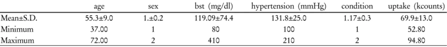
Table 1. Frequency Analysis about Age, Gender, Blood Sugar, Patient Condition, Scenium Mean Uptake Mean Value for 23 Target
Patientsage sex bst (mg/dl) hypertension (mmHg) condition uptake (kcounts) Mean±S.D.
Minimum Maximum
55.3±9.0 37.00 72.00
1.±0.2 1 2
119.09±74.4 80 410
131.8±25.0 100 210
1.17±0.3 1 2
69.9±13.0 52.80 94.80
99mTc-HMPAO 방사성 의약품을 사용하여 화학적 안전성을 확인하였다. 대상환자 23명은 본원 핵의학과 판독의가 판단 하여 99mTc-HMPAO 뇌조직 추출이 떨어져 영상의 질이 현 저하게 떨어지는 경우 및 Scenium Mean Uptake 수치가(평 균: 156±12.6) 100 이하였던 환자를 뇌조직 추출이 떨어졌던 환자로 선정하였다(Fig. 1, Fig. 2). Scenium Mean Uptake 수 치는 뇌혈류 SPECT 관심영역(Regions of Interest, ROIs)의 평균 계수치를 의미한다.4)
2. 검사장비
장비는 Symbia Gamma Camera (Siemens, Germany), 조준 기는 저에너지 고분해능 콜리메이터를 사용하였고 검사 프 로토콜은 뇌혈류 SPECT acquisition을 사용하여 Number of Views 32, Time per View 25 sec 설정하여 검사하였다. 영상 재구성 방법으로는 뇌혈류 Simense Singo SPECT processing 과 Simense Singo 뇌혈류 Scenium procssing을 사용하였다.
3. 검사방법
99mTc-HMPAO 뇌혈류 SPECT 검사를 동시에 시행할 환 자 2명을 대기시킨 후 HMPAO 1 vial에 99mTc 2960-3700 MBq (80~100 mCi)를 안정제와 합성하여 99mTc-HMPAO 방 사성 의약품을 만든 후에 925 MBq (25 mCi)씩 두 개의 주사 기에 담아 각각 준비한다. 첫 번째 환자에게 99mTc-HMPAO 925 MBq (25 mCi)를 정맥주사하고, 5분 후 검사를 진행하였 다. 첫 번째 환자 검사 시작 후 5분 후에 두 번째 환자에게
99mTc-HMPAO 925 MBq (25 mCi)를 정맥주사하고 같은 방 법으로 검사를 진행하였다. 획득된 raw data를 영상 재구성 하여 뇌혈류 영상을 구현하였다. 사용된 99mTc-HMPAO는 사용과 합성 시(용출된 지 30분된 신선한 99mTc을 사용하였 고 합성 시 안정제를 첨가하여 99mTc-HMPAO의 화학적 안 정성을 높임) 특히 주의하였고 환자의 몸무게 및 특성{소아 는 270~410 MBq (7.4~11.1 mCi)}에 따라 투여된 양은 925±185 MBq (25 mCi)로 99mTc-HMPAO 방사성 의약품에 의한 검사 영향을 최소화하였다.
4. 분석방법
대상환자 23명에 연령, 성별, 혈압, 당뇨 수치, 검사 시 사 용 약물, 검사 시 환자상태, 이 전 검사 시 CT/MRI 조영제 부작용 이력, 전과 후 검사 영상에 대한 8가지 사항을 선정하 여 99mTc-HMPAO 뇌혈류 SPECT 검사 시 뇌조직 추출에 영 향을 미치는 인자에 대해 분석하였다.⁵⁾ 검사 시 사용약물에 대한 분석은 뇌혈관질환 및 대뇌경색으로 본원에 입원하여
99mTc-HMPAO 뇌혈류 SPECT 검사를 받는 환자들에게 공 통으로 복용 및 투약 되었던 약물인 Aspirin, Pentaspan, Xanbon, Simvalord, Actigen, Cerezine, CLOPD를 제외하고 검사 당시 50% 이상 공통으로 복용 및 투약 되었던 약물을 분석하였다.⁶⁾ 검사 시 환자상태에 대한 분석은 ①명료(alert)
②기면(drowsy) ③혼미(stuporus) ④반혼수(semi-coma) ⑤혼 수(coma) 5단계로 구분하여 분석하였다. 대상환자 23명 중
99mTc-HMPAO 뇌혈류 SPECT검사를 2번 이상 받았던 환자 12명에 뇌혈류 SPECT 검사영상을 전과 후로 나누어 Scenium Mean Uptake 수치를 이용하여 영상을 분석하였고 Scenium Mean Uptake 평균 수치는 아래의 공식을 이용하였다.
Δm = Scenium Mean Uptake 평균 수치
Δs = Scenium Mean Uptake 각 ROI 계수치의 합 Δt = Scenium Mean Uptake 전체 ROI 수
5. 통계분석
통계학적 분석은 SPSS Ver. 17 (SPSS Inc, USA)을 이용 하였으며 정상환자와 대상환자의 비교분석은 Independent t-test를 사용하였고 뇌조직 추출에 영향을 미치는 인자에 대 한 분석에서는 다중선형회귀분석(Multiple Linear Regression) 을 사용하였다. 99mTc-HMPAO 뇌혈류 SPECT 검사를 2번 시행한 환자의 영상분석에서는 Wilcoxon Matched-Pairs Signed-Ranks Test를 사용하였으며 모든 통계적 처리 기 준은 p<0.05 미만일 때 유의한 차이가 있는 것으로 인정하 였다.
Table 2. As for Gender Results
Frequency Valid Percent Men
Women Total
21 2 23
91.3 9 100
Table 3. Patient Condition Categorized into the 5 Stages
Frequency Valid Percent MenWomen Total
19 4 23
82.6 17.4 100
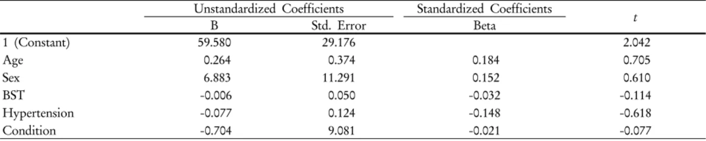
Table 4. Multiple Linear Regression about Factors that Affects on the Brain Extraction
Unstandardized Coefficients Standardized Coefficients
t
B Std. Error Beta
1 (Constant) Age Sex BST Hypertension Condition
59.580 0.264 6.883 -0.006 -0.077 -0.704
29.176 0.374 11.291 0.050 0.124 9.081
0.184 0.152 -0.032 -0.148 -0.021
2.042 0.705 0.610 -0.114 -0.618 -0.077
*a. Dependent Variable: Uptake.
†Scenium Mean Uptake: p>0.05.
Table 5. Non Parametric Test that Based on the Frequency Analysis Result
N Mean ± S.D Minimum Maximum
A B
12 12
68.87 ± 12.67 77.6 ± 11.49
52.80 63.10
92.00 96.15 Test Statistics
B-A Z
Asymp. sig. (2-tailed)
-1.883 0.06
*a. Based on Negative Ranks.
†b. Wilcoxon Matched-Pairs Signed-Ranks Test
‡
p<0.05
결 과
1. 연령, 성별, 당뇨 수치, 혈압, 검사 시 환자상태, Scenium mean Uptake에 대한 Frequency analysis
대상환자 23명의 연령, 성별, 당뇨 수치, 혈압, 검사 시 환 자상태, Scenium Mean Uptake 수치를 Frequency analysis하 여 나타내었다. 단, 검사 시 공통적으로 복용 되었던 약은 없 었으며 이전 검사 시 CT/MRI 조영제 부작용이 나타났던 환 자는 없었다(Table 1). 성별은 남자 21명(91.3%), 여자 2명 (8.7%)이었고, 평균연령은 55.3±9세였다(Table 2). 검사 시 환 자상태는 ①명료 19명(82.6%) ②기면 4명(17.3%)으로 나타
났고 ④반혼수 ⑤혼수상태의 환자는 검사를 진행하지 않았 다(Table 3).
2. 뇌조직 추출에 영향을 미치는 인자에 대한 다중선형 회귀분석
Frequency analysis를 통하여 나온 결과 값을 바탕으로 뇌 조직 추출에 영향을 미치는 인자에 대하여 다중선형회귀분 석을 사용하여 나타내었다(Table 4). 연령과 성별, 당뇨 수치, 혈압, 검사 시 환자상태에 따라 미치는 영향은 유의미하지 않는 것으로 나타났다(p>0.05).
3. 99mTc-HMPAO 뇌혈류 SPECT 검사를 2번 이상 받 았던 환자 12명에 대한 영상분석
환자 12명에 뇌혈류 SPECT 검사 영상을 전과 후로 나누어 Scenium Mean Uptake 수치를 이용하여 Frequency analysis하 였다. Frequency analysis를 통하여 나온 결과 값을 바탕으로 Wilcoxon Matched-Pairs Signed-Ranks Test를 사용하여 검사 의 유의성을 확인하여 나타내었다(Table 5). 2번의 검사에서 모두 Scenium Mean Uptake의 값이 유의미한 차이를 나타내지 않았으며 과거 또는 후에 검사에서 비슷한 99mTc-HMPAO 뇌
⇒
A. 2010/3/1 Transverse image of patient a B. 2010/11/9 Transverse image of patient a
⇒
C. 2010/3/1 Sagittal image of patient a D. 2010/11/9 Sagittal image of patient a
Fig. 3. Images of before (A, C) and after (B, D) of patient a, who received 99mTc-HMPAO cerebral blood flow
SPECT scan more than 2 times shows minor differences, but shows similar cerebral blood flow SPECT image.혈류 영상이 나타났다(Fig. 3, Fig. 4, Fig. 5, Fig. 6).
결 론
대상환자 23명의 8가지 사항을 뇌조직 추출에 영향을 미 치는 인자에 대하여 다중선형회귀분석을 사용하여 분석한 결과 연령, 혈압, 당뇨 수치, 검사 시 사용 약물, 검사 시 환자 상태, 과거 검사 시 CT/MRI 조영제 부작용 환자에서는 공통 점 및 특이한 상관관계를 발견할 수 없었다. 그러나 성별에 서는 남성이 91.3%, 여성이 8.7%이며 상대적으로 남성에게 서 뇌조직 추출이 떨어지는 경향이 나타났고 대상환자 가운 데 99mTc-HMPAO 뇌혈류 SPECT를 2번 이상 받았던 환자 12명의 Scenium Mean Uptake 수치를 이용한 Wilcoxon Matched-Pairs Signed-Ranks Test에서도 모두 유의미한 차이 를 나타내지 않았다(p>0.05). 결과적으로 12명 환자에게서는 전과 후 Scenium Mean Uptake 수치에 차이가 없는 비슷한 뇌조직 추출을 보이는 것을 확인할 수 있었다.
현재까지 99mTc-HMPAO 뇌조직 추출이 떨어졌던 영상에 주된 이유는 99mTc-HMPAO의 합성과 사용에 부주의로 인하
여 화학적으로 불안정한 99mTc-HMPAO 방사성 의약품을 사 용하는 것이라고 생각하였다. 그러나 99mTc-HMPAO의 합성 과 사용이 정상적으로 이루어지고 99mTc-HMPAO가 화학적 으로 안정해도 환자의 개인적인 특성 및 체질에 따라 99mTc- HMPAO 뇌조직 추출이 떨어지는 것을 이번 연구로 알 수 있었다.
앞선 99mTc-HMPAO 뇌혈류 SPECT 관련 논문을 참고할 때 뇌혈관 질환 중 측부 순환로에 의한 요인이 추측되지만, 환자에 따라 뇌조직 추출이 떨어지는 정확한 이유는 알 수 없었다. 또한, 체내 역학적 측면에서 99mTc-HMPAO를 이용 한 뇌혈류 값의 절대적 정량화가 쉽지 않으며 정상으로 판독 된 뇌혈류 SPECT 중에서도 방사성 의약품을 어느 것을 사 용하느냐에 따라 분명한 혈류 분포의 차이를 나타내며
99mTc-HMPAO 방사성 의약품 투여 전에 화학적 순도측정을 객관적으로 검증하지 못하였기 때문에 이번 연구 결과가 가지 는 한계점이 있을 거라 생각한다.7,8) 하지만 99mTc-HMPAO SPECT 검사가 대뇌경색에 의한 뇌혈류 영상 및 허혈 부위 의 범위를 평가, 문합수술의 계획에 대한 경과, 기질성 정신 장애 환자 경과를 추적하는데 유용한 검사이며 추적 검사 시
⇒
⇒
A. 2010/3/1 Scenium Mean Uptake of patient a B. 2010/ 11/9 Scenium Mean Uptake of patient a
Fig. 4. Before (A) and after (B) values of Scenium Mean Uptake of patient a, who received
99mTc-HMPAO cerebral blood flow SPECT scan more than 2 times shows values of Scenium Mean Uptake of 100 which is relatively low.뇌조직 추출이 떨어지는 환자에 대한 정보를 검사에 반영 하여 정확한 검사와 판독에 도움을 줄 것이라 생각한다.9) 앞으로 위 결과를 토대로 뇌조직 추출이 떨어지는 환자를 추적 관찰하여 환자에 따라 뇌조직 추출이 떨어지는 정확 한 이유를 알 수 있는 추가적인 연구가 좀 더 필요할 것으로 사료된다.
요 약
[목적] 본 연구는 화학적으로 안정된 99mTc-HMPAO (d, l-hexamethylpropylene amine oxime) 방사성 의약품을 사용 한 환자 가운데 뇌조직 추출률이 떨어져 영상의 질이 저하되 었던 환자를 대상으로 99mTc-HMPAO 방사성 의약품 외의
⇒
A. 2010/6/1 Transverse image of patient b B. 2011/1/27 Transverse image of patient b
⇒
C. 2010/6/1 Sagittal image of patient b D. 2011/1/27 Sagittal image of patient b
Fig. 5. Brain tissue extraction Images of before (A, C) and after (B, D) of patient b, who received
99mTc-HMPAO cerebral blood flow SPECT scan more than 2 times shows minor differences, but shows similar cerebral blood flow SPECT image.요인으로 뇌조직 추출률에 변화가 일어나는 요인을 알아보 고자 한다. [대상 및 방법] 2010년 1월부터 2010년 12월까지 대뇌경색(Cerebral infarction, CI)에 의한 99mTc-HMPAO 뇌 혈류 SPECT 검사를 시행한 환자 272명 가운데 뇌조직 추출 률이 떨어졌던 환자 23명(연령 55.3±9세, 남 21명, 여 3명)을 대상으로 하였다. 대상환자는 정상 뇌조직 추출률을 보인 환 자와 검사를 동시에 진행하여 같은 99mTc-HMPAO 방사성 의약품을 사용함으로써 화학적 안전성을 확인하였다. 대상 환자의 연령, 성별, 혈압, 당뇨 수치, 검사 시 사용 약물, 검사 시 환자상태, 과거 검사 시 CT/MRI 조영제 부작용 이력, 과 거 SPECT 검사를 시행한 환자의 전과 후 검사 영상을 비교 하여 뇌조직 추출률에 영향을 미치는 요인과의 상관관계를 분 석하였다. [결과] 다중선형회귀분석(Multiple Linear Regression) 의 결과, 성별 및 전, 후 검사 영상을 제외한 6가지 사항에서 는 특이한 상관관계를 발견할 수 없었다(p>0.05). 남성 91.3%, 여성 8.7%로 남성이 여성보다 뇌조직 추출률이 떨어 졌다고 보인다. 비모수 검증을 이용한 전과 후 검사 영상에 서는 유의미한 차이를 나타내지 않았다(p>0.05). 결과적으로
전과 후 검사 영상에서 비슷한 뇌조직 추출률을 나타냈으며 환자 개인에 따라 뇌조직 추출률에 변화가 있음을 확인하였 다. [결론] 화학적으로 안정된 99mTc-HMPAO 방사성 의약품 의 사용도 환자의 개인적인 특성 및 체질에 따라서 뇌조직 추출률이 떨어지는 유의한 요인임을 확인하였다. 앞선
99mTc-HMPAO 뇌혈류 SPECT 관련 논문을 참고할 때 뇌혈 관 질환 중 측부 순환로에 의한 요인이 추측되지만, 환자에 따라 뇌조직 추출률이 떨어지는 정확한 요인은 알 수 없었다. 그러나 99mTc-HMPAO SPECT 검사가 뇌질환에 따른 경과 추적에 유용한 검사이며 추적 검사 시 뇌조직 추출률이 떨어 지는 환자에 대한 정보를 검사에 반영하여 정확한 검사와 판 독에 도움을 줄 것으로 생각한다.
REFERENCES
1. 고창순 외. 제3판 핵의학. 고려의학; 2008. p. 395-465.
2. James H.Thrall. MD Nuclear Medicine THE REQUISITES IN RADIOLOGY Third Edition. 3rd ed. MOSBY; 2006-01-02.
p.419-449.
⇒
⇒
A. 2010/6/1 Scenium Mean Uptake of patient b B. 2011/1/27 Scenium Mean Uptake of patient b
Fig. 6. Before (A) and after (B) values of Scenium Mean Uptake of patient b, who received
99mTc-HMPAO cerebral blood flow SPECT scan more than 2 times shows values of Scenium Mean Uptake of 100 which is relatively low.3. Han HY. 99mTc-HMPAO regional cerebral blood flow spect in cerebral infarcts and ischemia. Korean journal of re- habilitation medicine 1992;19(3):265-271.
4. Siemens Medical Solutions USA, Inc. Operating Instructions Scenium Application with e.soft 2005. p.145-156.
5. Song HC. Change of Cerebral Blood Flow Distribution and Vascular Reserve according to Age in Koreans Measured by
Tc-99m HMPAO Brain SPECT. Nuclear Molecular Imaging 1999;33(3):247-261.
6. Springhouse Corporation. Clinical pharmacology made incred- ibly easy. 2nd ed. 군자출판; 2011. p.21-46: 47-86.
7. Park YH, Jung SG, Lee SY, Shin GS, Kim JW, Park YH.
Tc-99m-HMPAO Regional Cerebral Blood Flow SPECT in Cerebral Rete Mirabile. Nuclear Molecular Imaging 1988;22(2):
157-161.
8. Kim YN, Jung YA, Son HS, Kim SH ,Yoo IR, Jung SG.
Different Uptake of Tc-99m ECD and Tc-99m HMPAO in the Normal Brains: Analysis by Statistical Parametric Mapping.
Nuclear Molecular Imaging 2002;36(4):244-254.
9. Lee KW, Lee JJ, Song MH, Kang MH, Jee IS, Shin YT, No HG.
Tc-99m HMPAO Brain SPECT in Patients with Post-Traumatic Organic Mental Disorder. Nuclear Molecular Imaging 1994; 28(3):293-300.