∙ Received: March 8, 2012. Accepted: April 6, 2012.
∙ Corresponding author: Soon Sang Yoon
Department of Nuclear Medicine, Asan Medical Center, 388-1 Pungnap-2 dong, Songpa-gu, Seoul 138-736, Korea
Tel: +82-2-3010-4603, Fax: +82-2-3010-5429 E-mail: [email protected]
Original Article
게이트 심장 혈액풀 스캔에서 좌전사위상 각도의 변화에 따른 정량적 지표 비교서울아산병원 핵의학과
윤순상·남기표·류재광·김성환
The Comparison of Quantitative Indices by Changing an Angle of LAO View in Multi-Gated Cardiac Blood Pool Scan
Soon Sang Yoon, Ki Pyo Nam, Jae Kwang Ryu and Seong Hwan Kim Department of Nuclear Medicine, Asan Medical Center, Seoul, Korea
Purpose: The multi-gated cardiac blood pool scan is to evaluate the function of left ventricle (LV) and usefully observe a value of ejection fraction (EF) for a patient who is receiving chemotherapy. To calculate LVEF, we should adjust an angle of left anterior oblique (LAO) view to separate both ventricles. And by overlapped ventricles, it is possible to affect LVEF. The purpose of this study is to investigate and compare quantitative indices by changing an angle of LAO view. Materials and methods: We analyzed the 49 patients who were examined by multi-gated cardiac blood pool scan in department of nuclear medicine at Asan Medical Center from June to September 2011. Firstly, we acquired “Best septal” view. And then, we got images by addition and subtraction of angle for LAO view to anterior and lateral. We compared three LAO views for 20 people by 5 degrees and 39 people by 10 degrees. And we analyzed quantitative indices, EF, end diastole and end systole counts, by automated and manual region of interest (ROI) modes. Results: Firstly, we analyzed quantitative indices by automated ROI mode. In case of 5 degrees, the averages of EF are 61.0±7.5, 62.1±7.1, 60.9±6.7%
(p=0.841) in LAO, LAO -5o and LAO +5o respectively. And there is no difference in end diastole and end systole counts (p<0.05). In case of 10 degrees, the averages of EF are 62.4±9.5, 62.3±10.8, 61.6±.9.3% (p=0.938) in LAO, LAO -10o and LAO +10o respectively. And there is no difference in end diastole and end systole counts (p<0.05). Secondly, we analyzed quantitative indices by manual ROI mode. In case of 5 degrees, the averages of EF are 62.8±7.1, 63.6±7.5, 62.7±7.3% (p=0.903) in LAO, LAO -5o and LAO +5˚ respectively. And there is no difference in end diastole and end systole counts (p<0.05). In case of 10 degrees, the averages of EF are 65.5±9.0, 66.3±8.7, 63.5±.9.3% (p=0.473) in LAO, LAO -10o and LAO +10o respectively. And there is no difference in end diastole and end systole counts (p<0.05). Conclusion: When an image is nearly “Best septal” view, the difference of LAO angle would not affect to change LVEF. Although there was no difference in quantitative analysis, deviations could happen when to interpret wall motion qualitatively by reading physicians. (Korean J Nucl Med Technol 2012;16(1):57-61)
Key Words : Multi-gated cardiac blood pool scan, left anterior oblique (LAO), ejection fraction (EF)
PURPOSE
The multi-gated cardiac blood pool (MUGA) scan is a method to create images for change of blood pool using elec- trocardiogram as indices for contraction and relaxation dur- ing cardiac cycle.1) And, the MUGA scan is a procedure in which the patient’s red blood cells (RBCs) are radiolabeled and electrocardiograph (ECG)-gated cardiac scintigraphy is
Fig. 1. Correct positioning to image left ventricle. (A) LAO images that are optimal, too anterior, and too lateral in obliquity. (B) In an optimized LAO view the axis of the left ventricle should be vertical. In images that are too anterior, there is a rightward tilting of the axis from base to apex. In images that are too laterally positioned, there is a leftward tilting of the axis from base to apex.
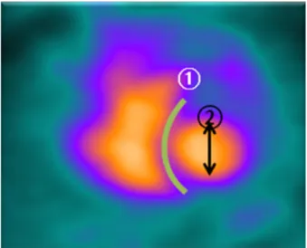
Fig. 2. Visualize the septum optimally ① Separate both ventricles,
② erect long axis vertically.
Fig. 5. EF processing by both ROI modes.
obtained. Data are collected from several hundred cardiac cy- cles to generate an image set of the beating heart that is pre- sented as a single, composite cardiac cycle.
The method can be used to assess regional and global wall motion, cardiac chamber size and morphology and ven- tricular systolic and diastolic function, including left and right ventricular ejection fractions (EF).2) Mostly, we ob- served left ventricle ejection fraction (LVEF) in left anterior oblique (LAO) view. Currently, there are a lot of examina- tions for a patient who is receiving chemotheraphy to ob-
serve LVEF because MUGA scan is non-invasive and reproducible. One of the most significant step is a determi- nation of patient position in MUGA scan. A wrong decision of position could bring errors of an evaluation for LV func- tion and an abnormality in wall motion.3) To calcultate LVEF, we are able to analyze LAO view. The LAO view is opimized to visualize the septum (“Best septal view” – usu- ally the 45° LAO, but the angle will depend on body habitus and cardiac orientation.) In the LAO view, the orientation should be such that the long axis of the ventricle is approx- imately vertical on the right side of the image (Fig. 1, 2).4)
Therefore, it is very important to adjust an angle to nearly
“Best septal view”. If both ventricles were not separate, we could hardly evaluate left ventricle’s own function. So by overlapped left ventricle, it is possible to affect EF. When a follow-up study, we usually followed an angle of previous study. However, it happen that previous study wasn’t “Best septal view”. In that case, we generally change an angle.
The purpose of this study is to investigate quantitative in- dices by changing an angle of LAO view.
MATERIALS AND METHODS
1. Patient population
We analyzed the 49 patients (8 men, 41 women) who were examined by MUGA scan in department of nuclear medicine at Asan Medical Center from June to September 2011. The study was established to patients who are receiving chemo- therapy with cardiotoxicity. The age range was 24-74 years
LAO LAO ‐5° LAO +5°
Fig. 3. Images of LAO view by 5 degree.
LAO LAO ‐10° LAO +10°
Fig. 4. Images of LAO view by 10 degree.
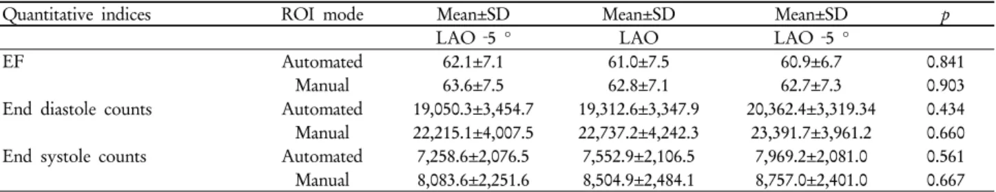
Table 1. Differences of indices in 5 degrees (one‐way Anova test) EF (%); End diastole counts, End systole counts (counts)
Quantitative indices ROI mode Mean±SD Mean±SD Mean±SD p
LAO ‐5 ° LAO LAO ‐5 °
EF Automated 62.1±7.1 61.0±7.5 60.9±6.7 0.841
Manual 63.6±7.5 62.8±7.1 62.7±7.3 0.903
End diastole counts Automated 19,050.3±3,454.7 19,312.6±3,347.9 20,362.4±3,319.34 0.434 Manual 22,215.1±4,007.5 22,737.2±4,242.3 23,391.7±3,961.2 0.660 End systole counts Automated 7,258.6±2,076.5 7,552.9±2,106.5 7,969.2±2,081.0 0.561 Manual 8,083.6±2,251.6 8,504.9±2,484.1 8,757.0±2,401.0 0.667
(mean=48.31±10.48).
2. Methods
All patients were examined three times. Firstly, we ac- quired “Best septal view”. And then, we got images by both adding and subtracting of fixed angle for LAO view to ante- rior and lateral (Fig. 3, 4). We compared three LAO views for
20 people by 5 degrees and 29 people by 10 degrees. And we analyzed quantitative indices, EF, end diastole and end sys- tole counts, by both automated and manual region of interest (ROI) modes (Fig.5).
For this study, a General Electric Infinia 90° angled du- al-headed scintillation camera equipped with gen- eral-purpose collimators was used for all images. A MUGA scan was performed with 24 frames and an irregular beat ac-
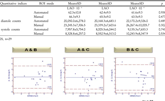
Table 2. Differences of indices in 10 degrees (one‐way Anova test) EF (%); End diastole counts, End systole counts (counts)
Quantitative indices ROI mode Mean±SD Mean±SD Mean±SD p
LAO ‐10 ° LAO LAO ‐10 °
EF Automated 62.3±10.8 62.4±9.5 61.6±9.3 0.938
Manual 66.3±9.3 65.5±9.0 63.5±9.3 0.473
End diastole counts Automated 20,050.0±6,278.0 20,168.3±6,683.1 22,170.2±9,326.0 0.489 Manual 23,245.3±7,306.5 23,339.2±7,603.6 26,267.4±10,005.7 0.302 End systole counts Automated 7,957.8±5,754.0 8,005.5±6,044.0 9,135.3±7,653.3 0.741 Manual 8,328.8±6,257.0 8,552.9±6,513.0 10,243.9±8,347.9 0.534 p>0.05, n=29
Fig. 6. The Bland-Altman graphs of agreement among three technologists.
ceptance window at 20% of the average R-R interval obtained. Data were acquired in 64×64 format with a zoom of 2.5 (pixel size, 3.5 mm) until a total counts density of 9 mil- lion counts were reached. And we used in vivo labeling meth- od and injected 925 MBq of technetium 99m–labeled red blood cell. For quantitative analysis, at first we acquired three LAO views. And then using Xeleris software from GE, we computed LVEF, end diastole and end systole counts by both ROI modes. Lastly, we analyzed quantitative indices by one-way Anova analysis.
RESULTS
Firstly, we analyzed quantitative indices by automated ROI mode. In case of 5 degrees, the averages of EF are 61.0±7.5, 62.1±7.1, 60.9±6.7% (p=0.841) in LAO, LAO -5˚
and LAO +5˚ respectively. And there is no difference in end diastole and end systole counts (p<0.05). In case of 10 de- grees, the averages of EF are 62.4±9.5, 62.3±10.8, 61.6±.9.3%
(p=0.938) in LAO, LAO -10˚ and LAO +10˚ respectively.
And there is no difference in end diastole and end systole
countg (p<0.05). Secondly, we analyzed quantitative indices by manual ROI mode. In case of 5 degrees, the averages of EF are 62.8±7.1, 63.6±7.5, 62.7±7.3% (p=0.903) in LAO, LAO -5˚ and LAO +5˚ respectively. And there is no difference in end diastole and end systole counts (p<0.05, Table 1). In case of 10 degrees, the averages of EF are 65.5±9.0, 66.3±8.7, 63.5±.9.3% (p=0.473) in LAO, LAO -10˚ and LAO +10˚
respectively. And there is no difference in end diastole and end systole counts (p<0.05, Table 2).
To investigate personal deviations, we compared agree- ment with 20 people. As you can see these graphs, almost all points are in the 95% confidence interval range (Fig. 6).
DISCUSSION
This study investigated the use of MUGA scan imaging for the assessment of left ventricular function in a group of pa- tients with heart failure. A MUGA scan imaging is the gold standard for the assessment of LVEF, largely because of the excellent repeatability.5) When follow up study, we usually followed an angle of previous study. However, it happen that
previous study wasn’t “Best septal view”. In that case, we generally change an angle by approximately five to ten degrees. That’s why we decided specific angles, 5 and 10 degrees.
This study has some limitations. In general, we use a func- tion of tilt for separating ventricle and atrium of LV.
However, GE’s gamma camera hasn’t a function of tilt, we didn’t use a function of caudal tilt. And deviations could hap- pen when to interpret wall-motion qualitatively by reading physicians. Because we simply compared quantitative indices and we had no consideration wall-motion of left ventricle.
Even if there is no statistically deference in quantitative in- dices, difference in regional wall-motion would occur. So, we usually acquired three views, LAO, ANT and LAT view for to analyze every regional ventricle.
CONCLUSION
When an image is nearly “Best septal view”, the difference of LAO angle more and less 5 to 10 degrees would not affect to change LVEF. If previous image wasn’t “Best septal view”, we have to change an angle. Despite changing an angle of 5 to10 degrees, there is no significant difference from quantita- tive indices. So, we have to examine patient with “Best septal view” for more reliable and accurate examination.
요 약
게이트 심장 혈액풀 스캔(Gated cardiac blood pool scan, MUGA)은 좌심실의 기능을 평가하는 데 유용한 검사로 심근독성 항암제를 투여 받는 환자에서 심박출 계수 (Ejection Fraction, EF)를 추적, 감시하는데 이용되어 많 은 검사가 시행되고 있다. EF를 산출하기 위해 좌전사위 상(Left Anterior Oblique, LAO)에서 좌우 심실이 분리되 도록 각도를 조절해야 하는데 이때 LAO 각도의 설정은 정량분석 값에 영향을 미칠 가능성이 있어, 이에 본 연구 는 LAO 각도의 변화가 정량분석 값에 영향을 미치는지 살펴보고자 한다. 2011년 06월부터 09월까지 본원에서
MUGA 검사를 시행한 환자 49명(남 8명, 여 41명)을 대 상으로 하였다. 먼저 최적 중격상(Best septal view)으로 촬영 후, 전면과 측면 측으로 LAO 각도를 가감하여 총 3회 촬영하였다. ±5°로 환자 20명, ±10°로 29명을 촬영하 였고, 관심영역을 자동 및 수동으로 각각 설정하여 좌심
실의 EF와 확장기말계수, 수축기말계수를 비교하였다.
먼저 관심영역을 자동으로 분석하였을 때, ±5°의 경우, LAO, LAO -5°, LAO +5°에서의 EF의 평균값은 각각 61.0±7.5%, 62.1±7.1%, 60.9±6.7% (p=0.841)이었고, 이때 의 확장기말계수, 수축기말계수 역시 통계적으로 유의한 차이가 없었다(p<0.05). ±10°의 경우, EF의 평균값은 각각 62.4±9.54%, 62.3±10.8%, 61.6±9.3% (p=0.938)이었고, 이 때의 확장기말계수, 수축기말계수 역시 통계적으로 유의 한 차이가 없었다(p<0.05). 또한 관심영역을 수동으로 분 석하였을 때, ±5°의 경우, EF의 평균값은 각각 62.8±7.1%, 63.6±7.5%, 62.7±7.3% (p=0.903)이었고, 이때의 확장기말 계수, 수축기말계수 역시 통계적으로 유의한 차이가 없었 다(p<0.05). ±10°의 경우, EF의 평균값은 각각 65.5±9.0%, 66.3±8.7%, 63.5±9.3% (p=0.473)이었고, 이때의 확장기말 계수, 수축기말계수 역시 통계적으로 유의한 차이가 없었 다(p<0.05). 최적 중격상을 기준으로 하였을 경우, ±5~10°
의 영상의 차이는 EF에 유의한 변화를 주지 않을 것으로 판단된다. 다만 최적 중격상에서 벗어난 영상은 좌심실의 벽 운동 등과 같은 심장의 운동상태를 정성적으로 평가함 에 있어서 판독자에 따라 편차가 발생할 수 있으므로 주 의해야 할 것이다.
REFERENCES
1. 이명철, 정준기. 심장핵의학. 고려의학 2002:33-42
2. Scheiner et al. Society of Nuclear Medicine Procedure Guideline for Gated Equilibrium Radionuclide Ventriculography. J Nucl Med 2012:1-7.
3. 고창순. 핵의학. 고려의학 1992:377-393.
4. Corbett et al. Equilibrium radionuclide angiocardiography.
Journal of Nuclear Cardiology 2008:1-25.
5. Wright et al. Left Ventricular Ejection Fraction and Volumes from Gated Blood-Pool SPECT: Comparison with Planar Gated Blood-Pool Imaging and Assessment of Repeatability in Patients with Heart Failure. J Nucl Med 2003;44:494-498.